Sports Biomechanics and Injury Prevention: From 'Twisted Ankles' to Rotator Cuff Management
From 'Twisted Ankles' to Rotator Cuff Management
Sports Medicine · Seminar week 44 · released October 26, 2026 · includes a discussion video
This seminar will delve into the biomechanics involved in common sports injuries like ankle sprains and rotator cuff tears. It leverages evidence from recent studies and…
Learning Objectives
- Analyze how external forces, joint position, neuromuscular control, and tissue capacity interact to produce acute and overuse injuries.
- Differentiate lateral ankle sprains from syndesmotic, deltoid, osteochondral, and fracture-related injuries using mechanism, examination, and selective imaging.
- Interpret rotator cuff imaging in the context of symptoms, functional limitations, tear characteristics, and patient goals.
- Integrate biological, biomechanical, psychological, environmental, and sociocultural factors into individualized injury-risk assessment.
- Select radiography, ultrasonography, CT, MRI, or MR arthrography according to a focused clinical question.
- Prescribe evidence-based prevention and rehabilitation programs with appropriate exercise dosage, progression, and return-to-sport criteria.
- Apply shared decision-making to complex sports-injury cases while avoiding overdiagnosis, premature return, and treatment driven solely by imaging.
Biomechanics of Sports Injuries: An Overview
%%FIG0%% Sports injuries occur when a tissue’s accumulated mechanical demand exceeds its capacity to tolerate and recover from that demand. The relevant “dose” includes not only peak force but also loading rate, repetition, direction, joint position, fatigue, recovery time, and the athlete’s recent training history. A single high-energy event may rupture a ligament whose instantaneous load exceeds its failure threshold; alternatively, thousands of individually tolerable repetitions may produce tendinopathy or bone stress injury when remodeling cannot keep pace.
External forces create internal joint moments. Ground-reaction force passing lateral to a planted ankle, for example, produces an inversion moment that must be resisted by ligament tension and rapidly activated fibularis musculature. At the shoulder, force generated by the legs and trunk normally transfers through the scapula and glenohumeral joint to the arm. Deficits anywhere in this kinetic chain can increase the workload demanded of the rotator cuff, although identifying a deficit does not prove that it caused an individual athlete’s pain.
Teaching Point: Injury is rarely explained by one “bad movement.” A useful model links exposure, movement strategy, tissue load, tissue capacity, and recovery. Neuromuscular control and proprioception influence how quickly and accurately an athlete modifies joint position after an unexpected perturbation; balance-based interventions can reduce recurrent ankle sprains (PMID: 25534579).
Joint stability has static and dynamic components. Static restraints include bone geometry, capsule, labrum, and ligaments. Dynamic stability depends on coordinated muscle activation, sensorimotor feedback, anticipatory control, and the athlete’s ability to absorb or redirect force. After an ankle sprain, altered afferent input, delayed fibularis activation, strength loss, and fear may persist even after swelling resolves. After shoulder pain, reduced cuff force or altered scapular motion may be a protective response rather than the original lesion.
Framework: Evaluate injury through five linked domains:
- Event: What force, direction, speed, contact, and joint position were involved?
- Tissue: Which structures were loaded, and under tension, compression, shear, or torsion?
- Athlete: What are the athlete’s strength, mobility, prior injuries, health, sleep, nutrition, and psychological state?
- Exposure: Did competition density, surface, footwear, technique, or recent workload change?
- Response: Are symptoms improving appropriately, or is there evidence of instability, fracture, neurovascular injury, or impaired healing?
This framework distinguishes risk factors from deterministic causes. Limited ankle dorsiflexion may alter landing mechanics, but its meaning differs between a symptomatic basketball player and an asymptomatic distance runner. Similarly, scapular dyskinesis is an observation—not a diagnosis—and occurs in athletes with and without shoulder symptoms.
Nuance: Screening tests have value when they identify a modifiable impairment that changes management. Their value is much lower when a single cutoff is used to label an otherwise healthy athlete “high risk.” Movement quality, strength ratios, limb symmetry, and wearable-derived workloads all have measurement error and should be interpreted alongside sport demands and longitudinal change.
Clinical reasoning begins by separating expected injury physiology from time-sensitive pathology. Pain, swelling, and guarded movement are common after trauma; disproportionate pain, deformity, progressive neurologic deficit, an absent pulse, an acutely locked joint, or inability to protect the injured area demands escalation.
MUST ACT: Before conducting elaborate biomechanical testing, exclude fracture-dislocation, compartment syndrome, neurovascular compromise, cervical cord or nerve-root disease, infection, and other conditions in which delay changes outcome.
Decision Point: Ask whether the primary problem is tissue damage, mechanical instability, load intolerance, altered motor control, or a combination. That answer—not the diagnostic label alone—should determine protection, exercise selection, imaging, and return criteria.
Audience Poll: Which variable most often changes your initial plan: injury mechanism, objective instability, recent workload, or the athlete’s functional goals?
Detailed Analysis of Ankle Sprains: Syndesmotic Injuries and Management
%%FIG1%% Most ankle sprains are lateral, usually involving the anterior talofibular ligament after plantar-flexion and inversion. Syndesmotic injuries are less common but typically cause longer disability and greater risk if occult instability is missed. The distal tibiofibular syndesmosis includes the anterior-inferior and posterior-inferior tibiofibular ligaments, interosseous ligament and membrane, and transverse ligament. These structures maintain the ankle mortise while permitting small physiologic motion.
A syndesmotic injury usually follows external rotation of the foot relative to the tibia, often with dorsiflexion, eversion, or a planted cleat. Pain may extend above the ankle rather than concentrating below the lateral malleolus. An associated deltoid injury, posterior malleolar fracture, proximal fibular fracture, or complete interosseous disruption can convert an apparently modest sprain into an unstable injury.
MUST ACT: Palpate the entire fibula, medial ankle, posterior malleolus, base of the fifth metatarsal, navicular, Achilles tendon, and proximal tibiofibular region. Proximal fibular pain with medial ankle or syndesmotic findings should trigger evaluation for a Maisonneuve injury.
The Ottawa Ankle Rules guide fracture radiography, not ligament grading. Obtain ankle radiographs when malleolar-zone pain accompanies posterior-edge or tip tenderness of either malleolus, or when the patient cannot take four steps both immediately after injury and during evaluation. Apply the corresponding navicular and fifth-metatarsal criteria for midfoot pain. A systematic review found that a negative rule has very high sensitivity for excluding clinically important fracture (PMID: 12595378). Reduced sensation, intoxication, distracting injury, unreliable examination, delayed presentation, or an atypical population lowers confidence in the rule.
Examine swelling and ecchymosis, active motion, neurovascular status, and ability to bear weight before provocative tests. Syndesmotic palpation and pain out of proportion to a routine lateral sprain raise suspicion. The dorsiflexion-external rotation stress test and squeeze test are relatively useful when positive, but neither is sufficiently sensitive to exclude injury when negative. Compare sides and interpret tests as a cluster. Acute pain may make anterior drawer and talar tilt unreliable; reassessment after several days can better characterize lateral instability.
Decision Point: The crucial distinction is not simply “high ankle sprain” versus “low ankle sprain,” but stable versus unstable syndesmotic injury. Widening of the medial clear space, tibiofibular diastasis, associated fracture, or instability under stress warrants urgent orthopedic or sports-surgery assessment.
Initial ankle radiographs should include anteroposterior, mortise, and lateral views. Weight-bearing or stress imaging may reveal instability when tolerable, but normal plain films do not exclude syndesmotic injury. MRI is helpful when examination remains suspicious, recovery is unexpectedly slow, or delineation of ligament, cartilage, tendon, or occult bone injury will change treatment. CT better defines subtle fractures and syndesmotic alignment, especially after fixation.
For uncomplicated lateral sprain, use compression, elevation, protected weight bearing, and a semirigid or lace-up brace. A grade III injury may benefit from brief immobilization—generally no more than about 7–10 days—followed by functional rehabilitation. Prolonged casting delays motion and neuromuscular recovery. Analgesia should be individualized; acetaminophen or a short NSAID course may reduce pain, but NSAIDs do not replace protection and rehabilitation.
Stable syndesmotic injuries often require a boot or brace initially, with weight bearing advanced according to gait and pain. Early rehabilitation avoids aggressive external rotation and forced dorsiflexion, then restores motion, calf strength, balance, and multidirectional control. Recovery commonly takes six weeks or longer. Unstable injuries may require screw or suture-button fixation; routine screw removal is not universally necessary and should be decided from symptoms, construct, and surgeon preference.
Teaching Point: Return to sport should assess the PAASS domains: pain, ankle impairments, athlete perception, sensorimotor control, and sport/functional performance. A pain-free jog alone is inadequate.
A home proprioceptive program reduced recurrent sprains after usual care in a randomized trial, with an approximate number needed to treat of nine (PMID: 19589822). Practical dosing is 10–15 minutes, three or more times weekly for at least eight weeks, progressing from stable double-leg tasks to single-leg reach, perturbation, landing, and sport-specific reaction. Bracing during high-risk sport provides additional protection for athletes with previous sprain.
Nuance: Persistent pain may reflect osteochondral injury, peroneal tendon pathology, occult fracture, impingement, or chronic instability—not merely “slow healing.”
Audience Poll: After normal radiographs, which finding most strongly prompts advanced imaging: syndesmotic tenderness, inability to hop, persistent effusion, or delayed functional progress?
Rotator Cuff Abnormalities: Clinical Significance and Management Strategies
%%FIG2%% The rotator cuff centers the humeral head while producing rotation and contributing to elevation. Supraspinatus, infraspinatus, teres minor, and subscapularis function with the deltoid, scapular stabilizers, trunk, and lower limbs. Cuff-related shoulder pain may reflect tendon load intolerance, partial or full-thickness tearing, bursal irritation, or a combination. Structural findings and symptoms overlap imperfectly.
Asymptomatic abnormalities become increasingly common with age. Population ultrasound studies show a marked age-related rise in full-thickness tears, many without pain (PMID: 19540777). Tendinosis, bursal fluid, acromioclavicular degeneration, and partial tearing are even more prevalent. The claim that an MRI “shows the pain generator” is therefore unsafe without clinical correlation.
Nuance: Imaging describes anatomy; it does not independently establish why the patient hurts, how disabled they are, or whether surgery will help. Avoid telling a patient that a tendon is “shredded” when the report describes common age-related change.
History should establish traumatic versus insidious onset, dominant arm, occupational and sport demands, night pain, weakness, instability, stiffness, neurologic symptoms, prior treatment, and goals. A sudden eccentric load followed by immediate weakness in a previously functional shoulder raises concern for an acute traumatic tear. Gradual lateral-arm pain aggravated by elevation or loading more often supports rotator cuff-related shoulder pain.
Examination includes cervical screening, active and passive range of motion, strength testing, scapular observation, and a focused neurologic assessment. Preserved passive motion with painful or weak active elevation favors cuff dysfunction; global restriction of active and passive motion suggests adhesive capsulitis or glenohumeral arthritis. True external-rotation lag, drop-arm findings, marked subscapularis weakness, or pseudoparalysis increases concern for a substantial tear. No single impingement maneuver reliably identifies a specific tendon lesion.
Framework: Before attributing symptoms to the cuff, consider cervical radiculopathy, suprascapular neuropathy, glenohumeral arthritis, adhesive capsulitis, instability, labral pathology, acromioclavicular pain, calcific tendinopathy, infection, inflammatory disease, and referred cardiopulmonary or visceral pain.
Most atraumatic cuff-related presentations begin with education and progressive exercise. Reduce provocative volume temporarily without prescribing complete rest. A practical program combines pain-limited range of motion, isometric or isotonic external rotation, scaption, rows, serratus work, and kinetic-chain conditioning. Begin with two to four sets of 6–15 repetitions, two or three days weekly, and progress resistance when symptoms return to baseline within approximately 24 hours. Some discomfort during exercise is acceptable when mild, predictable, and non-escalating.
The GRASP factorial randomized trial found no clinically important long-term advantage of a more intensive progressive-exercise program over a high-quality best-practice advice session for many adults with rotator cuff disorders. A corticosteroid injection produced only modest short-term improvement and no sustained 12-month benefit (PMID: 34265255). This supports accessible, well-taught exercise and realistic expectations rather than reflexive escalation.
Decision Point: Consider an injection when pain prevents sleep or meaningful rehabilitation, after discussing transient benefit, post-injection load management, hyperglycemia, skin changes, and the potential adverse effect of repeated injections on tendon health. Avoid repeated injections as a substitute for rehabilitation.
Surgical referral is more compelling for an acute traumatic full-thickness tear with substantial weakness, a reparable tear in an active patient whose function remains unacceptable after structured nonoperative care, or progressive functional loss. Tear size, retraction, fatty infiltration, tissue quality, age, smoking, diabetes, expectations, and rehabilitation capacity influence healing. Degenerative tears can improve without structural healing, while repaired tendons can retear without equivalent symptom recurrence.
Subacromial decompression should not be offered simply because an acromion is described as “hooked.” In the placebo-controlled CSAW trial, decompression did not provide a clinically important advantage over arthroscopy without decompression (PMID: 29169668). Repair is a different question from decompression, particularly for symptomatic full-thickness tears.
MUST ACT: Expedite evaluation for acute traumatic weakness, unreduced dislocation, fracture, infection, tumor, progressive neurologic deficit, or severe atraumatic pain accompanied by systemic or cardiopulmonary features.
Teaching Point: Treat the patient’s pain, function, and goals—not the scan in isolation.
Audience Poll: Which factor most influences your referral decision: traumatic onset, tear size, objective weakness, failed rehabilitation, or patient goals?
Integrating Biomechanical Analysis with Sociocultural Factors
%%FIG3%% Biomechanics occurs within a social system. Athletes do not receive equal access to coaching, strength facilities, medical assessment, safe playing surfaces, appropriately fitted equipment, nutrition, recovery time, or permission to report symptoms. These exposures can shape movement, tissue capacity, injury recognition, and recovery as powerfully as an isolated strength measurement.
Anterior cruciate ligament injury illustrates the need for careful interpretation. Female athletes in several comparable pivoting sports have a higher observed ACL-injury rate than male athletes, but “female sex” is not a complete mechanism. Potential contributors include anatomy, joint laxity, maturation, strength, neuromuscular strategy, sport rules, contact patterns, exposure, footwear-surface interaction, coaching, training history, and unequal access to preventive programs. The relative contribution varies across sports and individuals.
Teaching Point: Do not translate population-level association into individual biological destiny. A woman who tears an ACL did not necessarily land incorrectly, lack strength, or sustain injury because of hormones. Causal language should match the evidence.
Gender socialization may influence which sports children enter, how they are coached, whether strength training is encouraged, and how confidently they occupy space during contact or landing tasks. Stereotype threat can alter attention and motor performance in experimental contexts, but its direct contribution to clinical injury incidence remains emerging rather than proven. It should prompt curiosity about context, not become another unvalidated risk label.
Economic conditions affect early presentation and adherence. An athlete who cannot attend physical therapy three times weekly may succeed with one supervised session, a written progression, inexpensive resistance bands, school-based support, and telehealth review. Conversely, prescribing an idealized program that conflicts with work, caregiving, transport, or insurance constraints converts a system barrier into apparent “noncompliance.”
Framework: Organize sociocultural assessment at four levels:
- Individual: health literacy, prior injury, sleep, nutrition, menstrual health, disability, beliefs, and goals.
- Interpersonal: family expectations, teammate norms, coach pressure, psychological safety, and language.
- Sport environment: playing surface, schedule, equipment, rules, medical staffing, and selection pressure.
- System: cost, geography, racism, sexism, ableism, insurance, and access to specialist care.
Relative Energy Deficiency in Sport should be considered when recurrent bone stress injury, menstrual disturbance, reduced libido, fatigue, declining performance, mood change, or frequent illness accompanies training. RED-S affects athletes of all genders. Management requires confidential assessment, adequate energy availability, and coordinated medical, nutritional, and psychological support; merely reducing impact without addressing fueling leaves the driver intact.
Para-athletes may face sport-specific risks related to prosthetic fit, wheelchair configuration, altered sensation, autonomic dysfunction, skin integrity, or repetitive upper-extremity loading. Standard tests and “normal” symmetry targets may be inappropriate. Ask how the athlete performs the sport before interpreting movement.
Decision Point: When an athlete misses rehabilitation, determine whether the barrier is pain, fear, misunderstanding, cost, transport, unsafe coaching expectations, or a program that lacks relevance. Each requires a different intervention.
Communication also affects outcome. Use neutral language, ask the athlete’s preferred terminology, explain uncertainty, and include them in return-to-play decisions. Adolescents may need private time away from parents or coaches to disclose disordered eating, pressure, substance use, or fear of losing selection.
MUST ACT: Protect clinical autonomy when a coach, team, parent, or employer pressures an athlete to return before objective and psychological readiness. The clinician’s duty is to the patient, not the competition schedule.
Nuance: Cultural humility is an ongoing clinical behavior, not a demographic checklist. The aim is to understand the athlete’s actual constraints and resources without stereotyping.
Audience Poll: Which system-level factor most often delays recovery in your setting: cost, scheduling, coach pressure, equipment, or access to rehabilitation?
Diagnostic Imaging Protocols for Sports Injuries
%%FIG4%% Imaging should answer a specific question that will change management. The sequence is clinical assessment, probability estimate, targeted modality, and interpretation in context—not “MRI because pain persists.” Imaging too early can identify incidental abnormalities, intensify fear, and lead to procedures unrelated to the athlete’s symptoms.
Framework: Before ordering a study, document four elements: the suspected structure, the dangerous alternative being excluded, why the selected modality is appropriate, and what management will change for each plausible result.
For acute ankle trauma, apply the Ottawa Ankle Rules when valid. If indicated, obtain anteroposterior, mortise, and lateral ankle radiographs; add foot or proximal fibular views according to tenderness. Weight-bearing views can better demonstrate alignment when safe and tolerable. Radiographic assessment includes malleolar fracture, talar shift, medial clear-space widening, tibiofibular overlap and clear space, osteochondral fragments, and associated proximal injury.
Ultrasound can evaluate peroneal tendon subluxation, tendon tears, effusion, and selected ligament injuries dynamically. Its performance depends strongly on operator skill and equipment. MRI is preferred when persistent symptoms raise concern for syndesmotic disruption, osteochondral lesion, occult fracture, tendon injury, impingement, or another diagnosis not resolved by examination and radiographs. CT is superior for complex fracture geometry, small posterior malleolar fragments, subtle cortical injury, and postoperative syndesmotic alignment.
MUST ACT: Normal ankle radiographs do not exclude unstable ligament injury, osteochondral damage, or a nondisplaced fracture. Escalate when pain location, mechanism, instability, or failure to progress conflicts with the initial diagnosis.
Shoulder imaging begins with radiographs when trauma, significant loss of motion, suspected arthritis, calcific tendinopathy, instability, or persistent symptoms make bony information relevant. A typical series includes a true anteroposterior or Grashey view, axillary view, and scapular-Y or outlet view. The axillary view is particularly valuable for glenohumeral alignment.
Diagnostic ultrasound and conventional MRI both perform well for full-thickness rotator cuff tears when interpreted by experienced clinicians. Ultrasound is relatively inexpensive, permits dynamic assessment, compares sides, and evaluates the long-head biceps tendon and subacromial bursa. Limitations include operator dependence and restricted assessment of the labrum, cartilage, deep bone, muscle quality, and some intra-articular structures.
MRI offers a broader anatomic survey and better characterization of tear retraction, muscle atrophy, fatty infiltration, marrow, and associated joint pathology. It is most useful when surgery is being considered, marked weakness follows trauma, symptoms remain unexplained, or the result will alter rehabilitation. MR arthrography may improve evaluation of labral tears, capsular injury, subtle articular-sided cuff tears, and instability-related pathology, but it is invasive and unnecessary for routine cuff-related pain.
Decision Point: In an atraumatic shoulder with preserved strength and no red flags, begin appropriate rehabilitation before advanced imaging. Image earlier when there is acute traumatic weakness, instability, suspected fracture, systemic illness, or another finding that changes urgency.
Imaging reports require active reconciliation with the examination. “Tendinosis,” “partial tearing,” and “degenerative labral change” commonly coexist with normal function. Conversely, a technically normal scan does not invalidate pain. Ask whether the finding is anatomically concordant, mechanically plausible, and severe enough to explain the functional loss.
Nuance: Repeating MRI to “see whether the tear healed” is rarely useful when symptoms and function are improving. Structural change and clinical recovery do not occur on identical timelines.
Pitfalls include using non-weight-bearing ankle films to declare the syndesmosis stable, ordering ultrasound where local expertise is limited, treating acromial shape as an indication for decompression, overlooking cervical disease on a shoulder-focused pathway, and allowing radiology language to replace shared clinical reasoning.
Teaching Point: The best imaging test is not the one with the most detail; it is the least burdensome test that reliably resolves the decision at hand.
Audience Poll: When both are locally available, what most often determines ultrasound versus MRI for suspected cuff tear: diagnostic question, operator expertise, cost, or surgical planning?
Evidence-Based Prevention Strategies
%%FIG5%% Effective prevention is exposure-specific, progressive, and repeated often enough to change capacity and behavior. A program that works in a trial can fail in practice when athletes perform it irregularly, coaches remove difficult components, or exercises never progress. Adherence is therefore part of the intervention, not a background variable.
The FIFA 11+ is a structured warm-up combining running, strength, balance, landing, and cutting control. In the landmark cluster-randomized trial among young female footballers, teams using the program had approximately one-third fewer overall injuries and substantially fewer severe injuries (PMID: 19066253). Implementation usually requires about 20 minutes at least twice weekly, with correct technique and progression. Effects are greater when coaches understand the rationale, perform the program with the team, and maintain it throughout the season.
Teaching Point: Neuromuscular warm-ups are not static stretching sessions. Their active ingredients are repeated high-quality strength, deceleration, balance, landing, and change-of-direction exposures.
Athletes with a previous ankle sprain should receive balance and proprioceptive training, because prior injury is one of the strongest predictors of recurrence. Begin with single-leg stance and reach tasks, then add unstable surfaces selectively, perturbations, catching or decision tasks, hops, and sport-specific contact. Dose approximately 10–15 minutes at least three times weekly for eight weeks, followed by in-season maintenance. The home-based randomized trial by Hupperets and colleagues demonstrated meaningful recurrence reduction (PMID: 19589822).
A lace-up or semirigid ankle brace can further reduce sprain risk during basketball, volleyball, soccer, and similar sports, particularly in previously injured athletes. Bracing does not appear to create clinically important long-term “weakness” when combined with training. Fit, skin tolerance, footwear compatibility, sport rules, and athlete preference determine feasibility.
ACL-prevention programs should begin in preseason and continue two or three times weekly. Components include landing and deceleration technique, trunk and hip control, lower-extremity strength, plyometrics, agility, and progressive cutting under realistic cognitive demands. Programs should not be restricted to athletes labeled high risk by a screening test; team-wide delivery is more practical and avoids the limited predictive accuracy of isolated movement screens.
Hamstring prevention requires high-force eccentric exposure. A Nordic hamstring program introduced progressively over 8–10 weeks and maintained weekly can substantially reduce acute hamstring injury; a cluster-randomized football trial demonstrated preventive efficacy (PMID: 21825112). A reasonable progression moves from one set of three to five repetitions toward three sets of 8–12, adjusting for delayed-onset soreness, match density, and the athlete’s existing sprint exposure. High-speed running must also be prepared progressively rather than eliminated.
For groin problems, progressive hip-adductor strengthening—including Copenhagen adduction variants—can be performed two or three times weekly, beginning with short-lever exercises before long-lever loading. Prevention trials in football have reported fewer groin problems when an adductor-strengthening program is implemented consistently. Exercise selection should accommodate hip symptoms and training age.
Shoulder prevention in overhead athletes is less reducible to one universal program. Address throwing or serving volume, sudden workload spikes, cuff and scapular endurance, thoracic and shoulder mobility where restricted, lower-limb contribution, and recovery. Pitch counts and rest rules protect youth throwers only when followed across teams and showcases. There is no validated acute-to-chronic workload ratio that can guarantee safety.
Nuance: Fatigue is task-specific. An athlete may pass isolated strength testing yet lose trunk control, external-rotation capacity, or decision quality late in competition. Prevention should eventually reproduce fatigue, speed, uncertainty, and contact without exceeding recoverable load.
Equipment and rules matter. Appropriate footwear-surface interaction, mouthguards, breakaway bases, tackle restrictions, heat policies, and safe progression after concussion can alter risk without changing the athlete. Prevention should therefore combine athlete preparation with environmental modification.
Decision Point: When time is limited, prioritize high-yield exercises that address common injuries and can be embedded into warm-up. A consistently performed 15-minute program is more protective than an elaborate program completed once monthly.
MUST ACT: Investigate recurrent bone stress injuries, repeated muscle injuries, or declining performance for sleep deficit, inadequate energy availability, anemia, medication effects, and training-system problems. Adding another exercise does not correct an unrecognized medical driver.
Audience Poll: What most limits prevention-program success in your environment: time, adherence, coaching skill, athlete buy-in, or lack of progression?
Rehabilitation Frameworks for Optimal Recovery
%%FIG6%% Rehabilitation is a progression of capacity, not a countdown. Tissue biology sets boundaries, but symptoms, objective function, sport exposure, confidence, and healing risk determine advancement. The same diagnosis can require different timelines in a recreational runner, a professional goalkeeper, and a worker who repeatedly lifts overhead.
Framework: Organize rehabilitation into protection, restoration, capacity building, sport integration, and return-to-performance phases. Entry and exit criteria should be explicit even when phases overlap.
During the protection phase, control excessive load while preserving safe movement. After a lateral ankle sprain, use compression, elevation, a brace, and weight bearing as tolerated with a near-normal gait. Begin ankle pumps, gentle dorsiflexion, and isometric contraction early. Severe sprains may require brief immobilization, but avoid unnecessary non-weight bearing and prolonged casting.
For a stable syndesmotic injury, a boot may be needed until walking no longer produces significant pain or compensatory gait. Avoid forced external rotation and aggressive end-range dorsiflexion initially. Progress to calf raises, resisted inversion and eversion, knee-to-wall mobility, and balance once irritability falls. Unstable injuries follow fixation-specific restrictions and require communication with the surgeon.
The ankle capacity phase includes heavy calf work, fibularis and tibialis strengthening, single-leg balance, multidirectional reach, hopping, landing, acceleration, deceleration, and cutting. Progress from predictable to reactive tasks. A useful session might include three sets of 8–12 loaded calf raises, three sets of resisted eversion, and several short blocks of single-leg reach or perturbation. Volume should rise before maximal speed and chaotic decision-making.
Decision Point: Advance ankle running when walking and low-impact loading are tolerated, swelling is stable, motion is near functional range, and repeated single-leg calf raises are controlled. Advance cutting only after linear acceleration, hopping, and deceleration are acceptable.
Cuff-related shoulder rehabilitation starts with education, activity modification, and a load the patient can recover from. Isometrics may provide a tolerable entry point, but isotonic and eventually higher-load exercises are required. Train external rotation, scaption or elevation, rowing, serratus function, and relevant kinetic-chain tasks. Two or three sessions weekly with two to four sets of 6–15 repetitions is a reasonable starting dose. Progress resistance by approximately 5–10% when technique is maintained and symptoms settle to baseline by the next day.
Pain monitoring should guide rather than prohibit exercise. Mild pain during rehabilitation may be acceptable; sharply escalating pain, progressive night pain, loss of motion, or weakness lasting beyond the expected recovery window should prompt reassessment. Patients vary in what they consider tolerable, so use both numeric ratings and the 24-hour response.
Following rotator cuff repair, early priorities are protection of fixation, hand and elbow motion, and surgeon-directed passive shoulder movement. Many protocols use a sling for roughly four to six weeks, introduce active motion around six weeks, and begin strengthening near 10–12 weeks, but tear size, tissue quality, repair construct, associated procedures, and surgeon preference matter. Heavy overhead loading commonly requires six to nine months or longer. Aggressive early stretching or strengthening can threaten healing; excessive protection can contribute to stiffness.
Nuance: Strength symmetry is helpful but can mislead when both limbs are deconditioned or the dominant overhead arm normally differs from the nondominant arm. Compare with baseline data, sport norms, movement quality, fatigue response, and task-specific output.
Return-to-sport testing should sample the demands the athlete will face. For the ankle, assess pain, swelling response, dorsiflexion, calf endurance, strength, balance, hopping, cutting, athlete confidence, and completion of a graded practice. For the shoulder, assess range, cuff strength and endurance, kinetic-chain performance, repeated throwing or serving, accuracy, velocity where relevant, and next-day response.
Teaching Point: “Return to sport” is not identical to “return to performance.” An athlete may participate before regaining previous workload tolerance, skill consistency, or confidence.
Psychological readiness deserves direct assessment. Fear of reinjury can cause stiff landing, guarded movement, avoidance, or premature self-testing. Use graded exposure, clear milestones, shared decisions, and sport practice rather than reassurance alone.
MUST ACT: Stop progression and reconsider the diagnosis when function deteriorates, swelling repeatedly accumulates, mechanical locking develops, neurologic symptoms emerge, systemic features appear, or the rehabilitation trajectory is inconsistent with the presumed injury.
Audience Poll: Which return criterion is most often under-assessed in your practice: reactive movement, fatigue tolerance, next-day response, or psychological readiness?
Case Studies and Future Directions in Sports Injury Management
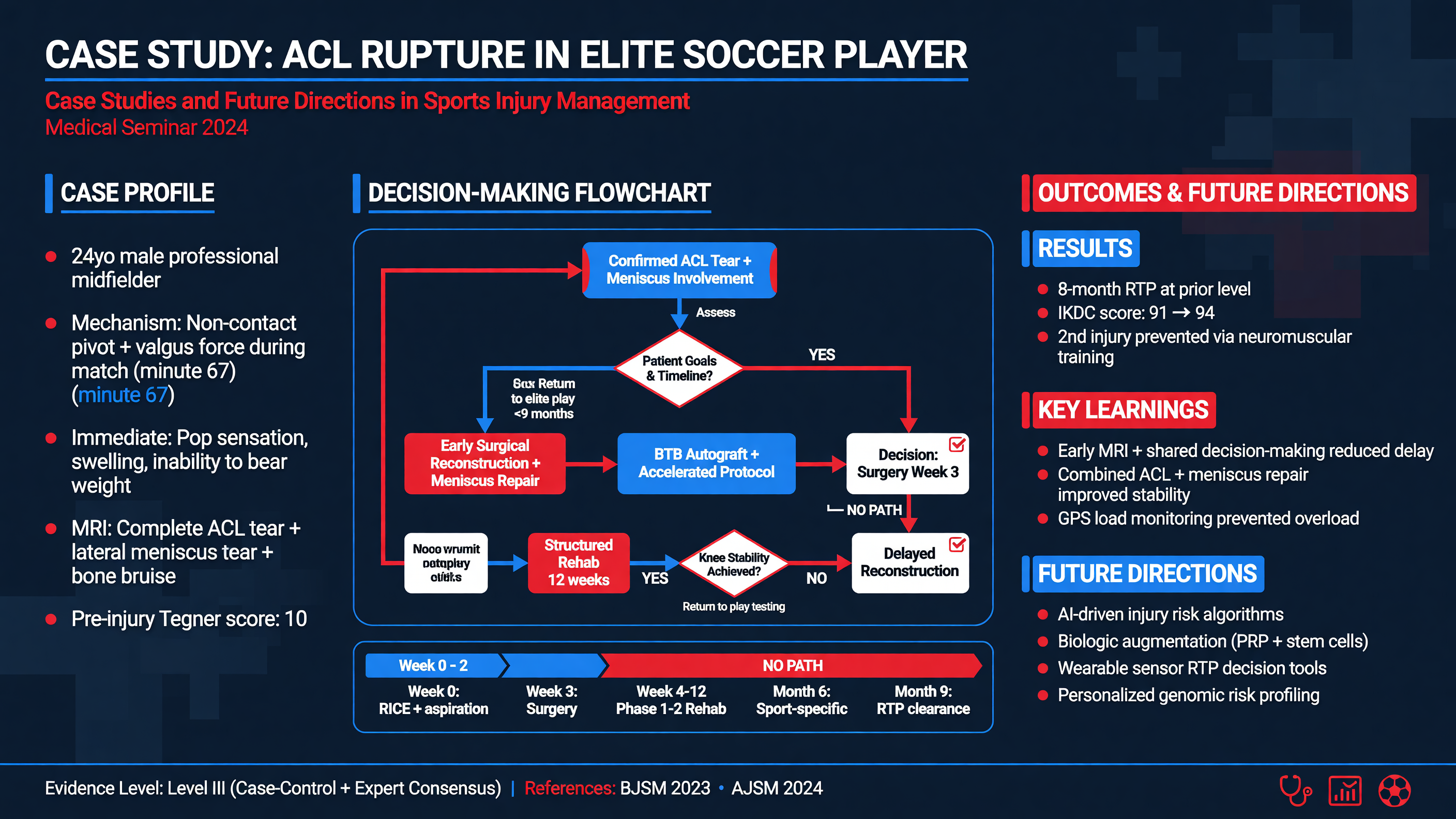
Integrated Clinical Case
A 22-year-old collegiate soccer midfielder is tackled while the right foot is planted and externally rotated. He feels pain above the ankle and cannot continue. The next morning he can take only two guarded steps. Examination shows tenderness over the anterior-inferior tibiofibular ligament extending 5 cm proximally, mild medial tenderness, pain with external-rotation stress, and a positive squeeze test. There is no deformity, neurovascular deficit, Achilles defect, or proximal fibular tenderness.
Audience Poll: Is the next best step functional rehabilitation, ankle radiographs, immediate MRI, or orthopedic fixation?
Because he cannot take four steps and has malleolar-zone pain, radiographs are indicated under the Ottawa Ankle Rules. Anteroposterior, mortise, and lateral views show no fracture or clear mortise widening. These findings reduce the probability of fracture but do not exclude syndesmotic disruption.
Decision Point: The mechanism, proximal tenderness, and clustered provocative findings sustain concern for a syndesmotic injury despite negative radiographs. The immediate question is whether the ankle is stable. Sports-medicine follow-up, protected weight bearing in a boot, and timely advanced or stress imaging are appropriate; simply relabeling this a lateral sprain and allowing play in several days would be unsafe.
MRI demonstrates anterior-inferior tibiofibular ligament disruption with interosseous edema but intact posterior structures and no deltoid disruption, osteochondral lesion, or occult fracture. Weight-bearing alignment remains normal. The injury is treated nonoperatively. During the first week, he uses a boot and crutches as needed to normalize gait. Swelling control, ankle pumps, and pain-limited motion begin immediately, without forced dorsiflexion or external rotation.
By week three, he walks without a limp and progresses to loaded calf raises, resisted ankle work, knee-to-wall mobility, and single-leg balance. Running begins only after brisk walking, repeated calf raises, and low-level hops are tolerated without next-day swelling. Cutting is reintroduced through planned angles, then reactive drills and team practice. He returns to unrestricted competition in week eight after completing the PAASS domains, including full practice under fatigue and reporting confidence in contact situations.
Teaching Point: The successful decision was not the MRI itself. It was recognizing that negative fracture radiographs did not resolve a clinically plausible instability question.
Now consider a second patient: a 52-year-old recreational tennis player with three months of atraumatic lateral shoulder pain. MRI obtained before rehabilitation reports a partial-thickness supraspinatus tear, acromial “hooking,” and acromioclavicular degeneration. He has painful elevation but nearly full passive motion, mild external-rotation weakness limited by pain, and no lag sign, pseudoparalysis, cervical deficit, or systemic features.
The imaging findings are anatomically plausible but not automatically surgical. Management begins with education, temporary reduction in high-volume serving, and progressive cuff, scapular, trunk, and lower-extremity work. A single injection may be discussed if pain prevents sleep or participation, but acromial morphology alone does not justify decompression. Surgical consultation becomes reasonable if a well-adhered rehabilitation trial fails, functional weakness progresses, or the patient’s goals remain unattainable after informed discussion.
Nuance: Both cases contain an imaging abnormality. In the ankle, imaging helps answer a stability question that changes protection. In the shoulder, imaging findings must be restrained by the high background prevalence of asymptomatic structural change.
Future practice will increasingly use wearable inertial sensors, force platforms, markerless motion capture, portable ultrasound, and machine-learning models. These tools may quantify cutting loads, limb acceleration, throwing volume, fatigue, or rehabilitation adherence outside a laboratory. Their value depends on reliability, validation in the relevant population, clinically meaningful thresholds, and proof that acting on the data improves outcomes.
Artificial intelligence may assist image interpretation and integrate longitudinal data, but it can amplify bias when trained on unrepresentative athletes or when diagnostic labels are treated as ground truth. A precise prediction is not necessarily an actionable one. Clinicians must ask whether a model performs across sex, age, skin tone, disability, sport, competitive level, and equipment environment.
Biologics remain an area of intense interest. Platelet-rich plasma may have selective roles in some tendinopathies, but preparation methods and trial results vary, and evidence does not support presenting it as a universal tendon-regeneration treatment. Cell therapies, exosomes, and many marketed “orthobiologics” lack sufficient evidence for routine sports-injury care. Cost and regulatory status should be discussed explicitly.
MUST ACT: Do not allow a proprietary score, wearable alert, AI interpretation, or commercial biologic claim to override examination, validated imaging criteria, patient preferences, or established red flags.
Framework: Adopt emerging technology only when it demonstrates measurement validity, incremental clinical value, feasible implementation, equity across populations, and a clear response pathway.
The future of sports medicine is therefore not merely more data. It is better integration of tissue biology, biomechanics, psychology, environment, and patient priorities—followed by decisions that remain explainable at the bedside.
Tonight on Shift
- Screen for emergencies first: document deformity, neurovascular status, compartment findings, proximal fibular tenderness, traumatic weakness, systemic illness, and other red flags before provocative testing.
- Use decision rules precisely: apply the Ottawa Ankle Rules to fracture imaging, but do not use a negative rule or normal radiograph to exclude syndesmotic instability.
- Correlate every image: ask whether an abnormality matches the mechanism, examination, functional deficit, and clinical question before assigning causation.
- Prescribe—not merely recommend—rehabilitation: specify exercise, sets, repetitions, weekly frequency, progression criteria, and acceptable symptom response.
- Prevent recurrence: provide balance training and bracing when appropriate after ankle sprain, and embed neuromuscular strength and landing work into regular team warm-ups.
- Make return criteria multidimensional: assess pain, motion, strength, sensorimotor control, sport-specific performance, fatigue response, psychological readiness, and full-practice tolerance.
Read this seminar as Markdown · All seminars · Lecture library · Question bank