# Advancements in ACL Reconstruction: BEAR Implant and Biomechanical Strategies
## Learning Objectives
- Differentiate ACL reconstruction, primary suture repair, and bridge-enhanced ACL repair by indication, biology, and biomechanics.
- Select appropriate candidates for BEAR implantation using injury pattern, tissue quality, skeletal maturity, timing, and patient goals.
- Interpret comparative BEAR and autograft outcomes without overstating current evidence.
- Construct a criterion-based rehabilitation plan that protects early ligament healing while restoring strength, movement quality, and sport capacity.
- Counsel patients and families about restrictions, reinjury risk, adherence, and realistic return-to-sport expectations.
- Evaluate long-term joint-health risks after ACL injury, including meniscal injury, recurrent instability, and post-traumatic osteoarthritis.
- Coordinate surgeons, physical therapists, athletic trainers, and performance staff around shared progression criteria.
---
## Introduction to ACL Injuries and Repair Techniques
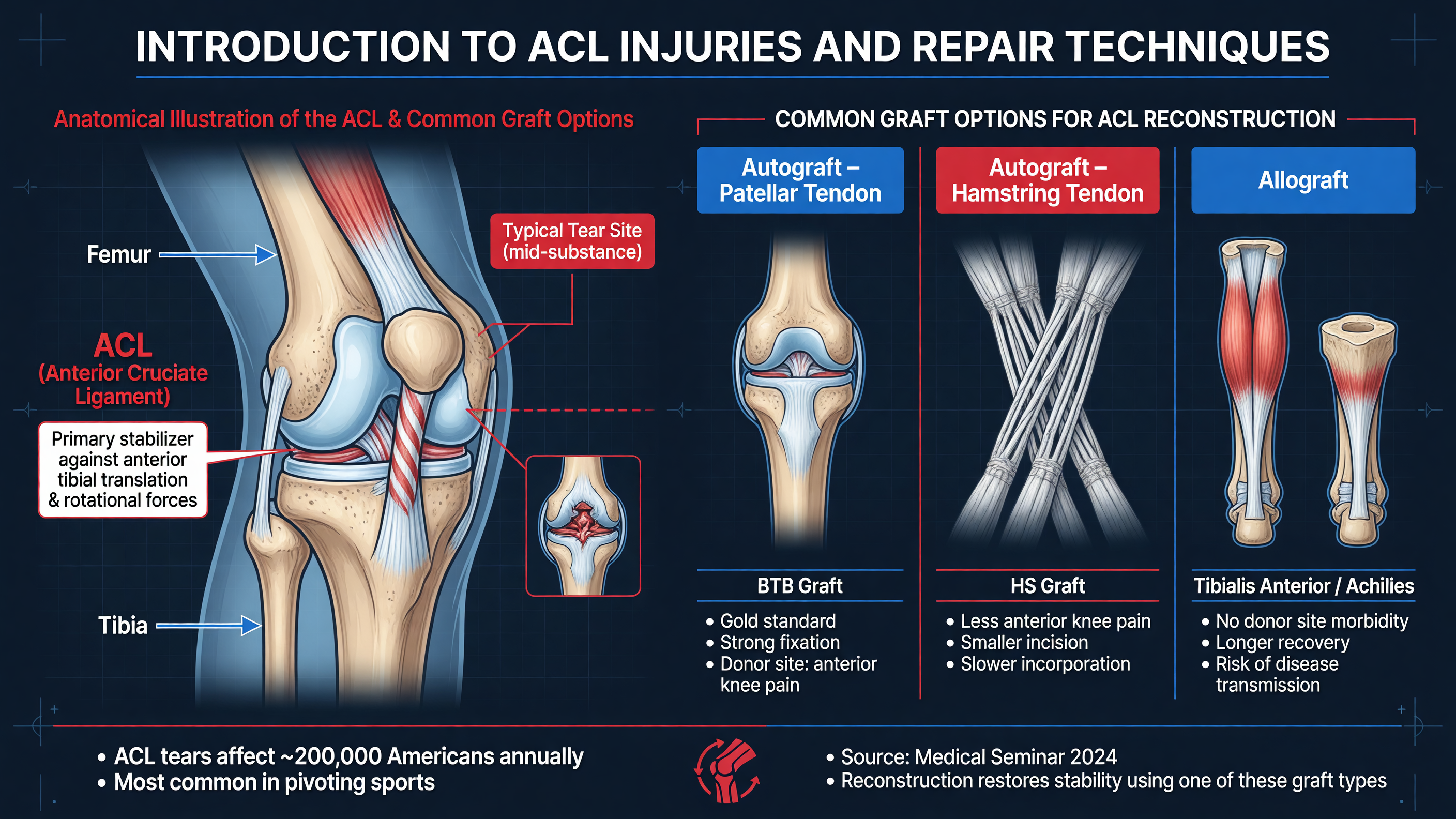 The anterior cruciate ligament is not simply a cord opposing anterior tibial translation. Its anteromedial and posterolateral fiber regions are recruited differently across knee flexion, and together they constrain anterior translation, coupled internal rotation, and the pivot-shift phenomenon. Injury usually follows a noncontact deceleration, cutting, landing, or valgus–internal-rotation event, although contact mechanisms are common. Patients may describe a pop, immediate swelling, and loss of confidence in the knee. Hemarthrosis within several hours strongly suggests an intra-articular injury but is not diagnostic of an ACL tear.
**MUST ACT:** An acutely locked knee, displaced bucket-handle meniscal tear, osteochondral fragment, extensor-mechanism disruption, knee dislocation, or neurovascular deficit takes priority over routine ACL planning. Document pulses, peroneal nerve function, collateral stability, and the ability to achieve extension.
The differential diagnosis includes patellar dislocation, tibial-spine avulsion in skeletally immature patients, posterior cruciate or posterolateral-corner injury, meniscal root injury, osteochondral fracture, and isolated collateral-ligament injury. Lachman testing is generally the most sensitive bedside maneuver, while the pivot shift is highly informative for functional rotational instability but may be limited by pain or guarding. MRI confirms tear location and associated pathology; it does not replace examination under anesthesia, direct assessment of tissue quality, or evaluation of alignment and generalized laxity.
**Framework:** Management begins with the patient rather than the MRI. Consider age, skeletal maturity, desired activity, instability episodes, occupation, sport, meniscal status, tissue pattern, hyperextension, coronal alignment, and willingness to complete rehabilitation. Structured nonoperative care can be reasonable for selected patients without recurrent instability who accept activity modification. Repeated giving-way in a pivoting athlete, however, threatens the menisci and articular cartilage.
Traditional ACL reconstruction removes or bypasses the torn ligament and substitutes a tendon graft. Bone–patellar tendon–bone autograft offers bone-to-bone healing and reliable stability but may produce anterior-knee or kneeling pain. Hamstring autograft avoids patellar harvest morbidity but can leave flexion-strength deficits and has fixation and graft-diameter considerations. Quadriceps-tendon autograft provides a large, versatile graft with or without a bone block, although quadriceps recovery requires deliberate attention. Allograft avoids harvest morbidity but carries a clinically important failure disadvantage in young, highly active patients; contemporary graft-choice literature continues to favor autograft in this population (Briem et al., *Knee Surg Sports Traumatol Arthrosc*, 2026; PMID: 40178127).
Primary suture repair is different from reconstruction. It is most defensible for selected acute, proximal avulsions with excellent tissue quality, sometimes with suture augmentation. BEAR—bridge-enhanced ACL repair—addresses complete tears by supplying a biologic bridge between the residual ligament ends. It therefore preserves native insertions rather than substituting a tendon.
**Nuance:** “Anatomic” reconstruction is still a graft approximation of native anatomy. Conversely, preserving the ACL does not guarantee normal mechanics if the repair heals elongated, rotational stabilizers remain deficient, or rehabilitation exposes the bridge to excessive load.
**Decision Point:** Surgery should generally occur after swelling improves and extension is restored unless associated pathology creates urgency. Operating on an inflamed, motion-limited knee increases arthrofibrosis risk; waiting too long may make a time-sensitive repair strategy unavailable.
**Audience Poll:** Which finding most changes your treatment recommendation: high-grade pivot shift, repairable meniscal tear, open physes, generalized laxity, or participation in cutting sport?
---
## BEAR Implant Mechanism and Biomechanical Principles
The anterior cruciate ligament is not simply a cord opposing anterior tibial translation. Its anteromedial and posterolateral fiber regions are recruited differently across knee flexion, and together they constrain anterior translation, coupled internal rotation, and the pivot-shift phenomenon. Injury usually follows a noncontact deceleration, cutting, landing, or valgus–internal-rotation event, although contact mechanisms are common. Patients may describe a pop, immediate swelling, and loss of confidence in the knee. Hemarthrosis within several hours strongly suggests an intra-articular injury but is not diagnostic of an ACL tear.
**MUST ACT:** An acutely locked knee, displaced bucket-handle meniscal tear, osteochondral fragment, extensor-mechanism disruption, knee dislocation, or neurovascular deficit takes priority over routine ACL planning. Document pulses, peroneal nerve function, collateral stability, and the ability to achieve extension.
The differential diagnosis includes patellar dislocation, tibial-spine avulsion in skeletally immature patients, posterior cruciate or posterolateral-corner injury, meniscal root injury, osteochondral fracture, and isolated collateral-ligament injury. Lachman testing is generally the most sensitive bedside maneuver, while the pivot shift is highly informative for functional rotational instability but may be limited by pain or guarding. MRI confirms tear location and associated pathology; it does not replace examination under anesthesia, direct assessment of tissue quality, or evaluation of alignment and generalized laxity.
**Framework:** Management begins with the patient rather than the MRI. Consider age, skeletal maturity, desired activity, instability episodes, occupation, sport, meniscal status, tissue pattern, hyperextension, coronal alignment, and willingness to complete rehabilitation. Structured nonoperative care can be reasonable for selected patients without recurrent instability who accept activity modification. Repeated giving-way in a pivoting athlete, however, threatens the menisci and articular cartilage.
Traditional ACL reconstruction removes or bypasses the torn ligament and substitutes a tendon graft. Bone–patellar tendon–bone autograft offers bone-to-bone healing and reliable stability but may produce anterior-knee or kneeling pain. Hamstring autograft avoids patellar harvest morbidity but can leave flexion-strength deficits and has fixation and graft-diameter considerations. Quadriceps-tendon autograft provides a large, versatile graft with or without a bone block, although quadriceps recovery requires deliberate attention. Allograft avoids harvest morbidity but carries a clinically important failure disadvantage in young, highly active patients; contemporary graft-choice literature continues to favor autograft in this population (Briem et al., *Knee Surg Sports Traumatol Arthrosc*, 2026; PMID: 40178127).
Primary suture repair is different from reconstruction. It is most defensible for selected acute, proximal avulsions with excellent tissue quality, sometimes with suture augmentation. BEAR—bridge-enhanced ACL repair—addresses complete tears by supplying a biologic bridge between the residual ligament ends. It therefore preserves native insertions rather than substituting a tendon.
**Nuance:** “Anatomic” reconstruction is still a graft approximation of native anatomy. Conversely, preserving the ACL does not guarantee normal mechanics if the repair heals elongated, rotational stabilizers remain deficient, or rehabilitation exposes the bridge to excessive load.
**Decision Point:** Surgery should generally occur after swelling improves and extension is restored unless associated pathology creates urgency. Operating on an inflamed, motion-limited knee increases arthrofibrosis risk; waiting too long may make a time-sensitive repair strategy unavailable.
**Audience Poll:** Which finding most changes your treatment recommendation: high-grade pivot shift, repairable meniscal tear, open physes, generalized laxity, or participation in cutting sport?
---
## BEAR Implant Mechanism and Biomechanical Principles
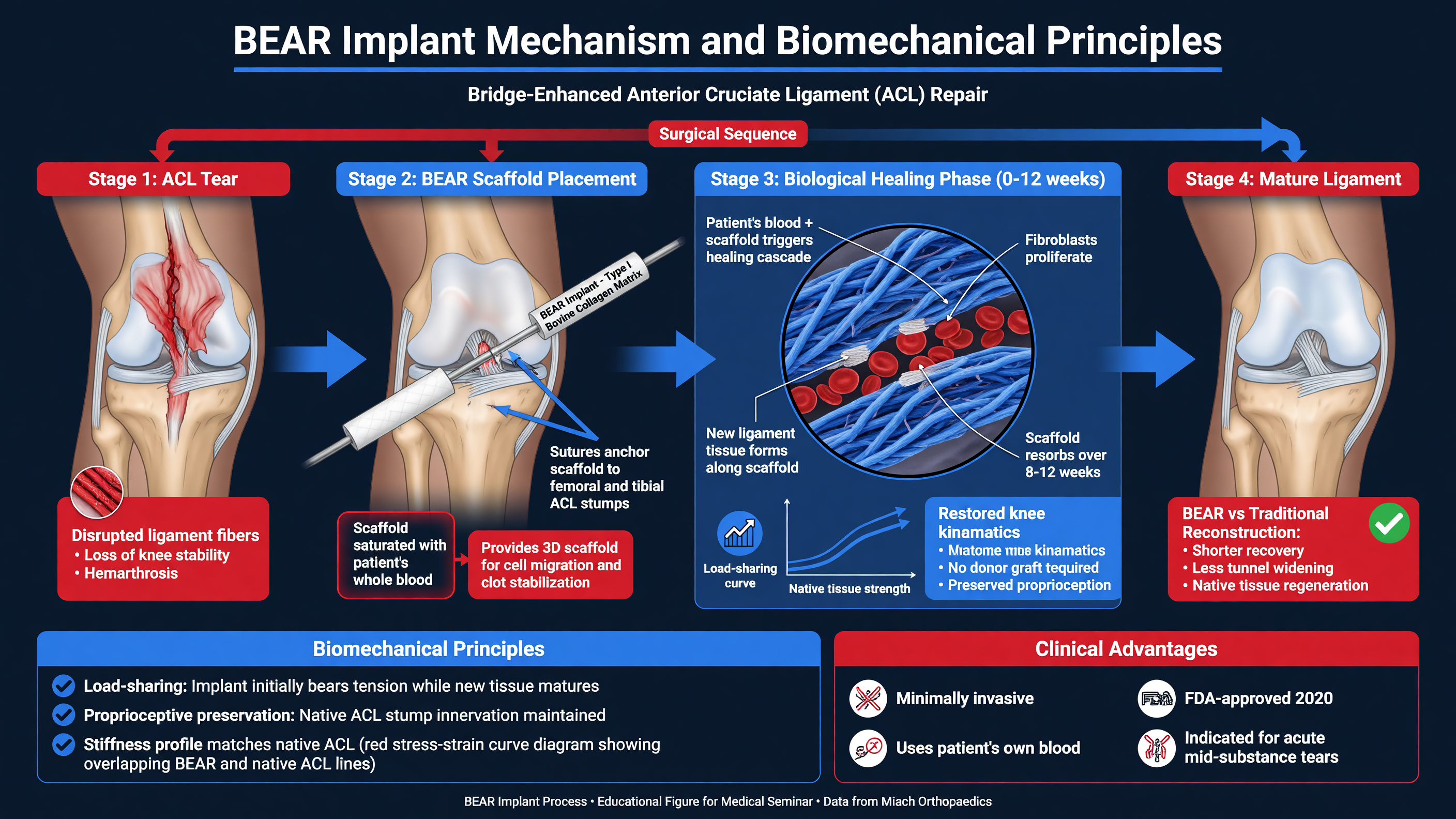 The native ACL has limited spontaneous healing despite retaining viable cells after many tears. Unlike an extra-articular medial collateral ligament injury, an ACL rupture is bathed in synovial fluid. The provisional fibrin clot that normally connects disrupted tissue is diluted and broken down, leaving a persistent gap. Retraction, mechanical motion, variable vascularity, and an inflammatory intra-articular environment further impair organized scar formation.
The BEAR implant is a porous, resorbable bovine extracellular-matrix scaffold placed between the torn ACL ends and saturated with the patient’s autologous whole blood. Sutures approximate the residual tissue and stabilize the construct while the blood–scaffold composite provides a provisional matrix. Platelets, cytokines, and migrating host cells populate the bridge; collagen is deposited and remodeled while the implant is gradually resorbed. The device is therefore neither a permanent synthetic ligament nor a tendon graft.
**Teaching Point:** BEAR does not “regrow a brand-new ACL” instantaneously. It facilitates healing of the patient’s residual ligament through an initially vulnerable repair continuum: inflammation, cellular proliferation, matrix deposition, collagen alignment, and prolonged remodeling.
Preservation of the femoral and tibial insertions may retain native fiber orientation, vascular remnants, and mechanoreceptor-containing tissue. Those properties could support more physiologic kinematics and sensorimotor function, but clinical proof of superior proprioception or prevention of osteoarthritis remains incomplete. A healed ligament can also become enlarged, heterogeneous on MRI, or mechanically lax without causing symptoms; imaging appearance must be interpreted alongside examination and function.
**Framework:** Successful bridge-enhanced repair requires three elements: biologic substrate, mechanical protection, and appropriate patient selection. The surgeon needs residual tissue capable of participating in healing, stable fixation that reduces the gap, and a postoperative environment that avoids both destructive overload and prolonged immobilization.
Current device labeling should be verified at the time of treatment. Core labeled considerations have included a complete MRI-confirmed ACL rupture, age of at least 14 years with skeletal maturity, a tibial stump adequate to facilitate restoration, and implantation during the defined acute postinjury window. Bovine-material hypersensitivity, active infection, poor tissue, chronic deficiency, and injury patterns outside labeling can redirect treatment toward reconstruction. Skeletal maturity deserves explicit confirmation in adolescents rather than assumption based on chronological age alone.
At surgery, the tissue is assessed arthroscopically, associated meniscal or chondral lesions are treated, and the implant is positioned between the ligament ends. Autologous blood is added immediately before final deployment. Sutures hold the construct while the knee is positioned according to the operative technique. Small differences in tunnel or suture placement, stump length, tension, and concomitant procedures can alter local strain.
**MUST ACT:** Do not apply a standard ACL-reconstruction rehabilitation order automatically. During early healing, the BEAR construct depends on protected approximation of tissue rather than tendon-to-bone integration inside mature tunnels. Range, weight-bearing, and brace instructions must match the surgeon’s repair and any meniscal procedure.
Biomechanically, a reconstruction seeks immediate graft continuity but undergoes graft necrosis, revascularization, cellular repopulation, and “ligamentization.” BEAR begins with native tissue connected by a biologic bridge whose early strength is limited. Both procedures therefore require protection, but for different reasons. Neither can tolerate an early pivoting episode simply because pain has resolved.
**Nuance:** Avoid equating less graft-harvest morbidity with a minor operation. The patient still has a repaired intra-articular ligament, bone tunnels or fixation sites, postoperative inhibition, and meaningful risks of stiffness, failure, infection, thrombosis, and reoperation.
**Decision Point:** When tissue eligibility is uncertain, counsel the patient preoperatively about a contingency plan. Arthroscopy may reveal that reconstruction is safer than attempting repair, and graft preference should be discussed before anesthesia.
**Audience Poll:** Which matters most for BEAR candidacy in your practice: tear location, tissue quality, injury-to-surgery interval, age and activity, or anticipated adherence?
---
## Clinical Outcomes and Comparative Studies
The native ACL has limited spontaneous healing despite retaining viable cells after many tears. Unlike an extra-articular medial collateral ligament injury, an ACL rupture is bathed in synovial fluid. The provisional fibrin clot that normally connects disrupted tissue is diluted and broken down, leaving a persistent gap. Retraction, mechanical motion, variable vascularity, and an inflammatory intra-articular environment further impair organized scar formation.
The BEAR implant is a porous, resorbable bovine extracellular-matrix scaffold placed between the torn ACL ends and saturated with the patient’s autologous whole blood. Sutures approximate the residual tissue and stabilize the construct while the blood–scaffold composite provides a provisional matrix. Platelets, cytokines, and migrating host cells populate the bridge; collagen is deposited and remodeled while the implant is gradually resorbed. The device is therefore neither a permanent synthetic ligament nor a tendon graft.
**Teaching Point:** BEAR does not “regrow a brand-new ACL” instantaneously. It facilitates healing of the patient’s residual ligament through an initially vulnerable repair continuum: inflammation, cellular proliferation, matrix deposition, collagen alignment, and prolonged remodeling.
Preservation of the femoral and tibial insertions may retain native fiber orientation, vascular remnants, and mechanoreceptor-containing tissue. Those properties could support more physiologic kinematics and sensorimotor function, but clinical proof of superior proprioception or prevention of osteoarthritis remains incomplete. A healed ligament can also become enlarged, heterogeneous on MRI, or mechanically lax without causing symptoms; imaging appearance must be interpreted alongside examination and function.
**Framework:** Successful bridge-enhanced repair requires three elements: biologic substrate, mechanical protection, and appropriate patient selection. The surgeon needs residual tissue capable of participating in healing, stable fixation that reduces the gap, and a postoperative environment that avoids both destructive overload and prolonged immobilization.
Current device labeling should be verified at the time of treatment. Core labeled considerations have included a complete MRI-confirmed ACL rupture, age of at least 14 years with skeletal maturity, a tibial stump adequate to facilitate restoration, and implantation during the defined acute postinjury window. Bovine-material hypersensitivity, active infection, poor tissue, chronic deficiency, and injury patterns outside labeling can redirect treatment toward reconstruction. Skeletal maturity deserves explicit confirmation in adolescents rather than assumption based on chronological age alone.
At surgery, the tissue is assessed arthroscopically, associated meniscal or chondral lesions are treated, and the implant is positioned between the ligament ends. Autologous blood is added immediately before final deployment. Sutures hold the construct while the knee is positioned according to the operative technique. Small differences in tunnel or suture placement, stump length, tension, and concomitant procedures can alter local strain.
**MUST ACT:** Do not apply a standard ACL-reconstruction rehabilitation order automatically. During early healing, the BEAR construct depends on protected approximation of tissue rather than tendon-to-bone integration inside mature tunnels. Range, weight-bearing, and brace instructions must match the surgeon’s repair and any meniscal procedure.
Biomechanically, a reconstruction seeks immediate graft continuity but undergoes graft necrosis, revascularization, cellular repopulation, and “ligamentization.” BEAR begins with native tissue connected by a biologic bridge whose early strength is limited. Both procedures therefore require protection, but for different reasons. Neither can tolerate an early pivoting episode simply because pain has resolved.
**Nuance:** Avoid equating less graft-harvest morbidity with a minor operation. The patient still has a repaired intra-articular ligament, bone tunnels or fixation sites, postoperative inhibition, and meaningful risks of stiffness, failure, infection, thrombosis, and reoperation.
**Decision Point:** When tissue eligibility is uncertain, counsel the patient preoperatively about a contingency plan. Arthroscopy may reveal that reconstruction is safer than attempting repair, and graft preference should be discussed before anesthesia.
**Audience Poll:** Which matters most for BEAR candidacy in your practice: tear location, tissue quality, injury-to-surgery interval, age and activity, or anticipated adherence?
---
## Clinical Outcomes and Comparative Studies
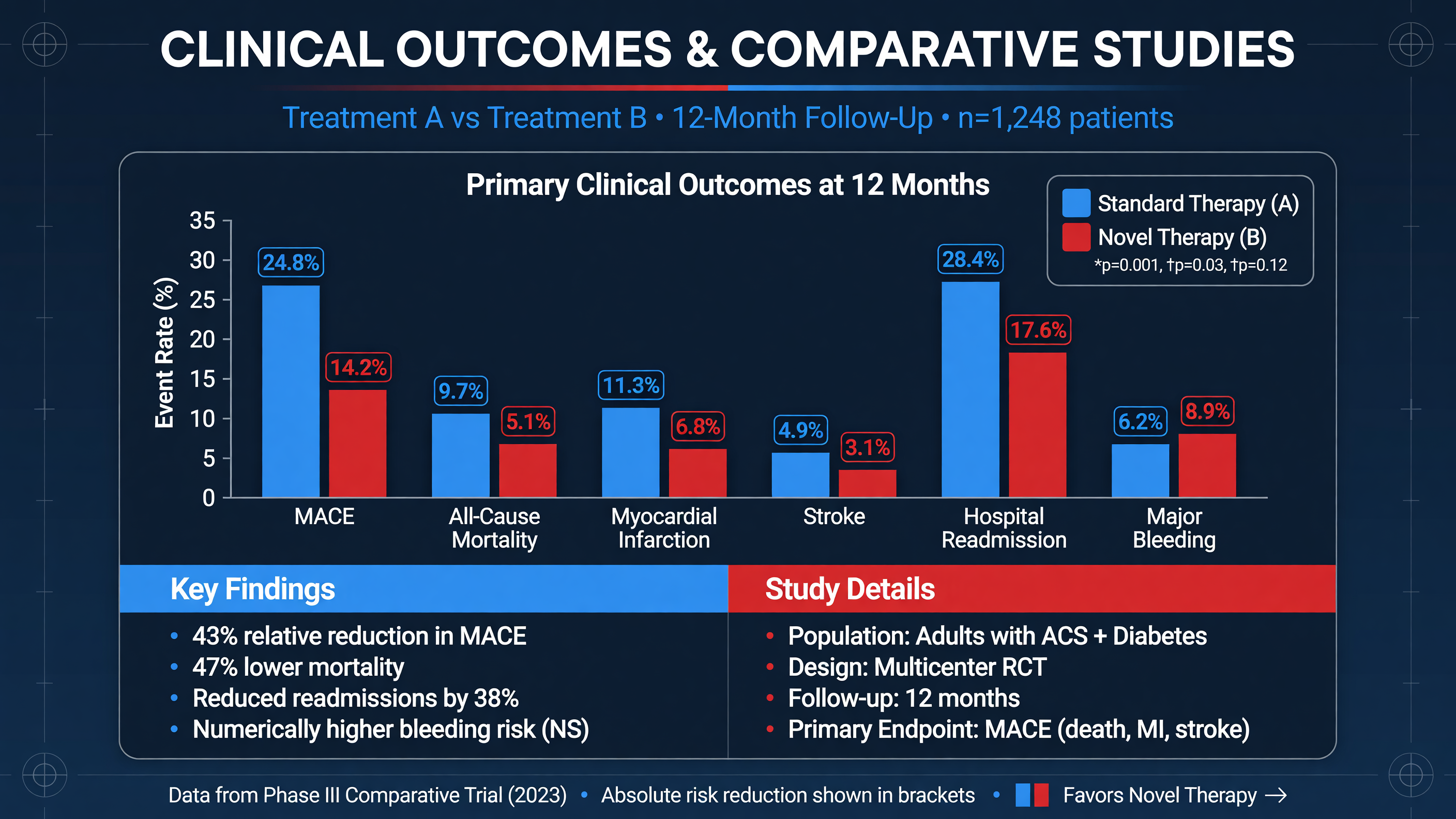 The central comparative evidence comes from BEAR II, a prospective randomized trial of 100 young patients with complete midsubstance ACL injuries who were allocated approximately 2:1 to BEAR or autograft reconstruction. At two years, BEAR met prespecified noninferiority criteria for the International Knee Documentation Committee subjective score and instrumented anteroposterior laxity. Mean IKDC scores were approximately 89 after BEAR and 85 after reconstruction, while side-to-side anteroposterior differences were approximately 1.6 and 1.8 mm, respectively (Murray et al., 2020; PMID: 32298131).
**Teaching Point:** Noninferiority means the observed result did not cross a predefined clinically unacceptable margin. It does not establish that BEAR is superior, equivalent for every outcome, or preferable for every patient.
The most striking strength difference in BEAR II involved hamstring strength, not quadriceps strength. The mean hamstring-strength index favored BEAR, approximately 98% versus 63%, largely reflecting avoidance of hamstring harvest in reconstruction patients. This finding should not be generalized to comparisons with quadriceps-tendon or patellar-tendon grafts. Quadriceps inhibition remains common after either procedure and can persist even when patient-reported function is high.
Ipsilateral reinjury requiring another operation occurred numerically more often after BEAR—approximately 14% versus 6% after reconstruction—but the trial was not powered to establish a definitive failure-rate difference. The absolute numbers were small and the difference was not statistically significant. That uncertainty is especially important when counseling adolescents and cutting-sport athletes, whose baseline risk of ipsilateral or contralateral ACL injury is already high.
**Framework:** Interpret outcomes in five domains: symptoms and self-reported function; objective laxity; strength and hop performance; return to the desired activity; and structural survival without revision. A favorable IKDC score cannot compensate for recurrent pivoting, and a normal KT-1000 measurement cannot establish readiness for chaotic sport.
Early BEAR cohorts and follow-up studies generally show substantial improvements in pain, function, activity, and stability. They also demonstrate that failure is possible and that revision reconstruction may be required. Revision after BEAR is conceptually feasible because no tendon autograft has been harvested initially, but prior tunnels, fixation, scar, meniscal status, and the reason for failure still influence complexity. “Saving the graft for later” should never be presented as evidence that an avoidable first failure is acceptable.
Comparisons are further complicated by graft choice and rehabilitation. A hamstring-harvest deficit makes BEAR look different from hamstring autograft but says little about quadriceps-tendon autograft. Likewise, outcomes from expert investigational centers may not fully predict performance across broad community practice. Surgeon learning, patient selection, protocol adherence, and loss to follow-up can influence registry findings.
**MUST ACT:** When discussing evidence, provide absolute event rates and the duration of follow-up. A two-year study cannot answer whether BEAR prevents osteoarthritis at 15 years, and a small midterm cohort cannot exclude uncommon complications.
Practical selection therefore remains preference-sensitive. A patient may prioritize native-tissue preservation and avoidance of harvest morbidity; another may prefer the longer performance history of autograft reconstruction. A high-risk young pivoting athlete, revision case, chronic tear, poor stump, multiligament injury, or patient unlikely to follow protection restrictions may favor reconstruction. A recently injured, appropriately labeled patient with suitable tissue and strong adherence may reasonably consider BEAR.
**Nuance:** Return-to-sport rate is not synonymous with successful return. The clinically meaningful outcome is sustained participation at the intended level without reinjury, unacceptable symptoms, or recurrent effusion.
**Decision Point:** Use the same rigorous return-to-sport criteria after BEAR as after reconstruction until procedure-specific evidence justifies otherwise.
**Audience Poll:** Would a possible reduction in donor-site morbidity outweigh uncertainty in long-term reinjury and osteoarthritis outcomes for a 17-year-old pivoting athlete?
---
## Rehabilitation Protocols for BEAR Implants
The central comparative evidence comes from BEAR II, a prospective randomized trial of 100 young patients with complete midsubstance ACL injuries who were allocated approximately 2:1 to BEAR or autograft reconstruction. At two years, BEAR met prespecified noninferiority criteria for the International Knee Documentation Committee subjective score and instrumented anteroposterior laxity. Mean IKDC scores were approximately 89 after BEAR and 85 after reconstruction, while side-to-side anteroposterior differences were approximately 1.6 and 1.8 mm, respectively (Murray et al., 2020; PMID: 32298131).
**Teaching Point:** Noninferiority means the observed result did not cross a predefined clinically unacceptable margin. It does not establish that BEAR is superior, equivalent for every outcome, or preferable for every patient.
The most striking strength difference in BEAR II involved hamstring strength, not quadriceps strength. The mean hamstring-strength index favored BEAR, approximately 98% versus 63%, largely reflecting avoidance of hamstring harvest in reconstruction patients. This finding should not be generalized to comparisons with quadriceps-tendon or patellar-tendon grafts. Quadriceps inhibition remains common after either procedure and can persist even when patient-reported function is high.
Ipsilateral reinjury requiring another operation occurred numerically more often after BEAR—approximately 14% versus 6% after reconstruction—but the trial was not powered to establish a definitive failure-rate difference. The absolute numbers were small and the difference was not statistically significant. That uncertainty is especially important when counseling adolescents and cutting-sport athletes, whose baseline risk of ipsilateral or contralateral ACL injury is already high.
**Framework:** Interpret outcomes in five domains: symptoms and self-reported function; objective laxity; strength and hop performance; return to the desired activity; and structural survival without revision. A favorable IKDC score cannot compensate for recurrent pivoting, and a normal KT-1000 measurement cannot establish readiness for chaotic sport.
Early BEAR cohorts and follow-up studies generally show substantial improvements in pain, function, activity, and stability. They also demonstrate that failure is possible and that revision reconstruction may be required. Revision after BEAR is conceptually feasible because no tendon autograft has been harvested initially, but prior tunnels, fixation, scar, meniscal status, and the reason for failure still influence complexity. “Saving the graft for later” should never be presented as evidence that an avoidable first failure is acceptable.
Comparisons are further complicated by graft choice and rehabilitation. A hamstring-harvest deficit makes BEAR look different from hamstring autograft but says little about quadriceps-tendon autograft. Likewise, outcomes from expert investigational centers may not fully predict performance across broad community practice. Surgeon learning, patient selection, protocol adherence, and loss to follow-up can influence registry findings.
**MUST ACT:** When discussing evidence, provide absolute event rates and the duration of follow-up. A two-year study cannot answer whether BEAR prevents osteoarthritis at 15 years, and a small midterm cohort cannot exclude uncommon complications.
Practical selection therefore remains preference-sensitive. A patient may prioritize native-tissue preservation and avoidance of harvest morbidity; another may prefer the longer performance history of autograft reconstruction. A high-risk young pivoting athlete, revision case, chronic tear, poor stump, multiligament injury, or patient unlikely to follow protection restrictions may favor reconstruction. A recently injured, appropriately labeled patient with suitable tissue and strong adherence may reasonably consider BEAR.
**Nuance:** Return-to-sport rate is not synonymous with successful return. The clinically meaningful outcome is sustained participation at the intended level without reinjury, unacceptable symptoms, or recurrent effusion.
**Decision Point:** Use the same rigorous return-to-sport criteria after BEAR as after reconstruction until procedure-specific evidence justifies otherwise.
**Audience Poll:** Would a possible reduction in donor-site morbidity outweigh uncertainty in long-term reinjury and osteoarthritis outcomes for a 17-year-old pivoting athlete?
---
## Rehabilitation Protocols for BEAR Implants
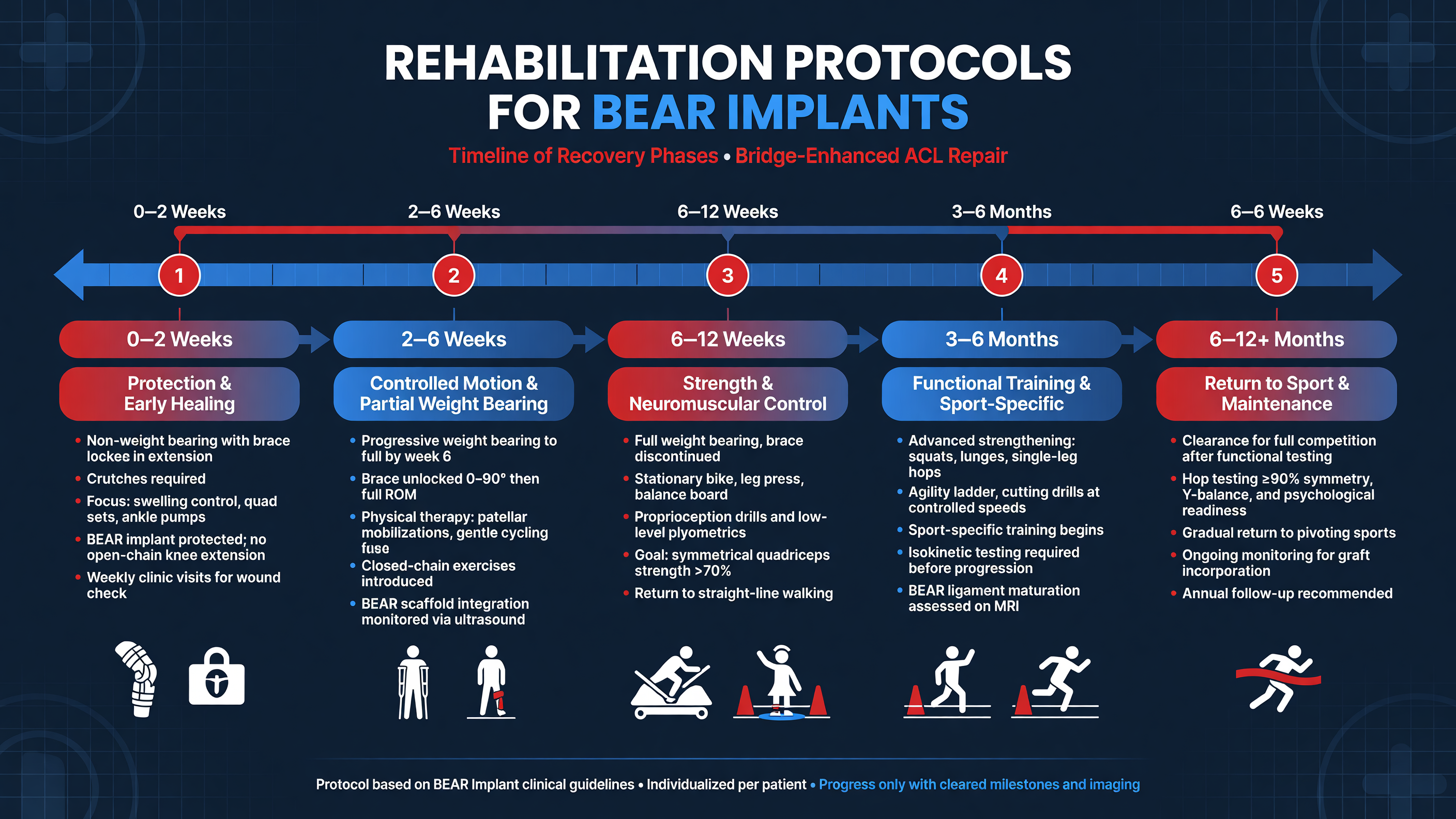 BEAR rehabilitation balances two competing harms: excessive early strain can elongate or disrupt the healing bridge, while excessive protection promotes arthrofibrosis, quadriceps inhibition, gait dysfunction, and deconditioning. The operative report is the starting document. Meniscal repair, root fixation, cartilage restoration, collateral injury, or an extension deficit may supersede the isolated-BEAR timeline.
**MUST ACT:** Confirm four orders before the first session: permitted weight-bearing, brace settings, allowed range of motion, and restrictions created by concomitant procedures. When instructions conflict, contact the surgeon rather than averaging protocols.
During approximately weeks 0–2, priorities are wound protection, edema control, full passive extension, safe transfers, patellar mobility, quadriceps activation, and adherence to brace and crutch instructions. Trial-era BEAR protocols commonly used partial weight-bearing, a brace locked in extension for ambulation, and flexion limited to roughly 45–50 degrees initially. Contemporary protocols vary, so these values are not universal prescriptions. Avoid placing pillows continuously behind the knee. Short heel-prop or prone-hang sessions—often 5–10 minutes several times daily if permitted—can address extension without aggressive force. Quadriceps sets, straight-leg raises without lag, ankle pumps, and hip exercises are typical early components.
From roughly weeks 2–6, flexion is progressively advanced, historically toward 90 degrees in protected protocols. Weight-bearing and brace unlocking depend on gait quality, quadriceps control, effusion, and surgeon authorization. Closed-chain exercises may include controlled sit-to-stand, mini-squats, low step-ups, calf raises, and balance tasks within allowed angles. Neuromuscular electrical stimulation can supplement volitional training when activation failure persists; it should produce a strong visible contraction, not merely sensory tingling.
**Teaching Point:** Full extension is an early priority; flexion progression is deliberate. A patient who reaches excessive flexion rapidly but walks with a flexed-knee gait is not ahead of schedule.
During weeks 6–12, goals usually include normalized gait, near-full motion, minimal or trace effusion, improved single-leg control, and progressive lower-extremity strength. Stationary cycling begins when motion and wound status permit. Resistance is advanced according to tissue response, typically using two to four sets of 6–15 repetitions across squats, split squats, step variations, hip hinges, hamstring work, and calf training. Open-chain knee extension is not inherently forbidden, but load, arc, and timing must follow the treating team because anterior tibial shear changes across the range.
The three-to-six-month phase emphasizes unilateral strength, eccentric control, landing mechanics, trunk and hip strategy, aerobic capacity, and preparation for running. A running progression should not begin because the calendar reaches 12 weeks. Common prerequisites include full motion, no instability, minimal effusion, pain-free hopping preparation, acceptable single-leg squat mechanics, and a meaningful quadriceps-strength threshold—often at least 70–80% limb symmetry before initial running, with the exact standard determined locally.
Later rehabilitation incorporates deceleration, plyometrics, cutting, reactive tasks, and sport-specific workload. Neurocognitive training matters because many ACL injuries occur while attention is divided. Visual perturbations, unanticipated direction changes, dual-task drills, opponent cues, and decision-making should progress after basic mechanics are secure; contemporary work supports integrating cognitive–motor demands rather than relying exclusively on rehearsed movements (Delvaux et al., 2025; PMID: 40518903).
**Framework:** Progress by tissue irritability and capability: no increase in effusion, no loss of extension, no instability, adequate strength, acceptable movement quality, and successful exposure to the previous workload.
Return to unrestricted pivoting sport commonly requires at least 9–12 months and should be criterion-based. Assess quadriceps and hamstring strength, hop testing, rate of force development where available, cutting mechanics, fatigue response, sport workload, and psychological readiness such as the ACL–Return to Sport after Injury scale. A 90% limb-symmetry index is a floor, not proof of normality; both limbs may be weak after prolonged deconditioning.
**MUST ACT:** Escalate calf swelling, dyspnea, fever, drainage, rapidly increasing pain, true giving-way, mechanical locking, or progressive loss of extension. Persistent effusion is a biologic load signal, not a nuisance to train through.
**Audience Poll:** Which criterion most often delays safe return in your setting: quadriceps force, reactive movement quality, persistent effusion, psychological readiness, or sport-conditioning exposure?
---
## Patient Education and Adherence Strategies
BEAR rehabilitation balances two competing harms: excessive early strain can elongate or disrupt the healing bridge, while excessive protection promotes arthrofibrosis, quadriceps inhibition, gait dysfunction, and deconditioning. The operative report is the starting document. Meniscal repair, root fixation, cartilage restoration, collateral injury, or an extension deficit may supersede the isolated-BEAR timeline.
**MUST ACT:** Confirm four orders before the first session: permitted weight-bearing, brace settings, allowed range of motion, and restrictions created by concomitant procedures. When instructions conflict, contact the surgeon rather than averaging protocols.
During approximately weeks 0–2, priorities are wound protection, edema control, full passive extension, safe transfers, patellar mobility, quadriceps activation, and adherence to brace and crutch instructions. Trial-era BEAR protocols commonly used partial weight-bearing, a brace locked in extension for ambulation, and flexion limited to roughly 45–50 degrees initially. Contemporary protocols vary, so these values are not universal prescriptions. Avoid placing pillows continuously behind the knee. Short heel-prop or prone-hang sessions—often 5–10 minutes several times daily if permitted—can address extension without aggressive force. Quadriceps sets, straight-leg raises without lag, ankle pumps, and hip exercises are typical early components.
From roughly weeks 2–6, flexion is progressively advanced, historically toward 90 degrees in protected protocols. Weight-bearing and brace unlocking depend on gait quality, quadriceps control, effusion, and surgeon authorization. Closed-chain exercises may include controlled sit-to-stand, mini-squats, low step-ups, calf raises, and balance tasks within allowed angles. Neuromuscular electrical stimulation can supplement volitional training when activation failure persists; it should produce a strong visible contraction, not merely sensory tingling.
**Teaching Point:** Full extension is an early priority; flexion progression is deliberate. A patient who reaches excessive flexion rapidly but walks with a flexed-knee gait is not ahead of schedule.
During weeks 6–12, goals usually include normalized gait, near-full motion, minimal or trace effusion, improved single-leg control, and progressive lower-extremity strength. Stationary cycling begins when motion and wound status permit. Resistance is advanced according to tissue response, typically using two to four sets of 6–15 repetitions across squats, split squats, step variations, hip hinges, hamstring work, and calf training. Open-chain knee extension is not inherently forbidden, but load, arc, and timing must follow the treating team because anterior tibial shear changes across the range.
The three-to-six-month phase emphasizes unilateral strength, eccentric control, landing mechanics, trunk and hip strategy, aerobic capacity, and preparation for running. A running progression should not begin because the calendar reaches 12 weeks. Common prerequisites include full motion, no instability, minimal effusion, pain-free hopping preparation, acceptable single-leg squat mechanics, and a meaningful quadriceps-strength threshold—often at least 70–80% limb symmetry before initial running, with the exact standard determined locally.
Later rehabilitation incorporates deceleration, plyometrics, cutting, reactive tasks, and sport-specific workload. Neurocognitive training matters because many ACL injuries occur while attention is divided. Visual perturbations, unanticipated direction changes, dual-task drills, opponent cues, and decision-making should progress after basic mechanics are secure; contemporary work supports integrating cognitive–motor demands rather than relying exclusively on rehearsed movements (Delvaux et al., 2025; PMID: 40518903).
**Framework:** Progress by tissue irritability and capability: no increase in effusion, no loss of extension, no instability, adequate strength, acceptable movement quality, and successful exposure to the previous workload.
Return to unrestricted pivoting sport commonly requires at least 9–12 months and should be criterion-based. Assess quadriceps and hamstring strength, hop testing, rate of force development where available, cutting mechanics, fatigue response, sport workload, and psychological readiness such as the ACL–Return to Sport after Injury scale. A 90% limb-symmetry index is a floor, not proof of normality; both limbs may be weak after prolonged deconditioning.
**MUST ACT:** Escalate calf swelling, dyspnea, fever, drainage, rapidly increasing pain, true giving-way, mechanical locking, or progressive loss of extension. Persistent effusion is a biologic load signal, not a nuisance to train through.
**Audience Poll:** Which criterion most often delays safe return in your setting: quadriceps force, reactive movement quality, persistent effusion, psychological readiness, or sport-conditioning exposure?
---
## Patient Education and Adherence Strategies
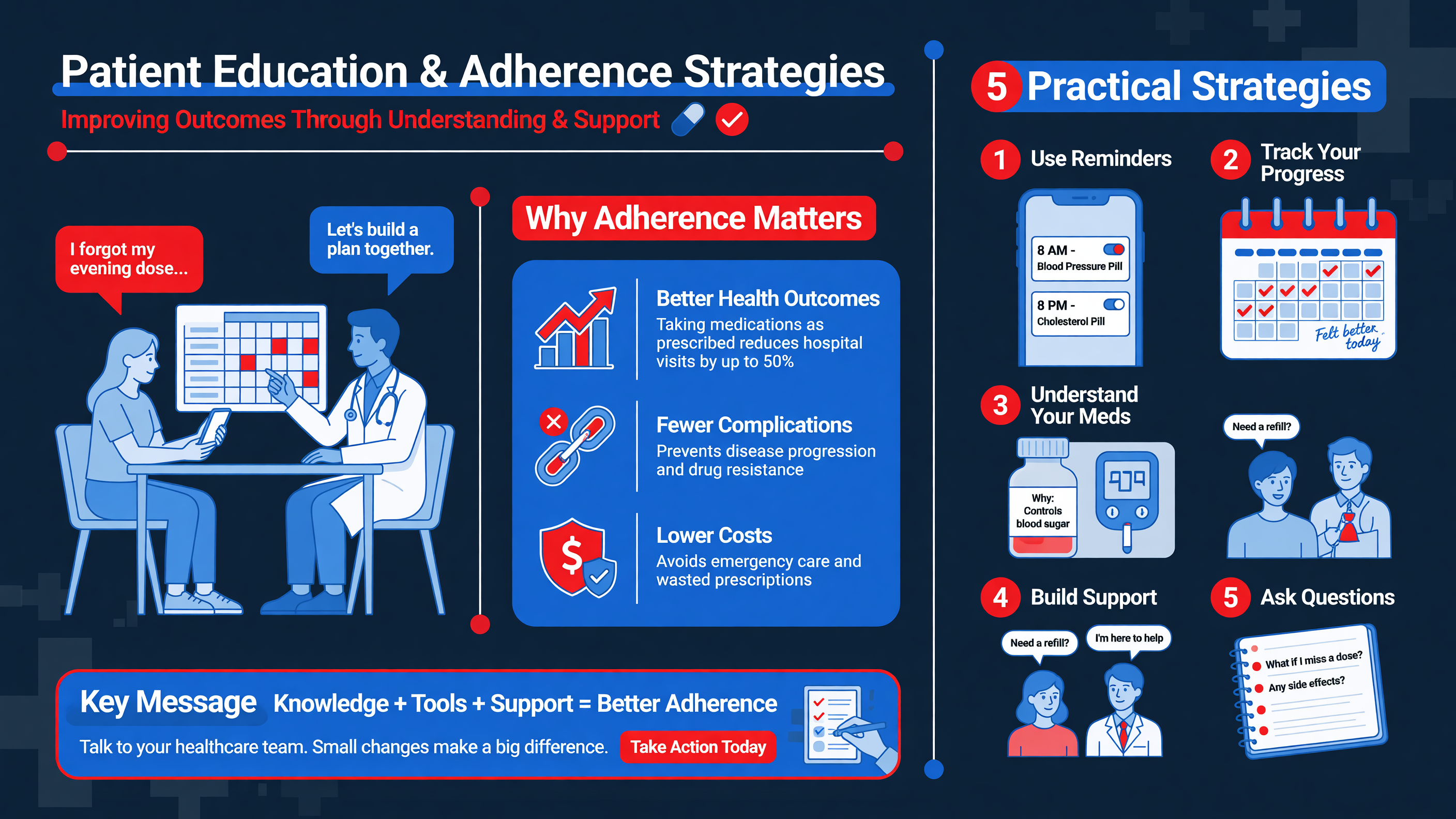 Patients often interpret decreasing pain as evidence that the ligament has healed. BEAR makes that misconception particularly hazardous because symptoms can improve long before the collagen bridge has matured. Education should begin preoperatively, continue at discharge, and be repeated at every transition in rehabilitation.
**Teaching Point:** A useful analogy is that early repair tissue resembles wet, disorganized strands of spaghetti: biologically active but poorly aligned and mechanically vulnerable. Loading helps those fibers organize only when the dose is appropriate.
Use teach-back rather than asking whether the patient understands. The patient should explain, in their own words, how to wear the brace, how much weight to place through the limb, what range is allowed, which exercises are scheduled, and which symptoms require a call. Provide a one-page plan with current restrictions, the next milestones, contact information, and a medication list. Adolescents need the same explanation delivered to a parent or guardian without removing the athlete from decision-making.
**Framework:** Build adherence around capability, opportunity, and motivation. Capability includes knowing how to perform exercises. Opportunity includes transportation, insurance visits, school accommodations, and access to equipment. Motivation includes confidence, perceived benefit, fear, and competing sport pressure. Labeling a patient “noncompliant” without identifying the failed component rarely improves behavior.
Concrete dosing is more useful than “exercise regularly.” For example, prescribe the number of sessions per day, repetitions, hold time, and stopping rule. Use a checklist or app to record brace use, exercises, pain, and effusion. Rehabilitation appointments should conclude with no more than three priority behaviors for the next interval. If swelling rises or extension declines after a progression, the patient needs a pre-agreed load-reduction rule rather than improvisation.
Pain control should support movement without creating unsafe expectations. Multimodal regimens commonly combine ice, elevation, and surgeon-directed nonopioid and short-course opioid medications. Acetaminophen exposure must account for combination products and liver disease; nonsteroidal anti-inflammatory drugs and thromboprophylaxis should follow the surgeon’s plan rather than generic advice. Patients must not drive while impaired by opioids or when the brace prevents safe pedal control.
**MUST ACT:** Teach urgent warning signs explicitly: chest pain or dyspnea, painful calf swelling, fever, spreading erythema, purulent drainage, uncontrolled pain, new numbness or weakness, a cold foot, or a traumatic pop followed by swelling or instability.
Nutrition messaging should be practical. Encourage sufficient total energy, protein distributed across meals, hydration, and correction of documented deficiencies. Athletes in intensive rehabilitation often target approximately 1.2–1.6 g/kg/day of protein when medically appropriate. Nicotine cessation is essential because smoking and vaping can impair tissue healing. Supplements, high-dose vitamin D, collagen products, platelet-rich plasma, or “stem-cell boosters” should not be presented as substitutes for adequate diet and progressive loading.
**Nuance:** Fear and overconfidence can both impair recovery. The fearful patient may avoid necessary loading; the confident patient may remove the brace or test cutting prematurely. Motivational interviewing should connect restrictions to the patient’s stated goal rather than relying on threats.
**Decision Point:** If adherence repeatedly fails, simplify the plan and remove logistical barriers before advancing. A biologically elegant operation cannot succeed without a workable home and school environment.
**Audience Poll:** What most commonly undermines adherence in your patients: unclear instructions, pain, transportation, early symptom improvement, family dynamics, or pressure to return?
---
## Longitudinal Studies on Joint Health Post-Repair
Patients often interpret decreasing pain as evidence that the ligament has healed. BEAR makes that misconception particularly hazardous because symptoms can improve long before the collagen bridge has matured. Education should begin preoperatively, continue at discharge, and be repeated at every transition in rehabilitation.
**Teaching Point:** A useful analogy is that early repair tissue resembles wet, disorganized strands of spaghetti: biologically active but poorly aligned and mechanically vulnerable. Loading helps those fibers organize only when the dose is appropriate.
Use teach-back rather than asking whether the patient understands. The patient should explain, in their own words, how to wear the brace, how much weight to place through the limb, what range is allowed, which exercises are scheduled, and which symptoms require a call. Provide a one-page plan with current restrictions, the next milestones, contact information, and a medication list. Adolescents need the same explanation delivered to a parent or guardian without removing the athlete from decision-making.
**Framework:** Build adherence around capability, opportunity, and motivation. Capability includes knowing how to perform exercises. Opportunity includes transportation, insurance visits, school accommodations, and access to equipment. Motivation includes confidence, perceived benefit, fear, and competing sport pressure. Labeling a patient “noncompliant” without identifying the failed component rarely improves behavior.
Concrete dosing is more useful than “exercise regularly.” For example, prescribe the number of sessions per day, repetitions, hold time, and stopping rule. Use a checklist or app to record brace use, exercises, pain, and effusion. Rehabilitation appointments should conclude with no more than three priority behaviors for the next interval. If swelling rises or extension declines after a progression, the patient needs a pre-agreed load-reduction rule rather than improvisation.
Pain control should support movement without creating unsafe expectations. Multimodal regimens commonly combine ice, elevation, and surgeon-directed nonopioid and short-course opioid medications. Acetaminophen exposure must account for combination products and liver disease; nonsteroidal anti-inflammatory drugs and thromboprophylaxis should follow the surgeon’s plan rather than generic advice. Patients must not drive while impaired by opioids or when the brace prevents safe pedal control.
**MUST ACT:** Teach urgent warning signs explicitly: chest pain or dyspnea, painful calf swelling, fever, spreading erythema, purulent drainage, uncontrolled pain, new numbness or weakness, a cold foot, or a traumatic pop followed by swelling or instability.
Nutrition messaging should be practical. Encourage sufficient total energy, protein distributed across meals, hydration, and correction of documented deficiencies. Athletes in intensive rehabilitation often target approximately 1.2–1.6 g/kg/day of protein when medically appropriate. Nicotine cessation is essential because smoking and vaping can impair tissue healing. Supplements, high-dose vitamin D, collagen products, platelet-rich plasma, or “stem-cell boosters” should not be presented as substitutes for adequate diet and progressive loading.
**Nuance:** Fear and overconfidence can both impair recovery. The fearful patient may avoid necessary loading; the confident patient may remove the brace or test cutting prematurely. Motivational interviewing should connect restrictions to the patient’s stated goal rather than relying on threats.
**Decision Point:** If adherence repeatedly fails, simplify the plan and remove logistical barriers before advancing. A biologically elegant operation cannot succeed without a workable home and school environment.
**Audience Poll:** What most commonly undermines adherence in your patients: unclear instructions, pain, transportation, early symptom improvement, family dynamics, or pressure to return?
---
## Longitudinal Studies on Joint Health Post-Repair
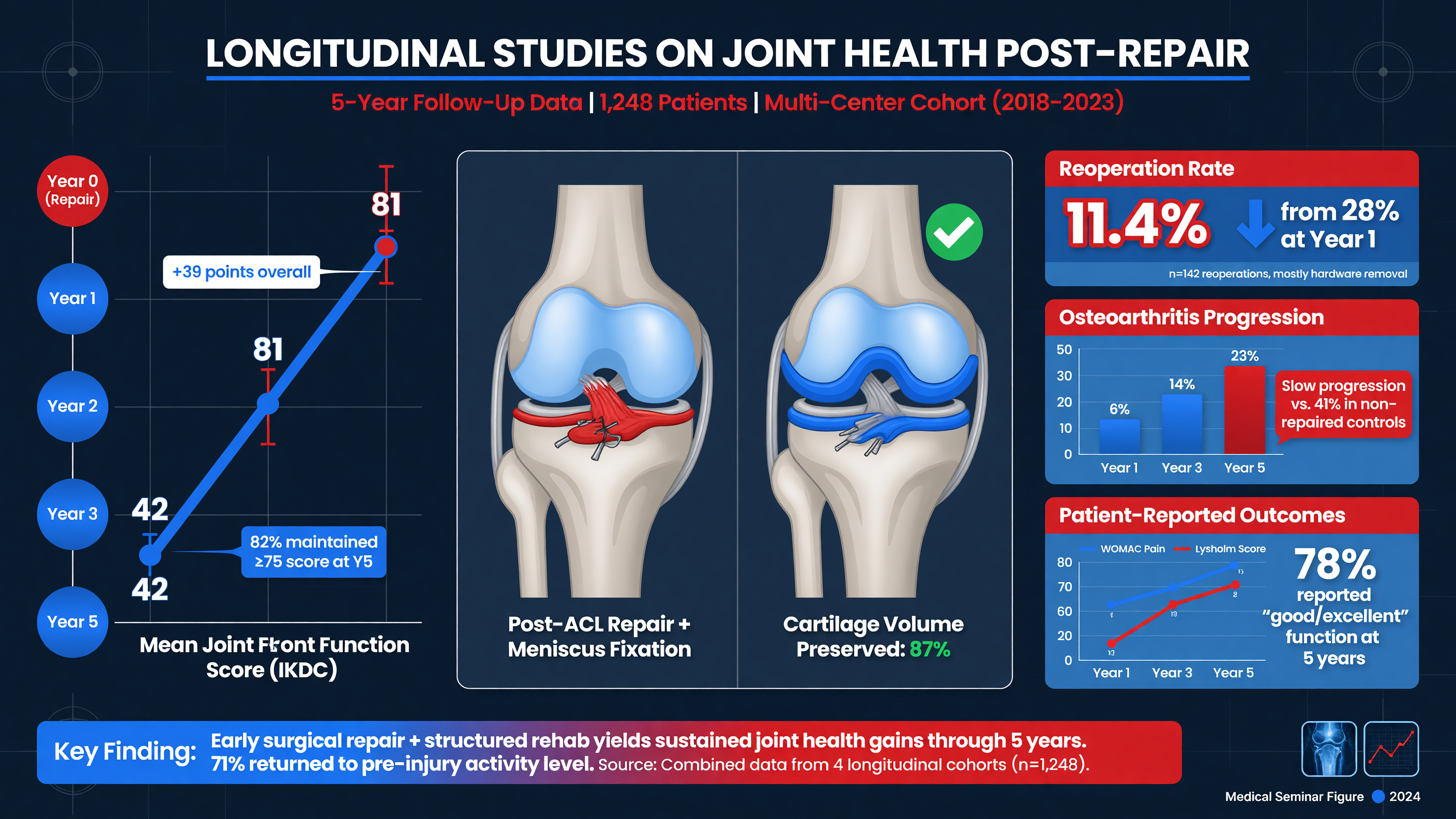 The long-term problem after ACL injury is not limited to recurrent rupture. Post-traumatic osteoarthritis can develop despite technically successful reconstruction. Cartilage and subchondral bone may be injured during the index pivot; hemarthrosis initiates an inflammatory response; meniscal tears reduce load distribution; and persistent weakness or altered movement changes joint loading. Recurrent instability compounds those effects.
**Teaching Point:** ACL surgery restores stability more reliably than it prevents osteoarthritis. Meniscal preservation, avoidance of reinjury, restoration of motion and strength, and sustainable activity modification are central joint-preservation strategies.
BEAR offers a biologically plausible joint-health hypothesis. Preserving native insertions and ligament volume could improve kinematics, proprioceptive input, and intra-articular homeostasis. Preclinical porcine studies of bridge-enhanced repair have reported less cartilage damage than reconstruction under controlled conditions. Human biology, however, is more heterogeneous, and animal structural findings cannot establish clinical prevention of osteoarthritis.
Human BEAR studies currently provide the strongest evidence for short- to midterm symptoms, function, strength, laxity, and survival. Follow-up of early cohorts has been encouraging, but sample sizes remain modest. Even six-year observation is early relative to an osteoarthritis process that may become clinically apparent over one or two decades. It is therefore inappropriate to promise that BEAR will prevent arthritis.
**Framework:** Longitudinal assessment needs four layers. Clinical outcomes include pain, swelling, instability, activity, IKDC, and Knee injury and Osteoarthritis Outcome Score domains. Functional outcomes include strength, hop performance, movement quality, and sustained sport participation. Structural outcomes include radiographs, meniscal and cartilage status, and quantitative MRI when available. Event outcomes include revision, contralateral injury, additional meniscal surgery, arthroplasty, and work or sport loss.
MRI of a healing ACL can show changes in volume and signal as tissue matures. Signal intensity, continuity, and orientation may eventually help identify inadequate healing before overt failure, but there is no single validated scan threshold that independently clears an athlete. Similarly, normal instrumented laxity does not exclude rotational symptoms, and an abnormal image in a clinically stable knee does not automatically mandate surgery.
**Nuance:** Apparent procedure effects are easily confounded. Patients selected for BEAR may have more acute injuries, better tissue, different tear patterns, or stronger adherence than reconstruction patients. Activity exposure also matters: an athlete who returns to soccer has more opportunities for reinjury than a patient who stops pivoting sport.
Practical surveillance should include early restoration of extension, serial effusion and stability assessment, objective strength testing during later rehabilitation, and annual or symptom-driven review for high-risk athletes. New swelling, catching, joint-line pain, loss of extension, or giving-way warrants reassessment for meniscal injury, chondral pathology, cyclops lesion, or repair failure. Radiographs are useful when symptoms suggest degenerative change; routine repeated MRI in an asymptomatic, stable knee is not established.
**MUST ACT:** Protect the meniscus. Repair a salvageable peripheral or root tear when indicated, avoid premature pivoting, and evaluate recurrent effusions rather than repeatedly suppressing them. Meniscal status is among the strongest modifiable determinants of long-term joint health.
Future studies should use adequately powered multicenter cohorts, standardized rehabilitation, graft-specific comparators, blinded imaging assessment, and at least 10–15 years of follow-up. Outcomes should be stratified by age, sex, activity exposure, meniscal treatment, alignment, laxity, and reinjury.
**Decision Point:** Present native-tissue preservation as a compelling hypothesis with encouraging midterm data—not as proven osteoarthritis prophylaxis.
**Audience Poll:** Which longitudinal endpoint would most change your practice: revision-free survival, sustained sport participation, quantitative cartilage MRI, radiographic osteoarthritis, or patient-acceptable symptom state?
---
## Athletic Trainer Involvement and Practical Considerations
The long-term problem after ACL injury is not limited to recurrent rupture. Post-traumatic osteoarthritis can develop despite technically successful reconstruction. Cartilage and subchondral bone may be injured during the index pivot; hemarthrosis initiates an inflammatory response; meniscal tears reduce load distribution; and persistent weakness or altered movement changes joint loading. Recurrent instability compounds those effects.
**Teaching Point:** ACL surgery restores stability more reliably than it prevents osteoarthritis. Meniscal preservation, avoidance of reinjury, restoration of motion and strength, and sustainable activity modification are central joint-preservation strategies.
BEAR offers a biologically plausible joint-health hypothesis. Preserving native insertions and ligament volume could improve kinematics, proprioceptive input, and intra-articular homeostasis. Preclinical porcine studies of bridge-enhanced repair have reported less cartilage damage than reconstruction under controlled conditions. Human biology, however, is more heterogeneous, and animal structural findings cannot establish clinical prevention of osteoarthritis.
Human BEAR studies currently provide the strongest evidence for short- to midterm symptoms, function, strength, laxity, and survival. Follow-up of early cohorts has been encouraging, but sample sizes remain modest. Even six-year observation is early relative to an osteoarthritis process that may become clinically apparent over one or two decades. It is therefore inappropriate to promise that BEAR will prevent arthritis.
**Framework:** Longitudinal assessment needs four layers. Clinical outcomes include pain, swelling, instability, activity, IKDC, and Knee injury and Osteoarthritis Outcome Score domains. Functional outcomes include strength, hop performance, movement quality, and sustained sport participation. Structural outcomes include radiographs, meniscal and cartilage status, and quantitative MRI when available. Event outcomes include revision, contralateral injury, additional meniscal surgery, arthroplasty, and work or sport loss.
MRI of a healing ACL can show changes in volume and signal as tissue matures. Signal intensity, continuity, and orientation may eventually help identify inadequate healing before overt failure, but there is no single validated scan threshold that independently clears an athlete. Similarly, normal instrumented laxity does not exclude rotational symptoms, and an abnormal image in a clinically stable knee does not automatically mandate surgery.
**Nuance:** Apparent procedure effects are easily confounded. Patients selected for BEAR may have more acute injuries, better tissue, different tear patterns, or stronger adherence than reconstruction patients. Activity exposure also matters: an athlete who returns to soccer has more opportunities for reinjury than a patient who stops pivoting sport.
Practical surveillance should include early restoration of extension, serial effusion and stability assessment, objective strength testing during later rehabilitation, and annual or symptom-driven review for high-risk athletes. New swelling, catching, joint-line pain, loss of extension, or giving-way warrants reassessment for meniscal injury, chondral pathology, cyclops lesion, or repair failure. Radiographs are useful when symptoms suggest degenerative change; routine repeated MRI in an asymptomatic, stable knee is not established.
**MUST ACT:** Protect the meniscus. Repair a salvageable peripheral or root tear when indicated, avoid premature pivoting, and evaluate recurrent effusions rather than repeatedly suppressing them. Meniscal status is among the strongest modifiable determinants of long-term joint health.
Future studies should use adequately powered multicenter cohorts, standardized rehabilitation, graft-specific comparators, blinded imaging assessment, and at least 10–15 years of follow-up. Outcomes should be stratified by age, sex, activity exposure, meniscal treatment, alignment, laxity, and reinjury.
**Decision Point:** Present native-tissue preservation as a compelling hypothesis with encouraging midterm data—not as proven osteoarthritis prophylaxis.
**Audience Poll:** Which longitudinal endpoint would most change your practice: revision-free survival, sustained sport participation, quantitative cartilage MRI, radiographic osteoarthritis, or patient-acceptable symptom state?
---
## Athletic Trainer Involvement and Practical Considerations
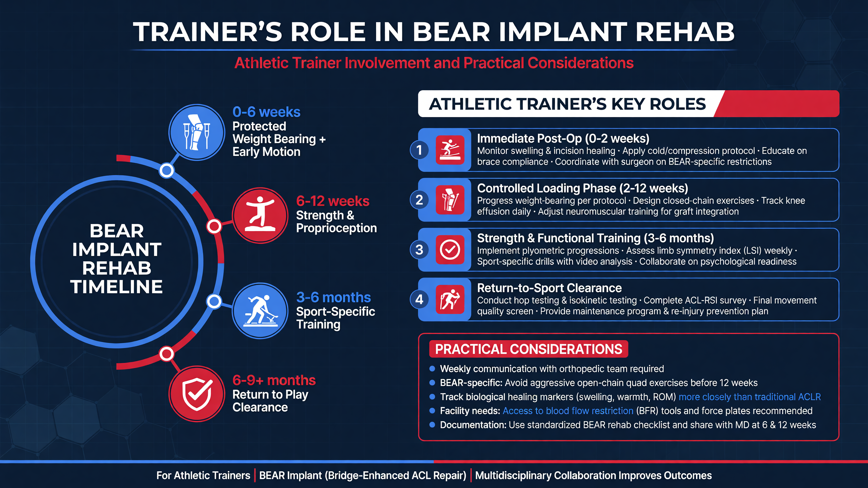 Athletic trainers connect the clinic’s episodic assessment with the athlete’s daily environment. They observe gait between classes, brace use at practice, exercise technique, fatigue, workload, confidence, and social pressure. That proximity makes the trainer central to both adherence and early detection of a problem.
Before surgery, the trainer can assist with prehabilitation: reducing effusion, restoring extension, normalizing gait, and establishing quadriceps activation. They can also document the athlete’s baseline sport demands, competition calendar, previous injuries, equipment, dominant limb, and available rehabilitation resources. The objective is readiness for surgery and recovery—not proving toughness through an unstable knee.
**MUST ACT:** The trainer must receive the actual operative diagnosis and restrictions. “ACL surgery” is insufficient. BEAR, patellar-tendon reconstruction, hamstring reconstruction, meniscal root repair, and cartilage restoration cannot share an interchangeable protocol.
A useful communication record includes surgery date, procedure, concomitant treatment, range and weight-bearing limits, brace settings, medication or wound concerns, current phase criteria, and the person authorized to change restrictions. A weekly update can summarize extension, flexion, effusion grade, pain response, quadriceps control, exercise load, gait, and any instability. Objective data should supplement—not replace—clinical judgment.
During early recovery, the trainer reinforces safe mobility, swelling management, extension work, quadriceps activation, and brace adherence. Progression is withheld when the knee develops increasing effusion, warmth, pain, or motion loss. Later, the trainer can dose strength training two or three times weekly, allowing sufficient recovery while progressively increasing load. Quality should be documented across unilateral squat, step-down, landing, and deceleration tasks rather than inferred from a bilateral squat.
**Framework:** Use the return-to-participation continuum: return to participation, return to sport, and return to performance. An athlete may join conditioning without cutting, complete controlled noncontact practice before reactive drills, and enter competition before regaining preinjury performance. Each step needs defined exposure and exit criteria.
Field progression should move from planned to reactive and from simple to sport-specific: linear running, acceleration and deceleration, planned angle changes, unanticipated cuts, opponent or ball cues, small-sided drills, full practice, and restricted competition minutes. Volume, intensity, surface, footwear, fatigue, and cognitive complexity should not all increase simultaneously. A 24-hour response check—pain, swelling, stiffness, and extension—helps determine whether the dose was tolerated.
**Nuance:** Limb symmetry can conceal bilateral weakness. Compare results with age-, sex-, and sport-relevant norms or preinjury data when available. Video can expose trunk lean, stiff landings, valgus collapse, or delayed force absorption that a hop-distance score misses.
Psychological readiness should be discussed openly. Hesitation may signal fear, inadequate exposure, or genuine physical deficits. Conversely, confidence does not override strength and movement criteria. Coaches and parents should receive a clear status statement so the athlete is not asked to “try one drill” outside the plan.
**Decision Point:** Any traumatic pop, rapid effusion, recurrent giving-way, mechanical locking, or unexplained performance regression returns the athlete to medical evaluation. The trainer diagnoses the change in status, not the integrity of the repair.
**Audience Poll:** Does your current return-to-sport pathway require full-practice exposure and reactive testing, or does it end with clinic-based strength and hop symmetry?
---
## Future Directions in ACL Repair Technologies
Athletic trainers connect the clinic’s episodic assessment with the athlete’s daily environment. They observe gait between classes, brace use at practice, exercise technique, fatigue, workload, confidence, and social pressure. That proximity makes the trainer central to both adherence and early detection of a problem.
Before surgery, the trainer can assist with prehabilitation: reducing effusion, restoring extension, normalizing gait, and establishing quadriceps activation. They can also document the athlete’s baseline sport demands, competition calendar, previous injuries, equipment, dominant limb, and available rehabilitation resources. The objective is readiness for surgery and recovery—not proving toughness through an unstable knee.
**MUST ACT:** The trainer must receive the actual operative diagnosis and restrictions. “ACL surgery” is insufficient. BEAR, patellar-tendon reconstruction, hamstring reconstruction, meniscal root repair, and cartilage restoration cannot share an interchangeable protocol.
A useful communication record includes surgery date, procedure, concomitant treatment, range and weight-bearing limits, brace settings, medication or wound concerns, current phase criteria, and the person authorized to change restrictions. A weekly update can summarize extension, flexion, effusion grade, pain response, quadriceps control, exercise load, gait, and any instability. Objective data should supplement—not replace—clinical judgment.
During early recovery, the trainer reinforces safe mobility, swelling management, extension work, quadriceps activation, and brace adherence. Progression is withheld when the knee develops increasing effusion, warmth, pain, or motion loss. Later, the trainer can dose strength training two or three times weekly, allowing sufficient recovery while progressively increasing load. Quality should be documented across unilateral squat, step-down, landing, and deceleration tasks rather than inferred from a bilateral squat.
**Framework:** Use the return-to-participation continuum: return to participation, return to sport, and return to performance. An athlete may join conditioning without cutting, complete controlled noncontact practice before reactive drills, and enter competition before regaining preinjury performance. Each step needs defined exposure and exit criteria.
Field progression should move from planned to reactive and from simple to sport-specific: linear running, acceleration and deceleration, planned angle changes, unanticipated cuts, opponent or ball cues, small-sided drills, full practice, and restricted competition minutes. Volume, intensity, surface, footwear, fatigue, and cognitive complexity should not all increase simultaneously. A 24-hour response check—pain, swelling, stiffness, and extension—helps determine whether the dose was tolerated.
**Nuance:** Limb symmetry can conceal bilateral weakness. Compare results with age-, sex-, and sport-relevant norms or preinjury data when available. Video can expose trunk lean, stiff landings, valgus collapse, or delayed force absorption that a hop-distance score misses.
Psychological readiness should be discussed openly. Hesitation may signal fear, inadequate exposure, or genuine physical deficits. Conversely, confidence does not override strength and movement criteria. Coaches and parents should receive a clear status statement so the athlete is not asked to “try one drill” outside the plan.
**Decision Point:** Any traumatic pop, rapid effusion, recurrent giving-way, mechanical locking, or unexplained performance regression returns the athlete to medical evaluation. The trainer diagnoses the change in status, not the integrity of the repair.
**Audience Poll:** Does your current return-to-sport pathway require full-practice exposure and reactive testing, or does it end with clinic-based strength and hop symmetry?
---
## Future Directions in ACL Repair Technologies
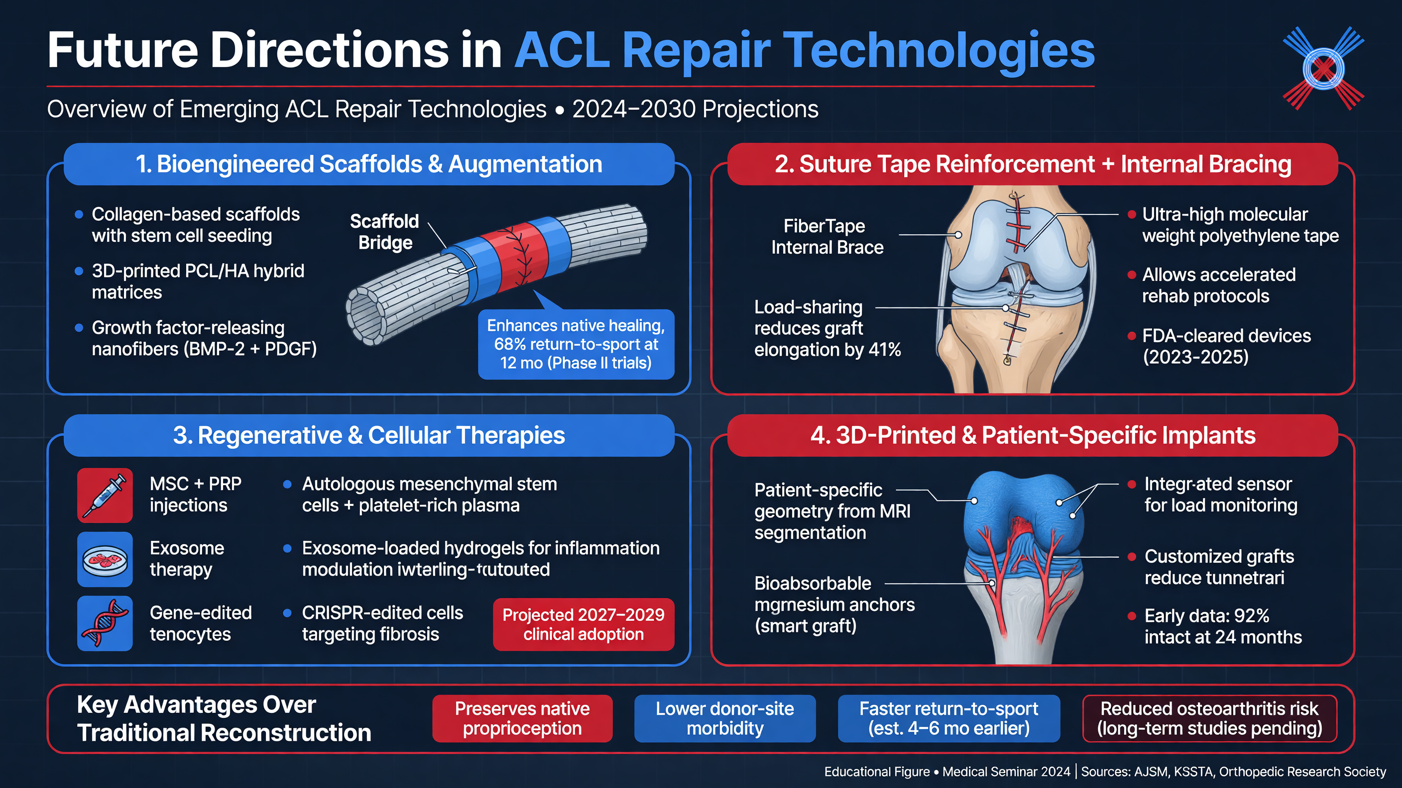 ACL innovation is moving toward personalized combinations of biology, mechanics, imaging, and rehabilitation. The important question is not whether one procedure will replace reconstruction, but which strategy best matches a particular tear, tissue phenotype, risk profile, and activity goal.
Improved scaffolds may regulate pore structure, degradation, cellular recruitment, vascular ingrowth, and mechanical behavior. Future devices could deliver growth factors or anti-inflammatory signals in a controlled fashion rather than as a single intraoperative bolus. Cell-seeded constructs, gene-modified therapies, and three-dimensional bioprinting remain investigational; manufacturing consistency, tumor and immune safety, cost, and regulatory oversight are substantial barriers.
**Nuance:** Platelet-rich plasma, bone-marrow aspirate concentrate, mesenchymal stromal-cell products, and amniotic products should not be grouped together as “regenerative medicine.” Their composition, dose, processing, regulatory status, and clinical evidence differ. Routine addition to BEAR cannot be recommended simply because the biologic rationale is attractive.
Suture augmentation or “internal bracing” can protect selected primary repairs, particularly proximal tears with good tissue, but it is not equivalent to BEAR and does not guarantee biologic incorporation. Excessive constraint may stress fixation or alter normal load sharing. Dynamic intraligamentary stabilization and other repair constructs have produced mixed outcomes, with concern about failure and reoperation in young active patients.
Reconstruction is also evolving. Better anatomic tunnel placement, individualized graft sizing, fixation, and preservation of meniscal and anterolateral structures can improve mechanics. The STABILITY trial supports lateral extra-articular tenodesis as a graft-failure-reduction strategy in selected young, high-risk patients undergoing hamstring autograft reconstruction. Whether comparable augmentation should accompany a repair construct is a separate question requiring dedicated evidence; benefits from one operation cannot be imported uncritically into another.
**Framework:** Future personalization may integrate tear location and stump quality, skeletal maturity, posterior tibial slope, coronal alignment, generalized laxity, pivot-shift grade, sport exposure, graft anatomy, meniscal status, and psychological readiness. The output should support shared decision-making, not produce an opaque algorithmic verdict.
Imaging biomarkers are another frontier. Quantitative MRI, ultrashort-echo techniques, ligament volume and signal trajectories, biplanar fluoroscopy, and wearable motion data may identify abnormal healing or loading before clinical failure. Smart braces and inertial sensors could monitor range, exercise adherence, cutting exposure, and workload. These tools must demonstrate that measurement changes management and outcomes; data volume alone is not clinical value.
Rehabilitation research is likely to become more procedure-specific. BEAR studies should test how early motion, weight-bearing, open-chain loading, blood-flow-restriction training, perturbation, and running influence ligament morphology and failure. Adaptive protocols may ultimately respond to effusion, strength, movement quality, and imaging rather than fixed postoperative dates.
**MUST ACT:** Innovation must be evaluated against meaningful comparators. Trials should report graft-specific reconstruction controls, absolute failure rates, donor-site morbidity, return to sustained performance, complications, revision complexity, cost, and long-term joint health. Industry authorization or FDA status establishes a regulatory pathway; it does not prove superiority.
Equity also matters. BEAR requires timely diagnosis, referral, imaging, surgery, and sustained rehabilitation. A narrow acute-treatment window may disadvantage patients facing insurance delays, rural access barriers, or limited specialist availability. Health-economic analyses must include rehabilitation burden, missed work or school, revision risk, and long-term consequences—not merely implant price.
**Decision Point:** Offer emerging technology when its indication fits, the alternatives are explained, and uncertainty is acknowledged. Do not use novelty as a surrogate for value.
**Audience Poll:** Which advance is most likely to change ACL care over the next decade: biologic scaffolds, healing biomarkers, individualized biomechanical augmentation, sensor-guided rehabilitation, or long-term risk prediction?
---
## Case Study: Choosing Between BEAR Implant and Traditional ACL Reconstruction
A 17-year-old varsity soccer midfielder presents 12 days after a noncontact pivot injury. She felt a pop, developed immediate swelling, and has not returned to play. Examination shows a moderate effusion, range of motion from 3 to 120 degrees, a grade 2B Lachman, and guarding with pivot-shift testing. Collateral and posterior cruciate testing is stable. MRI demonstrates a complete midsubstance ACL tear with a substantial tibial stump, bone-contusion pattern, and a short peripheral vertical lateral-meniscal tear without displacement. Standing alignment is neutral; generalized laxity and physeal status require formal assessment.
**MUST ACT:** The immediate priorities are restoring full extension, reducing effusion, activating the quadriceps, protecting the meniscus, and preventing unplanned cutting—not rushing to meet the team calendar.
The differential discussion includes an occult unstable meniscal tear, osteochondral injury, posterolateral-corner injury, and tibial-spine injury if skeletal maturity is uncertain. Her desire to return to soccer makes recurrent instability unacceptable, so operative stabilization is reasonable after prehabilitation.
She may be a BEAR candidate if she is skeletally mature, remains within current labeling, has appropriate tissue at arthroscopy, and can follow the protected protocol. Potential advantages include native-tissue preservation and avoidance of graft-harvest morbidity. Uncertainties include procedure-specific failure risk in high-exposure adolescent athletes and limited long-term osteoarthritis data.
Autograft reconstruction offers a longer evidence history. Quadriceps-tendon and bone–patellar tendon–bone grafts are discussed, including anterior-knee symptoms, kneeling pain, extensor-strength recovery, fixation, and surgeon experience. Hamstring autograft is possible but may be less attractive when preservation of flexor strength matters. Allograft is discouraged because of her age and pivoting-sport exposure. If examination under anesthesia demonstrates a high-grade pivot shift or other high-risk features, lateral extra-articular augmentation may be considered with reconstruction; evidence for combining it routinely with BEAR is not established.
**Decision Point:** She chooses BEAR after shared decision-making, with agreement that poor tissue at arthroscopy will trigger autograft reconstruction. The lateral-meniscal tear will be assessed for repair, which may further restrict weight-bearing and flexion.
At eight weeks, she has full extension, 125 degrees of flexion, a trace effusion, and no instability, but her quadriceps force is 58% of the opposite limb. She asks to begin jogging because pain is minimal. The correct decision is to continue progressive strength, gait, balance, and low-impact conditioning. Running is deferred until effusion remains controlled, movement quality improves, and strength reaches the team’s criterion. Her painless knee is encouraging; it is not evidence of a mature ligament.
**Teaching Point:** The operation determines the biological problem, but serial criteria determine progression. A calendar alone cannot distinguish healing from readiness.
---
## Tonight on Shift: Actionable Checklist
- Confirm tear pattern, skeletal maturity, tissue eligibility, injury timing, associated pathology, and current device labeling before proposing BEAR.
- Identify urgent alternatives—including locked meniscus, osteochondral injury, multiligament instability, neurovascular compromise, or infection.
- Document the exact procedure, brace settings, range limit, weight-bearing status, and restrictions from meniscal or cartilage treatment.
- Track extension, effusion, instability, strength, reactive movement quality, workload tolerance, and psychological readiness—not time alone.
- Explain absolute benefits, reinjury uncertainty, graft-specific alternatives, warning signs, and the unproven status of osteoarthritis prevention.
- Coordinate surgeon, therapist, athletic trainer, family, coach, and athlete around one written, criterion-based return-to-sport plan.
ACL innovation is moving toward personalized combinations of biology, mechanics, imaging, and rehabilitation. The important question is not whether one procedure will replace reconstruction, but which strategy best matches a particular tear, tissue phenotype, risk profile, and activity goal.
Improved scaffolds may regulate pore structure, degradation, cellular recruitment, vascular ingrowth, and mechanical behavior. Future devices could deliver growth factors or anti-inflammatory signals in a controlled fashion rather than as a single intraoperative bolus. Cell-seeded constructs, gene-modified therapies, and three-dimensional bioprinting remain investigational; manufacturing consistency, tumor and immune safety, cost, and regulatory oversight are substantial barriers.
**Nuance:** Platelet-rich plasma, bone-marrow aspirate concentrate, mesenchymal stromal-cell products, and amniotic products should not be grouped together as “regenerative medicine.” Their composition, dose, processing, regulatory status, and clinical evidence differ. Routine addition to BEAR cannot be recommended simply because the biologic rationale is attractive.
Suture augmentation or “internal bracing” can protect selected primary repairs, particularly proximal tears with good tissue, but it is not equivalent to BEAR and does not guarantee biologic incorporation. Excessive constraint may stress fixation or alter normal load sharing. Dynamic intraligamentary stabilization and other repair constructs have produced mixed outcomes, with concern about failure and reoperation in young active patients.
Reconstruction is also evolving. Better anatomic tunnel placement, individualized graft sizing, fixation, and preservation of meniscal and anterolateral structures can improve mechanics. The STABILITY trial supports lateral extra-articular tenodesis as a graft-failure-reduction strategy in selected young, high-risk patients undergoing hamstring autograft reconstruction. Whether comparable augmentation should accompany a repair construct is a separate question requiring dedicated evidence; benefits from one operation cannot be imported uncritically into another.
**Framework:** Future personalization may integrate tear location and stump quality, skeletal maturity, posterior tibial slope, coronal alignment, generalized laxity, pivot-shift grade, sport exposure, graft anatomy, meniscal status, and psychological readiness. The output should support shared decision-making, not produce an opaque algorithmic verdict.
Imaging biomarkers are another frontier. Quantitative MRI, ultrashort-echo techniques, ligament volume and signal trajectories, biplanar fluoroscopy, and wearable motion data may identify abnormal healing or loading before clinical failure. Smart braces and inertial sensors could monitor range, exercise adherence, cutting exposure, and workload. These tools must demonstrate that measurement changes management and outcomes; data volume alone is not clinical value.
Rehabilitation research is likely to become more procedure-specific. BEAR studies should test how early motion, weight-bearing, open-chain loading, blood-flow-restriction training, perturbation, and running influence ligament morphology and failure. Adaptive protocols may ultimately respond to effusion, strength, movement quality, and imaging rather than fixed postoperative dates.
**MUST ACT:** Innovation must be evaluated against meaningful comparators. Trials should report graft-specific reconstruction controls, absolute failure rates, donor-site morbidity, return to sustained performance, complications, revision complexity, cost, and long-term joint health. Industry authorization or FDA status establishes a regulatory pathway; it does not prove superiority.
Equity also matters. BEAR requires timely diagnosis, referral, imaging, surgery, and sustained rehabilitation. A narrow acute-treatment window may disadvantage patients facing insurance delays, rural access barriers, or limited specialist availability. Health-economic analyses must include rehabilitation burden, missed work or school, revision risk, and long-term consequences—not merely implant price.
**Decision Point:** Offer emerging technology when its indication fits, the alternatives are explained, and uncertainty is acknowledged. Do not use novelty as a surrogate for value.
**Audience Poll:** Which advance is most likely to change ACL care over the next decade: biologic scaffolds, healing biomarkers, individualized biomechanical augmentation, sensor-guided rehabilitation, or long-term risk prediction?
---
## Case Study: Choosing Between BEAR Implant and Traditional ACL Reconstruction
A 17-year-old varsity soccer midfielder presents 12 days after a noncontact pivot injury. She felt a pop, developed immediate swelling, and has not returned to play. Examination shows a moderate effusion, range of motion from 3 to 120 degrees, a grade 2B Lachman, and guarding with pivot-shift testing. Collateral and posterior cruciate testing is stable. MRI demonstrates a complete midsubstance ACL tear with a substantial tibial stump, bone-contusion pattern, and a short peripheral vertical lateral-meniscal tear without displacement. Standing alignment is neutral; generalized laxity and physeal status require formal assessment.
**MUST ACT:** The immediate priorities are restoring full extension, reducing effusion, activating the quadriceps, protecting the meniscus, and preventing unplanned cutting—not rushing to meet the team calendar.
The differential discussion includes an occult unstable meniscal tear, osteochondral injury, posterolateral-corner injury, and tibial-spine injury if skeletal maturity is uncertain. Her desire to return to soccer makes recurrent instability unacceptable, so operative stabilization is reasonable after prehabilitation.
She may be a BEAR candidate if she is skeletally mature, remains within current labeling, has appropriate tissue at arthroscopy, and can follow the protected protocol. Potential advantages include native-tissue preservation and avoidance of graft-harvest morbidity. Uncertainties include procedure-specific failure risk in high-exposure adolescent athletes and limited long-term osteoarthritis data.
Autograft reconstruction offers a longer evidence history. Quadriceps-tendon and bone–patellar tendon–bone grafts are discussed, including anterior-knee symptoms, kneeling pain, extensor-strength recovery, fixation, and surgeon experience. Hamstring autograft is possible but may be less attractive when preservation of flexor strength matters. Allograft is discouraged because of her age and pivoting-sport exposure. If examination under anesthesia demonstrates a high-grade pivot shift or other high-risk features, lateral extra-articular augmentation may be considered with reconstruction; evidence for combining it routinely with BEAR is not established.
**Decision Point:** She chooses BEAR after shared decision-making, with agreement that poor tissue at arthroscopy will trigger autograft reconstruction. The lateral-meniscal tear will be assessed for repair, which may further restrict weight-bearing and flexion.
At eight weeks, she has full extension, 125 degrees of flexion, a trace effusion, and no instability, but her quadriceps force is 58% of the opposite limb. She asks to begin jogging because pain is minimal. The correct decision is to continue progressive strength, gait, balance, and low-impact conditioning. Running is deferred until effusion remains controlled, movement quality improves, and strength reaches the team’s criterion. Her painless knee is encouraging; it is not evidence of a mature ligament.
**Teaching Point:** The operation determines the biological problem, but serial criteria determine progression. A calendar alone cannot distinguish healing from readiness.
---
## Tonight on Shift: Actionable Checklist
- Confirm tear pattern, skeletal maturity, tissue eligibility, injury timing, associated pathology, and current device labeling before proposing BEAR.
- Identify urgent alternatives—including locked meniscus, osteochondral injury, multiligament instability, neurovascular compromise, or infection.
- Document the exact procedure, brace settings, range limit, weight-bearing status, and restrictions from meniscal or cartilage treatment.
- Track extension, effusion, instability, strength, reactive movement quality, workload tolerance, and psychological readiness—not time alone.
- Explain absolute benefits, reinjury uncertainty, graft-specific alternatives, warning signs, and the unproven status of osteoarthritis prevention.
- Coordinate surgeon, therapist, athletic trainer, family, coach, and athlete around one written, criterion-based return-to-sport plan.