# Calcium Pyrophosphate Deposition Disease: Diagnostic Challenges and Treatment Innovations
## Learning Objectives
- Define the modern clinical phenotypes and nomenclature of calcium pyrophosphate deposition disease.
- Explain the molecular pathways responsible for CPP crystal formation and inflammation.
- Distinguish acute and chronic CPPD from septic arthritis, gout, rheumatoid arthritis, osteoarthritis, and polymyalgia rheumatica.
- Interpret synovial-fluid, radiographic, ultrasound, and CT findings in suspected CPPD.
- Select acute and preventive therapy according to joint distribution, comorbidity, and medication risk.
- Appraise the limited evidence for anakinra, tocilizumab, hydroxychloroquine, methotrexate, and crystal-directed therapies.
- Apply a practical diagnostic and management algorithm during acute care.
---
## Overview of Calcium Pyrophosphate Deposition Disease (CPPD)
 ### Duration: 10 minutes
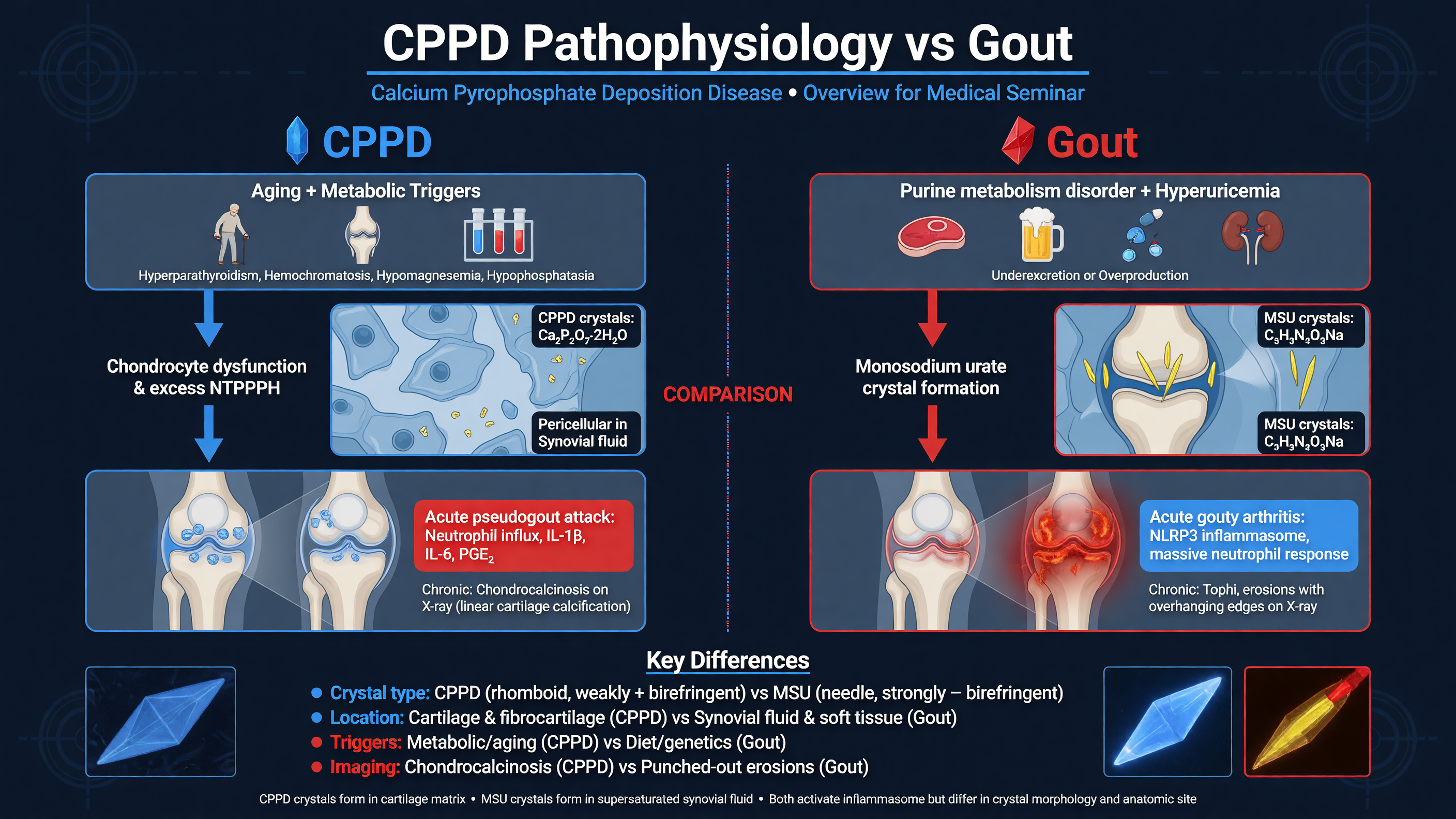
Calcium pyrophosphate deposition is not a single presentation. CPPD is the umbrella term encompassing asymptomatic CPP deposition, acute CPP crystal arthritis, osteoarthritis with CPPD, and chronic CPP crystal inflammatory arthritis. “Pseudogout” describes only the acute gout-like phenotype and is increasingly avoided because it obscures the broader disease spectrum. Likewise, chondrocalcinosis is an imaging or histologic description of cartilage calcification; it is neither synonymous with symptomatic CPPD nor completely specific for CPP crystals. A patient may have extensive chondrocalcinosis without inflammatory disease, while a patient with crystal-proven CPP arthritis may have unrevealing radiographs (PMID: [21216817](https://pubmed.ncbi.nlm.nih.gov/21216817/)).
**Teaching Point:** Separate three questions: Are CPP deposits present? Are they producing the current symptoms? Is another process—especially infection—better able to explain the presentation?
CPP crystals form extracellularly within hyaline cartilage, fibrocartilage, and areas of chondroid metaplasia. The contemporary model begins with ANKH-mediated ATP efflux from chondrocytes and matrix vesicles. Ectonucleotide pyrophosphatase/phosphodiesterase-1, or ENPP1, converts extracellular ATP to AMP and inorganic pyrophosphate. Tissue-nonspecific alkaline phosphatase, or TNAP, normally hydrolyzes pyrophosphate. When production exceeds clearance, extracellular pyrophosphate complexes with calcium and nucleates CPP crystals within an aging or injured cartilage matrix. This is more precise than the older shorthand that ANKH simply transports pyrophosphate (PMID: [35147247](https://pubmed.ncbi.nlm.nih.gov/35147247/)).
Once shed into the joint cavity, CPP crystals activate innate immunity through membrane interactions, Toll-like receptor signaling, and the NLRP3–ASC–caspase-1 inflammasome. Mature IL-1β promotes endothelial and synovial activation, while IL-8 and other mediators recruit neutrophils. Neutrophil phagocytosis, protease release, oxidative signaling, and extracellular-trap formation amplify the flare. IL-6 is produced downstream and provides a rationale—but not yet high-level clinical proof—for IL-6 blockade. This inflammatory cascade resembles gout, but gout originates from monosodium urate deposition driven by hyperuricemia. CPPD has no equivalent serum biomarker, treat-to-target concentration, or established crystal-depleting therapy (PMIDs: [39508814](https://pubmed.ncbi.nlm.nih.gov/39508814/), [39775910](https://pubmed.ncbi.nlm.nih.gov/39775910/)).
**Framework:** Think of CPPD as two linked problems: abnormal crystal formation in cartilage and episodic or persistent immune responses when those crystals interact with synovium.
Age is the dominant epidemiologic risk. Sporadic CPPD is unusual before age 60 and becomes progressively more common in later life. Prior trauma, meniscectomy, joint surgery, and osteoarthritis create a local environment favorable to deposition. Established systemic associations include hereditary hemochromatosis, primary or secondary hyperparathyroidism, persistent hypomagnesemia—including Gitelman syndrome—and hypophosphatasia. Low magnesium reduces TNAP activity; deficient TNAP activity in hypophosphatasia directly impairs pyrophosphate clearance. Familial early-onset disease may involve variants in ANKH or, more rarely, TNFRSF11B.
**MUST ACT:** In onset before approximately age 60, unusually extensive polyarticular disease, or a strong family history, investigate treatable contributors. A practical panel includes calcium, magnesium, phosphate, alkaline phosphatase, parathyroid hormone, ferritin, transferrin saturation, and renal function. Persistently low alkaline phosphatase should prompt consideration of hypophosphatasia; elevated transferrin saturation should trigger evaluation for hemochromatosis. Thyroid testing is reasonable when clinically indicated, but the association with hypothyroidism is less consistent.
Treating a metabolic disorder is important for general health and may reduce an ongoing driver, but it does not reliably remove established crystals. Similarly, the association between CPPD and osteoarthritis is real, but directionality remains uncertain: damaged cartilage may foster deposition, while crystals may amplify synovitis and cartilage catabolism. Current evidence does not justify telling patients that every calcified joint will inevitably undergo rapid destruction (PMID: [39089298](https://pubmed.ncbi.nlm.nih.gov/39089298/)).
**Nuance:** CPPD is common in older adults, but “age-related” should not become a diagnostic stopping point when the distribution, severity, or age of onset is atypical.
**Audience Poll:** Which finding would most strongly prompt a secondary-cause evaluation: a first knee flare at age 88, incidental meniscal calcification at age 82, recurrent polyarticular CPP arthritis at age 52, or isolated hand osteoarthritis at age 70?
---
## Clinical Presentation: From Acute to Chronic CPPD
### Duration: 10 minutes
Calcium pyrophosphate deposition is not a single presentation. CPPD is the umbrella term encompassing asymptomatic CPP deposition, acute CPP crystal arthritis, osteoarthritis with CPPD, and chronic CPP crystal inflammatory arthritis. “Pseudogout” describes only the acute gout-like phenotype and is increasingly avoided because it obscures the broader disease spectrum. Likewise, chondrocalcinosis is an imaging or histologic description of cartilage calcification; it is neither synonymous with symptomatic CPPD nor completely specific for CPP crystals. A patient may have extensive chondrocalcinosis without inflammatory disease, while a patient with crystal-proven CPP arthritis may have unrevealing radiographs (PMID: [21216817](https://pubmed.ncbi.nlm.nih.gov/21216817/)).
**Teaching Point:** Separate three questions: Are CPP deposits present? Are they producing the current symptoms? Is another process—especially infection—better able to explain the presentation?
CPP crystals form extracellularly within hyaline cartilage, fibrocartilage, and areas of chondroid metaplasia. The contemporary model begins with ANKH-mediated ATP efflux from chondrocytes and matrix vesicles. Ectonucleotide pyrophosphatase/phosphodiesterase-1, or ENPP1, converts extracellular ATP to AMP and inorganic pyrophosphate. Tissue-nonspecific alkaline phosphatase, or TNAP, normally hydrolyzes pyrophosphate. When production exceeds clearance, extracellular pyrophosphate complexes with calcium and nucleates CPP crystals within an aging or injured cartilage matrix. This is more precise than the older shorthand that ANKH simply transports pyrophosphate (PMID: [35147247](https://pubmed.ncbi.nlm.nih.gov/35147247/)).
Once shed into the joint cavity, CPP crystals activate innate immunity through membrane interactions, Toll-like receptor signaling, and the NLRP3–ASC–caspase-1 inflammasome. Mature IL-1β promotes endothelial and synovial activation, while IL-8 and other mediators recruit neutrophils. Neutrophil phagocytosis, protease release, oxidative signaling, and extracellular-trap formation amplify the flare. IL-6 is produced downstream and provides a rationale—but not yet high-level clinical proof—for IL-6 blockade. This inflammatory cascade resembles gout, but gout originates from monosodium urate deposition driven by hyperuricemia. CPPD has no equivalent serum biomarker, treat-to-target concentration, or established crystal-depleting therapy (PMIDs: [39508814](https://pubmed.ncbi.nlm.nih.gov/39508814/), [39775910](https://pubmed.ncbi.nlm.nih.gov/39775910/)).
**Framework:** Think of CPPD as two linked problems: abnormal crystal formation in cartilage and episodic or persistent immune responses when those crystals interact with synovium.
Age is the dominant epidemiologic risk. Sporadic CPPD is unusual before age 60 and becomes progressively more common in later life. Prior trauma, meniscectomy, joint surgery, and osteoarthritis create a local environment favorable to deposition. Established systemic associations include hereditary hemochromatosis, primary or secondary hyperparathyroidism, persistent hypomagnesemia—including Gitelman syndrome—and hypophosphatasia. Low magnesium reduces TNAP activity; deficient TNAP activity in hypophosphatasia directly impairs pyrophosphate clearance. Familial early-onset disease may involve variants in ANKH or, more rarely, TNFRSF11B.
**MUST ACT:** In onset before approximately age 60, unusually extensive polyarticular disease, or a strong family history, investigate treatable contributors. A practical panel includes calcium, magnesium, phosphate, alkaline phosphatase, parathyroid hormone, ferritin, transferrin saturation, and renal function. Persistently low alkaline phosphatase should prompt consideration of hypophosphatasia; elevated transferrin saturation should trigger evaluation for hemochromatosis. Thyroid testing is reasonable when clinically indicated, but the association with hypothyroidism is less consistent.
Treating a metabolic disorder is important for general health and may reduce an ongoing driver, but it does not reliably remove established crystals. Similarly, the association between CPPD and osteoarthritis is real, but directionality remains uncertain: damaged cartilage may foster deposition, while crystals may amplify synovitis and cartilage catabolism. Current evidence does not justify telling patients that every calcified joint will inevitably undergo rapid destruction (PMID: [39089298](https://pubmed.ncbi.nlm.nih.gov/39089298/)).
**Nuance:** CPPD is common in older adults, but “age-related” should not become a diagnostic stopping point when the distribution, severity, or age of onset is atypical.
**Audience Poll:** Which finding would most strongly prompt a secondary-cause evaluation: a first knee flare at age 88, incidental meniscal calcification at age 82, recurrent polyarticular CPP arthritis at age 52, or isolated hand osteoarthritis at age 70?
---
## Clinical Presentation: From Acute to Chronic CPPD
 ### Duration: 15 minutes
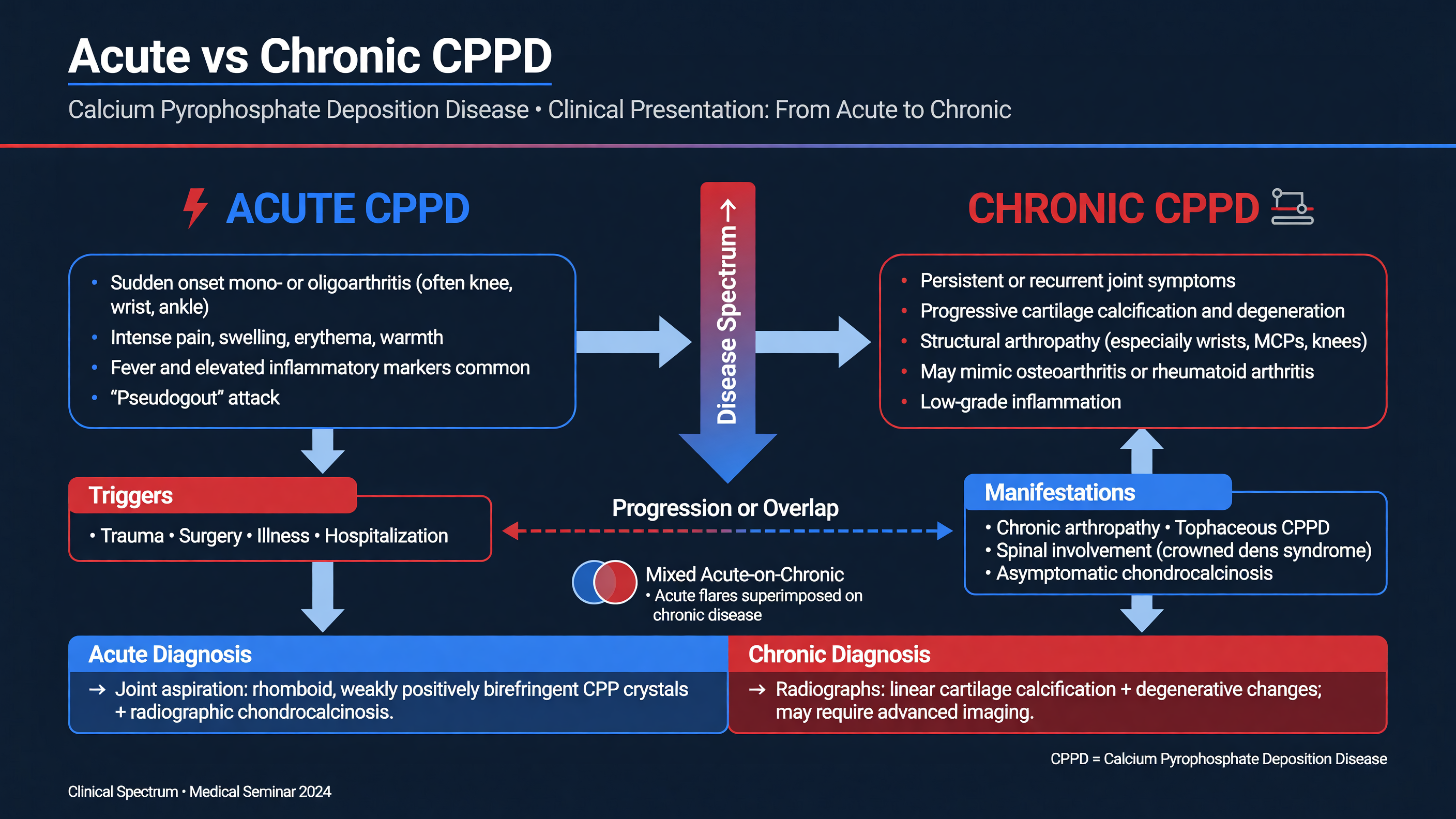
Acute CPP crystal arthritis usually presents with rapidly progressive pain, warmth, swelling, erythema, and loss of motion in one or several joints. The knee and wrist are signature locations; ankles, elbows, shoulders, and metacarpophalangeal joints are also affected. First-metatarsophalangeal disease is possible but less characteristic than in gout. Attacks may follow acute illness, hospitalization, surgery, trauma, or rapid calcium shifts such as those occurring after parathyroidectomy. Compared with gout, CPP flares may take longer to peak and can persist for days to several weeks, but tempo and joint location are only probabilistic clues.
Fever, delirium, peripheral leukocytosis, and striking CRP or ESR elevation are well-described, particularly in frail older adults. Synovial leukocyte counts can enter a range commonly associated with infection. Conversely, septic arthritis may occur without fever or an extreme cell count.
**MUST ACT:** Treat an acutely hot, swollen joint as potentially infected until septic arthritis has been reasonably excluded. When feasible, aspirate before antibiotics or intra-articular glucocorticoid and send cell count with differential, Gram stain, culture, and crystal analysis. Finding CPP crystals does not sterilize the joint.
**Decision Point:** A stereotyped recurrent wrist flare in a stable patient may be managed differently from a first febrile knee effusion in an immunosuppressed patient. Diagnostic certainty should rise with the consequence of being wrong.
Osteoarthritis with CPPD generally produces activity-related pain, stiffness, crepitus, and loss of function, sometimes punctuated by acute inflammatory attacks. Distribution is often more informative than severity. CPPD-associated structural disease may involve the radiocarpal or midcarpal wrist, second and third MCP joints, elbow, shoulder, ankle, or lateral and patellofemoral knee compartments—sites or patterns less typical of primary osteoarthritis. Scapholunate widening, scaphotrapeziotrapezoid degeneration, subchondral cysts, and hook-like metacarpal-head osteophytes are useful clues. Hook osteophytes at MCP2–3 should also trigger consideration of hemochromatosis.
Chronic CPP crystal inflammatory arthritis produces persistent or fluctuating synovitis, often involving wrists, MCP joints, knees, ankles, or shoulders. It may be symmetric, cause prolonged morning stiffness, and respond to glucocorticoids, thereby resembling late-onset seronegative rheumatoid arthritis or polymyalgia rheumatica. CPPD is underrecognized in precisely these chronic phenotypes (PMID: [36943699](https://pubmed.ncbi.nlm.nih.gov/36943699/)).
**Teaching Point:** Uncomplicated CPPD is not classically a marginally erosive arthritis. It can produce severe joint-space loss, remodeling, cysts, attrition, instability, and deformity, but sharply defined marginal erosions should prompt reconsideration of rheumatoid arthritis, gout, or infection.
Rheumatoid factor becomes less specific with age. Negative RF and anti-CCP tests do not prove CPPD, and CPPD can coexist with rheumatoid arthritis. Persistent symmetric small-joint synovitis, high-titer anti-CCP antibodies, marginal erosions, periarticular osteopenia, nodules, or another extra-articular RA feature strengthen the case for coexisting RA. Conversely, late onset, episodic attacks, knee or wrist predominance, characteristic cartilage deposits, and OA-pattern MCP or wrist damage favor CPPD.
Crowned dens syndrome is the characteristic axial presentation. Patients develop acute or subacute severe upper-cervical or occipital pain, markedly restricted rotation, elevated inflammatory markers, and sometimes fever. The syndrome requires matching symptoms with periodontoid calcification on CT; incidental calcification alone is insufficient. It may mimic meningitis, giant-cell arteritis, polymyalgia rheumatica, discitis, epidural infection, or cervical fracture. CPP deposits elsewhere in the spine can also cause facet-joint inflammation, radicular symptoms, or a retro-odontoid mass.
**Framework:** Classify the active phenotype before treating: acute mono/oligoarthritis, recurrent flares, persistent inflammatory arthritis, OA-dominant disease, or axial CPPD. A patient may move between categories.
**Nuance:** Glucocorticoid responsiveness does not distinguish CPPD from RA or PMR. It confirms steroid-responsive inflammation, not its cause.
**Audience Poll:** In an 80-year-old with six months of symmetric wrist and MCP synovitis, negative anti-CCP antibodies, and no erosions, what would most change your diagnosis: a positive rheumatoid factor, meniscal and triangular-fibrocartilage CPP deposits, a brisk prednisone response, or an elevated CRP?
---
## Imaging Modalities: Role of Ultrasound in CPPD
### Duration: 15 minutes
Acute CPP crystal arthritis usually presents with rapidly progressive pain, warmth, swelling, erythema, and loss of motion in one or several joints. The knee and wrist are signature locations; ankles, elbows, shoulders, and metacarpophalangeal joints are also affected. First-metatarsophalangeal disease is possible but less characteristic than in gout. Attacks may follow acute illness, hospitalization, surgery, trauma, or rapid calcium shifts such as those occurring after parathyroidectomy. Compared with gout, CPP flares may take longer to peak and can persist for days to several weeks, but tempo and joint location are only probabilistic clues.
Fever, delirium, peripheral leukocytosis, and striking CRP or ESR elevation are well-described, particularly in frail older adults. Synovial leukocyte counts can enter a range commonly associated with infection. Conversely, septic arthritis may occur without fever or an extreme cell count.
**MUST ACT:** Treat an acutely hot, swollen joint as potentially infected until septic arthritis has been reasonably excluded. When feasible, aspirate before antibiotics or intra-articular glucocorticoid and send cell count with differential, Gram stain, culture, and crystal analysis. Finding CPP crystals does not sterilize the joint.
**Decision Point:** A stereotyped recurrent wrist flare in a stable patient may be managed differently from a first febrile knee effusion in an immunosuppressed patient. Diagnostic certainty should rise with the consequence of being wrong.
Osteoarthritis with CPPD generally produces activity-related pain, stiffness, crepitus, and loss of function, sometimes punctuated by acute inflammatory attacks. Distribution is often more informative than severity. CPPD-associated structural disease may involve the radiocarpal or midcarpal wrist, second and third MCP joints, elbow, shoulder, ankle, or lateral and patellofemoral knee compartments—sites or patterns less typical of primary osteoarthritis. Scapholunate widening, scaphotrapeziotrapezoid degeneration, subchondral cysts, and hook-like metacarpal-head osteophytes are useful clues. Hook osteophytes at MCP2–3 should also trigger consideration of hemochromatosis.
Chronic CPP crystal inflammatory arthritis produces persistent or fluctuating synovitis, often involving wrists, MCP joints, knees, ankles, or shoulders. It may be symmetric, cause prolonged morning stiffness, and respond to glucocorticoids, thereby resembling late-onset seronegative rheumatoid arthritis or polymyalgia rheumatica. CPPD is underrecognized in precisely these chronic phenotypes (PMID: [36943699](https://pubmed.ncbi.nlm.nih.gov/36943699/)).
**Teaching Point:** Uncomplicated CPPD is not classically a marginally erosive arthritis. It can produce severe joint-space loss, remodeling, cysts, attrition, instability, and deformity, but sharply defined marginal erosions should prompt reconsideration of rheumatoid arthritis, gout, or infection.
Rheumatoid factor becomes less specific with age. Negative RF and anti-CCP tests do not prove CPPD, and CPPD can coexist with rheumatoid arthritis. Persistent symmetric small-joint synovitis, high-titer anti-CCP antibodies, marginal erosions, periarticular osteopenia, nodules, or another extra-articular RA feature strengthen the case for coexisting RA. Conversely, late onset, episodic attacks, knee or wrist predominance, characteristic cartilage deposits, and OA-pattern MCP or wrist damage favor CPPD.
Crowned dens syndrome is the characteristic axial presentation. Patients develop acute or subacute severe upper-cervical or occipital pain, markedly restricted rotation, elevated inflammatory markers, and sometimes fever. The syndrome requires matching symptoms with periodontoid calcification on CT; incidental calcification alone is insufficient. It may mimic meningitis, giant-cell arteritis, polymyalgia rheumatica, discitis, epidural infection, or cervical fracture. CPP deposits elsewhere in the spine can also cause facet-joint inflammation, radicular symptoms, or a retro-odontoid mass.
**Framework:** Classify the active phenotype before treating: acute mono/oligoarthritis, recurrent flares, persistent inflammatory arthritis, OA-dominant disease, or axial CPPD. A patient may move between categories.
**Nuance:** Glucocorticoid responsiveness does not distinguish CPPD from RA or PMR. It confirms steroid-responsive inflammation, not its cause.
**Audience Poll:** In an 80-year-old with six months of symmetric wrist and MCP synovitis, negative anti-CCP antibodies, and no erosions, what would most change your diagnosis: a positive rheumatoid factor, meniscal and triangular-fibrocartilage CPP deposits, a brisk prednisone response, or an elevated CRP?
---
## Imaging Modalities: Role of Ultrasound in CPPD
 ### Duration: 15 minutes
Synovial-fluid identification of CPP crystals remains the most specific practical evidence for peripheral CPPD. Under compensated polarized-light microscopy, crystals are typically short rods, rhomboids, or parallelepipeds with weak positive birefringence; some appear nonbirefringent. They may be intracellular or extracellular. CPP crystals are smaller, less strongly birefringent, and more easily missed than monosodium urate crystals, so a negative examination—especially of scant fluid or by an inexperienced observer—does not exclude disease.
**MUST ACT:** Imaging can support CPPD, guide aspiration, and map disease, but it cannot exclude septic arthritis in an acutely inflamed joint. Culture the aspirate whenever infection is clinically plausible.
Conventional radiography remains inexpensive, widely available, and useful for showing the entire joint. Characteristic deposits are linear or punctate calcifications within hyaline cartilage or fibrocartilage. High-yield locations include the knee menisci and femoral cartilage, wrist triangular fibrocartilage complex and intrinsic ligaments, pubic symphysis, acetabular labrum, and MCP joints. Radiography also demonstrates associated joint-space loss, cysts, osteophytes, scapholunate instability, or atypically distributed OA.
Its major limitation is sensitivity. A comparative meta-analysis using synovial fluid or histology as reference found pooled sensitivity and specificity of approximately 0.47 and 0.95 for radiography versus 0.85 and 0.87 for ultrasound, although study heterogeneity was substantial (PMID: [33577959](https://pubmed.ncbi.nlm.nih.gov/33577959/)). Thus, a positive characteristic radiograph is persuasive, but a negative film is not a rule-out test.
**Teaching Point:** Ultrasound is most valuable when it answers a targeted question: Is there a deposit in cartilage or fibrocartilage? Is the joint actively inflamed? Can ultrasound guide aspiration or injection?
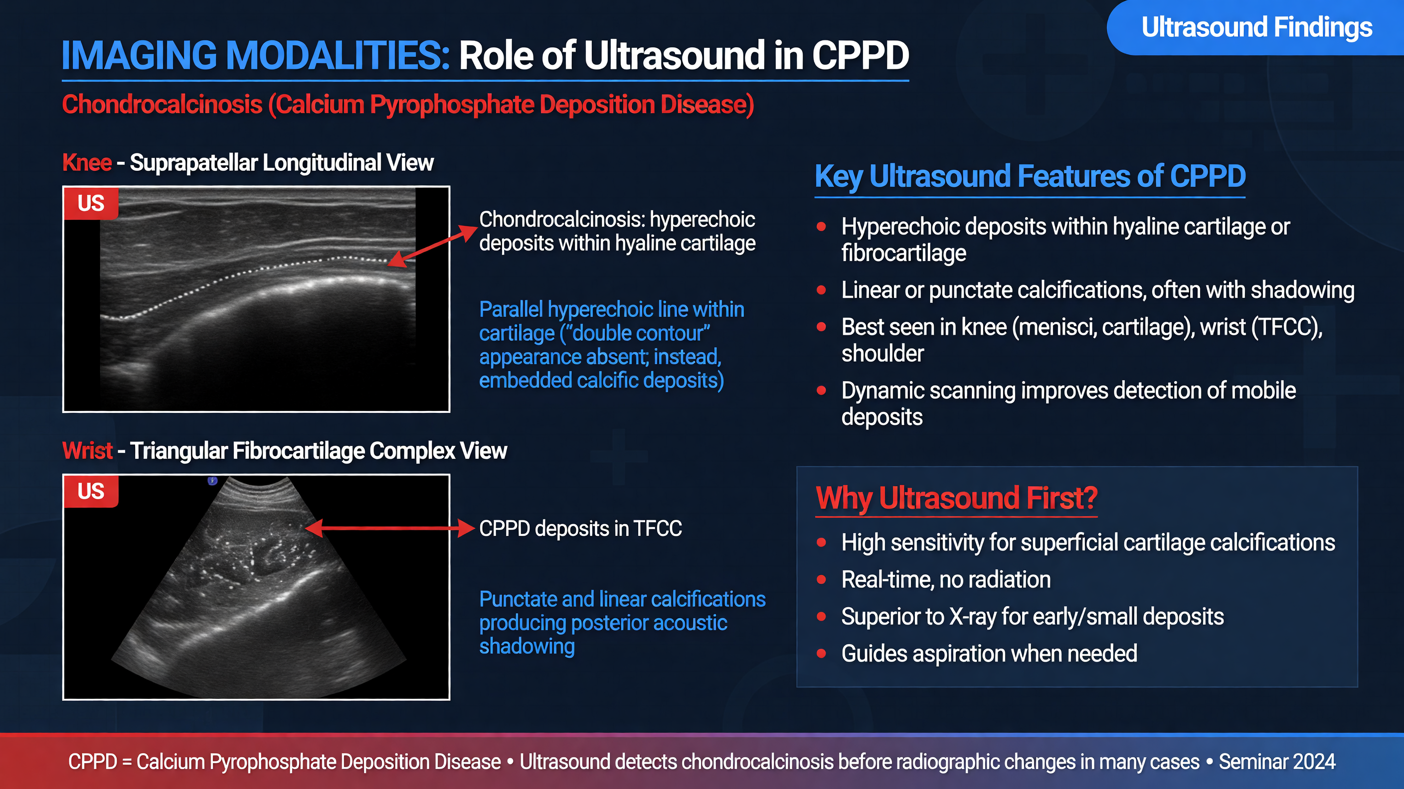
OMERACT definitions describe CPP deposits according to location, echogenicity, shape, and dynamic behavior. In fibrocartilage, such as a meniscus or triangular fibrocartilage complex, deposits appear as hyperechoic foci or bands embedded within the structure. In hyaline cartilage, deposits are typically within—not merely over—the cartilage and move with it during dynamic examination. Tendon deposits are usually linear echogenic structures within the tendon. Most small deposits do not produce posterior acoustic shadowing; large or dense aggregates may.
The knees and wrists are disease-specific target sites. A practical examination scans the symptomatic joint plus bilateral knees and wrists when the diagnosis remains uncertain. Ultrasound can also show effusion, synovial hypertrophy, and power Doppler activity, but these establish inflammation rather than its etiology. Validated definitions and targeted protocols have made ultrasound particularly useful for chronic CPPD masquerading as RA or PMR (PMIDs: [31860551](https://pubmed.ncbi.nlm.nih.gov/31860551/), [36943699](https://pubmed.ncbi.nlm.nih.gov/36943699/)).
**Nuance:** Gout classically creates a monosodium urate double-contour sign on the cartilage surface, whereas CPP deposits usually lie within cartilage. Static CPP deposits can nevertheless produce a pseudo-double contour. Dynamic scanning, correct gain, and assessment in two planes reduce misclassification. Anisotropy, cartilage interfaces, dense OA, and partially visualized menisci are common pitfalls.
International consensus definitions now standardize characteristic CPPD findings on radiography, conventional CT, dual-energy CT, and MRI (PMID: [35439343](https://pubmed.ncbi.nlm.nih.gov/35439343/)). Thin-section noncontrast CT centered on C1–C2 is the preferred test for crowned dens syndrome and is also valuable for other deep or axial deposits. CT shows linear or punctate calcification within cartilage, ligaments, capsule, synovium, or tendons. Dual-energy CT is promising but remains protocol- and scanner-dependent; its ability to distinguish CPP from basic calcium phosphate deposits is not sufficiently reliable to make it a universal clinical gold standard. MRI is relatively insensitive for calcific deposits. Its role is adjunctive—evaluating synovitis, marrow or soft-tissue disease, cord compression, tumor, discitis, or epidural infection rather than identifying CPP crystals themselves (PMIDs: [39415374](https://pubmed.ncbi.nlm.nih.gov/39415374/), [38320811](https://pubmed.ncbi.nlm.nih.gov/38320811/)).
The 2023 ACR/EULAR criteria should be framed as classification criteria for research, not a substitute for bedside diagnosis. After an entry history of joint pain, swelling, or tenderness and exclusion of a better explanation, crowned dens syndrome or CPP crystals from a symptomatic joint are sufficient for classification. Otherwise, a weighted score greater than 56 incorporates age, inflammatory episodes, joint distribution, associated metabolic disease, synovial-fluid results, and imaging. Validation sensitivity and specificity were 99.2% and 92.5%, respectively (PMID: [37495237](https://pubmed.ncbi.nlm.nih.gov/37495237/)).
**Decision Point:** Use radiography for accessible whole-joint structural assessment, ultrasound for sensitive peripheral detection and procedures, CT for axial disease, and MRI for complications or mimics.
**Audience Poll:** Which test should be obtained first for severe febrile upper-neck pain with restricted rotation and suspected crowned dens syndrome: cervical radiographs, ultrasound, thin-section CT, or MRI?
---
## Current Treatment Paradigms and Challenges
### Duration: 15 minutes
Synovial-fluid identification of CPP crystals remains the most specific practical evidence for peripheral CPPD. Under compensated polarized-light microscopy, crystals are typically short rods, rhomboids, or parallelepipeds with weak positive birefringence; some appear nonbirefringent. They may be intracellular or extracellular. CPP crystals are smaller, less strongly birefringent, and more easily missed than monosodium urate crystals, so a negative examination—especially of scant fluid or by an inexperienced observer—does not exclude disease.
**MUST ACT:** Imaging can support CPPD, guide aspiration, and map disease, but it cannot exclude septic arthritis in an acutely inflamed joint. Culture the aspirate whenever infection is clinically plausible.
Conventional radiography remains inexpensive, widely available, and useful for showing the entire joint. Characteristic deposits are linear or punctate calcifications within hyaline cartilage or fibrocartilage. High-yield locations include the knee menisci and femoral cartilage, wrist triangular fibrocartilage complex and intrinsic ligaments, pubic symphysis, acetabular labrum, and MCP joints. Radiography also demonstrates associated joint-space loss, cysts, osteophytes, scapholunate instability, or atypically distributed OA.
Its major limitation is sensitivity. A comparative meta-analysis using synovial fluid or histology as reference found pooled sensitivity and specificity of approximately 0.47 and 0.95 for radiography versus 0.85 and 0.87 for ultrasound, although study heterogeneity was substantial (PMID: [33577959](https://pubmed.ncbi.nlm.nih.gov/33577959/)). Thus, a positive characteristic radiograph is persuasive, but a negative film is not a rule-out test.
**Teaching Point:** Ultrasound is most valuable when it answers a targeted question: Is there a deposit in cartilage or fibrocartilage? Is the joint actively inflamed? Can ultrasound guide aspiration or injection?
OMERACT definitions describe CPP deposits according to location, echogenicity, shape, and dynamic behavior. In fibrocartilage, such as a meniscus or triangular fibrocartilage complex, deposits appear as hyperechoic foci or bands embedded within the structure. In hyaline cartilage, deposits are typically within—not merely over—the cartilage and move with it during dynamic examination. Tendon deposits are usually linear echogenic structures within the tendon. Most small deposits do not produce posterior acoustic shadowing; large or dense aggregates may.
The knees and wrists are disease-specific target sites. A practical examination scans the symptomatic joint plus bilateral knees and wrists when the diagnosis remains uncertain. Ultrasound can also show effusion, synovial hypertrophy, and power Doppler activity, but these establish inflammation rather than its etiology. Validated definitions and targeted protocols have made ultrasound particularly useful for chronic CPPD masquerading as RA or PMR (PMIDs: [31860551](https://pubmed.ncbi.nlm.nih.gov/31860551/), [36943699](https://pubmed.ncbi.nlm.nih.gov/36943699/)).
**Nuance:** Gout classically creates a monosodium urate double-contour sign on the cartilage surface, whereas CPP deposits usually lie within cartilage. Static CPP deposits can nevertheless produce a pseudo-double contour. Dynamic scanning, correct gain, and assessment in two planes reduce misclassification. Anisotropy, cartilage interfaces, dense OA, and partially visualized menisci are common pitfalls.
International consensus definitions now standardize characteristic CPPD findings on radiography, conventional CT, dual-energy CT, and MRI (PMID: [35439343](https://pubmed.ncbi.nlm.nih.gov/35439343/)). Thin-section noncontrast CT centered on C1–C2 is the preferred test for crowned dens syndrome and is also valuable for other deep or axial deposits. CT shows linear or punctate calcification within cartilage, ligaments, capsule, synovium, or tendons. Dual-energy CT is promising but remains protocol- and scanner-dependent; its ability to distinguish CPP from basic calcium phosphate deposits is not sufficiently reliable to make it a universal clinical gold standard. MRI is relatively insensitive for calcific deposits. Its role is adjunctive—evaluating synovitis, marrow or soft-tissue disease, cord compression, tumor, discitis, or epidural infection rather than identifying CPP crystals themselves (PMIDs: [39415374](https://pubmed.ncbi.nlm.nih.gov/39415374/), [38320811](https://pubmed.ncbi.nlm.nih.gov/38320811/)).
The 2023 ACR/EULAR criteria should be framed as classification criteria for research, not a substitute for bedside diagnosis. After an entry history of joint pain, swelling, or tenderness and exclusion of a better explanation, crowned dens syndrome or CPP crystals from a symptomatic joint are sufficient for classification. Otherwise, a weighted score greater than 56 incorporates age, inflammatory episodes, joint distribution, associated metabolic disease, synovial-fluid results, and imaging. Validation sensitivity and specificity were 99.2% and 92.5%, respectively (PMID: [37495237](https://pubmed.ncbi.nlm.nih.gov/37495237/)).
**Decision Point:** Use radiography for accessible whole-joint structural assessment, ultrasound for sensitive peripheral detection and procedures, CT for axial disease, and MRI for complications or mimics.
**Audience Poll:** Which test should be obtained first for severe febrile upper-neck pain with restricted rotation and suspected crowned dens syndrome: cervical radiographs, ultrasound, thin-section CT, or MRI?
---
## Current Treatment Paradigms and Challenges
 ### Duration: 15 minutes
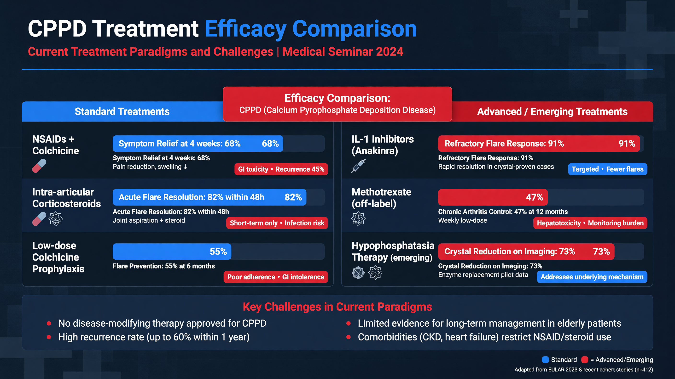
No currently established therapy dissolves CPP crystals or reliably prevents structural progression. Treatment therefore targets the active phenotype: terminate acute inflammation, prevent recurrent attacks, control persistent synovitis, and treat coexisting osteoarthritis. Asymptomatic chondrocalcinosis requires no pharmacologic treatment (PMIDs: [21257614](https://pubmed.ncbi.nlm.nih.gov/21257614/), [39775910](https://pubmed.ncbi.nlm.nih.gov/39775910/)).
**MUST ACT:** Before injecting or escalating immunosuppression, exclude septic arthritis to the degree required by the clinical context. Do not let a positive crystal result, chondrocalcinosis, or rapid steroid response terminate the infection assessment.
For an accessible monoarthritis, aspiration relieves pressure, establishes diagnostic material, and permits intra-articular glucocorticoid once infection is reasonably excluded. A typical large-knee dose is triamcinolone acetonide 40 mg; smaller joints receive lower, joint-specific doses. Ice and temporary rest are useful during peak inflammation, followed by early range-of-motion work to avoid deconditioning.
**Decision Point:** Local therapy is often preferable for one or two joints. Polyarticular disease, inaccessible joints, or inability to perform aspiration usually requires systemic treatment.
Low-dose colchicine should be started early. A commonly used regimen is 1.2 mg orally followed by 0.6 mg one hour later, then 0.5–0.6 mg once or twice daily for several days according to response and tolerance. The CPPD-specific COLCHICORT trial randomized older hospitalized patients to colchicine 1.5 mg on day 1 and 1 mg on day 2 or prednisone 30 mg daily for two days. Pain reduction at 24 hours was equivalent: 36 mm with colchicine and 38 mm with prednisone on a 100-mm scale. Diarrhea occurred in 22% of colchicine-treated patients; prednisone produced more hypertension and hyperglycemia (PMID: [38251496](https://pubmed.ncbi.nlm.nih.gov/38251496/)).
Colchicine requires medication reconciliation. Renal or hepatic impairment, frailty, and concomitant statins increase neuromyopathy risk. Strong CYP3A4 or P-glycoprotein inhibitors—including clarithromycin, azole antifungals, cyclosporine, and ritonavir-containing regimens—can produce life-threatening toxicity, particularly with renal or hepatic dysfunction. Dose reduction, avoidance, or pharmacist consultation may be safer than applying a standard regimen.
NSAIDs remain reasonable for selected low-risk patients despite the absence of robust CPPD-specific trials. Naproxen 250–500 mg twice daily, or an equivalent anti-inflammatory dose, may be used for approximately three to five days with rapid reassessment. Avoid or strongly reconsider NSAIDs in acute kidney injury, advanced CKD, decompensated heart failure, uncontrolled hypertension, active or recent gastrointestinal bleeding, anticoagulation, or high cardiovascular risk. Use the lowest effective dose for the shortest duration and add gastroprotection when indicated.
Prednisone or prednisolone 10–30 mg daily for three to seven days is a practical range, individualized to disease burden and comorbidity. COLCHICORT supports 30 mg daily for two days. A separate open-label randomized trial of 79 crystal-confirmed patients found no adjusted difference in resolution between 10 and 30 mg of prednisolone daily for seven days, supporting lower dosing in selected frail patients (PMID: [39412710](https://pubmed.ncbi.nlm.nih.gov/39412710/)). Monitor glucose, blood pressure, volume status, delirium, and infection risk; extend or taper only when persistent inflammation or rebound justifies additional exposure.
**Framework:** For recurrent attacks, first verify that episodes are truly inflammatory. Colchicine 0.5–0.6 mg once or twice daily can be considered when attacks are frequent, with renal and interaction review. In a classic ten-patient before-and-after study, 0.6 mg twice daily reduced attacks from 3.2 to 1.0 per patient-year, but this evidence is small and uncontrolled (PMID: [3772928](https://pubmed.ncbi.nlm.nih.gov/3772928/)).
Persistent inflammatory CPPD may require low-dose colchicine, intermittent intra-articular glucocorticoid, or the lowest feasible oral prednisone dose. Hydroxychloroquine has a signal from one small crossover trial but no adequate confirmatory study. If used, dosing should remain at or below 5 mg/kg/day of actual body weight with contemporary retinal screening. Methotrexate is not proven: a 26-patient double-blind crossover trial of 15 mg weekly found no significant improvement in pain, disease activity, or secondary outcomes (PMID: [25315665](https://pubmed.ncbi.nlm.nih.gov/25315665/)).
**Nuance:** OA-dominant CPPD needs ordinary OA care—strengthening, aerobic activity, weight management when applicable, assistive devices, topical analgesia, selective injections, and arthroplasty when indicated—not chronic immunosuppression.
**Audience Poll:** For an 86-year-old with an aspirated, culture-pending knee flare, stage 4 CKD, diabetes, and no bacteremia, which option has the most favorable initial risk profile: naproxen, full-dose colchicine, intra-articular glucocorticoid after infection is reasonably excluded, or prolonged oral prednisone?
---
## Emerging Therapies and Future Prospects
### Duration: 15 minutes
No currently established therapy dissolves CPP crystals or reliably prevents structural progression. Treatment therefore targets the active phenotype: terminate acute inflammation, prevent recurrent attacks, control persistent synovitis, and treat coexisting osteoarthritis. Asymptomatic chondrocalcinosis requires no pharmacologic treatment (PMIDs: [21257614](https://pubmed.ncbi.nlm.nih.gov/21257614/), [39775910](https://pubmed.ncbi.nlm.nih.gov/39775910/)).
**MUST ACT:** Before injecting or escalating immunosuppression, exclude septic arthritis to the degree required by the clinical context. Do not let a positive crystal result, chondrocalcinosis, or rapid steroid response terminate the infection assessment.
For an accessible monoarthritis, aspiration relieves pressure, establishes diagnostic material, and permits intra-articular glucocorticoid once infection is reasonably excluded. A typical large-knee dose is triamcinolone acetonide 40 mg; smaller joints receive lower, joint-specific doses. Ice and temporary rest are useful during peak inflammation, followed by early range-of-motion work to avoid deconditioning.
**Decision Point:** Local therapy is often preferable for one or two joints. Polyarticular disease, inaccessible joints, or inability to perform aspiration usually requires systemic treatment.
Low-dose colchicine should be started early. A commonly used regimen is 1.2 mg orally followed by 0.6 mg one hour later, then 0.5–0.6 mg once or twice daily for several days according to response and tolerance. The CPPD-specific COLCHICORT trial randomized older hospitalized patients to colchicine 1.5 mg on day 1 and 1 mg on day 2 or prednisone 30 mg daily for two days. Pain reduction at 24 hours was equivalent: 36 mm with colchicine and 38 mm with prednisone on a 100-mm scale. Diarrhea occurred in 22% of colchicine-treated patients; prednisone produced more hypertension and hyperglycemia (PMID: [38251496](https://pubmed.ncbi.nlm.nih.gov/38251496/)).
Colchicine requires medication reconciliation. Renal or hepatic impairment, frailty, and concomitant statins increase neuromyopathy risk. Strong CYP3A4 or P-glycoprotein inhibitors—including clarithromycin, azole antifungals, cyclosporine, and ritonavir-containing regimens—can produce life-threatening toxicity, particularly with renal or hepatic dysfunction. Dose reduction, avoidance, or pharmacist consultation may be safer than applying a standard regimen.
NSAIDs remain reasonable for selected low-risk patients despite the absence of robust CPPD-specific trials. Naproxen 250–500 mg twice daily, or an equivalent anti-inflammatory dose, may be used for approximately three to five days with rapid reassessment. Avoid or strongly reconsider NSAIDs in acute kidney injury, advanced CKD, decompensated heart failure, uncontrolled hypertension, active or recent gastrointestinal bleeding, anticoagulation, or high cardiovascular risk. Use the lowest effective dose for the shortest duration and add gastroprotection when indicated.
Prednisone or prednisolone 10–30 mg daily for three to seven days is a practical range, individualized to disease burden and comorbidity. COLCHICORT supports 30 mg daily for two days. A separate open-label randomized trial of 79 crystal-confirmed patients found no adjusted difference in resolution between 10 and 30 mg of prednisolone daily for seven days, supporting lower dosing in selected frail patients (PMID: [39412710](https://pubmed.ncbi.nlm.nih.gov/39412710/)). Monitor glucose, blood pressure, volume status, delirium, and infection risk; extend or taper only when persistent inflammation or rebound justifies additional exposure.
**Framework:** For recurrent attacks, first verify that episodes are truly inflammatory. Colchicine 0.5–0.6 mg once or twice daily can be considered when attacks are frequent, with renal and interaction review. In a classic ten-patient before-and-after study, 0.6 mg twice daily reduced attacks from 3.2 to 1.0 per patient-year, but this evidence is small and uncontrolled (PMID: [3772928](https://pubmed.ncbi.nlm.nih.gov/3772928/)).
Persistent inflammatory CPPD may require low-dose colchicine, intermittent intra-articular glucocorticoid, or the lowest feasible oral prednisone dose. Hydroxychloroquine has a signal from one small crossover trial but no adequate confirmatory study. If used, dosing should remain at or below 5 mg/kg/day of actual body weight with contemporary retinal screening. Methotrexate is not proven: a 26-patient double-blind crossover trial of 15 mg weekly found no significant improvement in pain, disease activity, or secondary outcomes (PMID: [25315665](https://pubmed.ncbi.nlm.nih.gov/25315665/)).
**Nuance:** OA-dominant CPPD needs ordinary OA care—strengthening, aerobic activity, weight management when applicable, assistive devices, topical analgesia, selective injections, and arthroplasty when indicated—not chronic immunosuppression.
**Audience Poll:** For an 86-year-old with an aspirated, culture-pending knee flare, stage 4 CKD, diabetes, and no bacteremia, which option has the most favorable initial risk profile: naproxen, full-dose colchicine, intra-articular glucocorticoid after infection is reasonably excluded, or prolonged oral prednisone?
---
## Emerging Therapies and Future Prospects
 ### Duration: 10 minutes
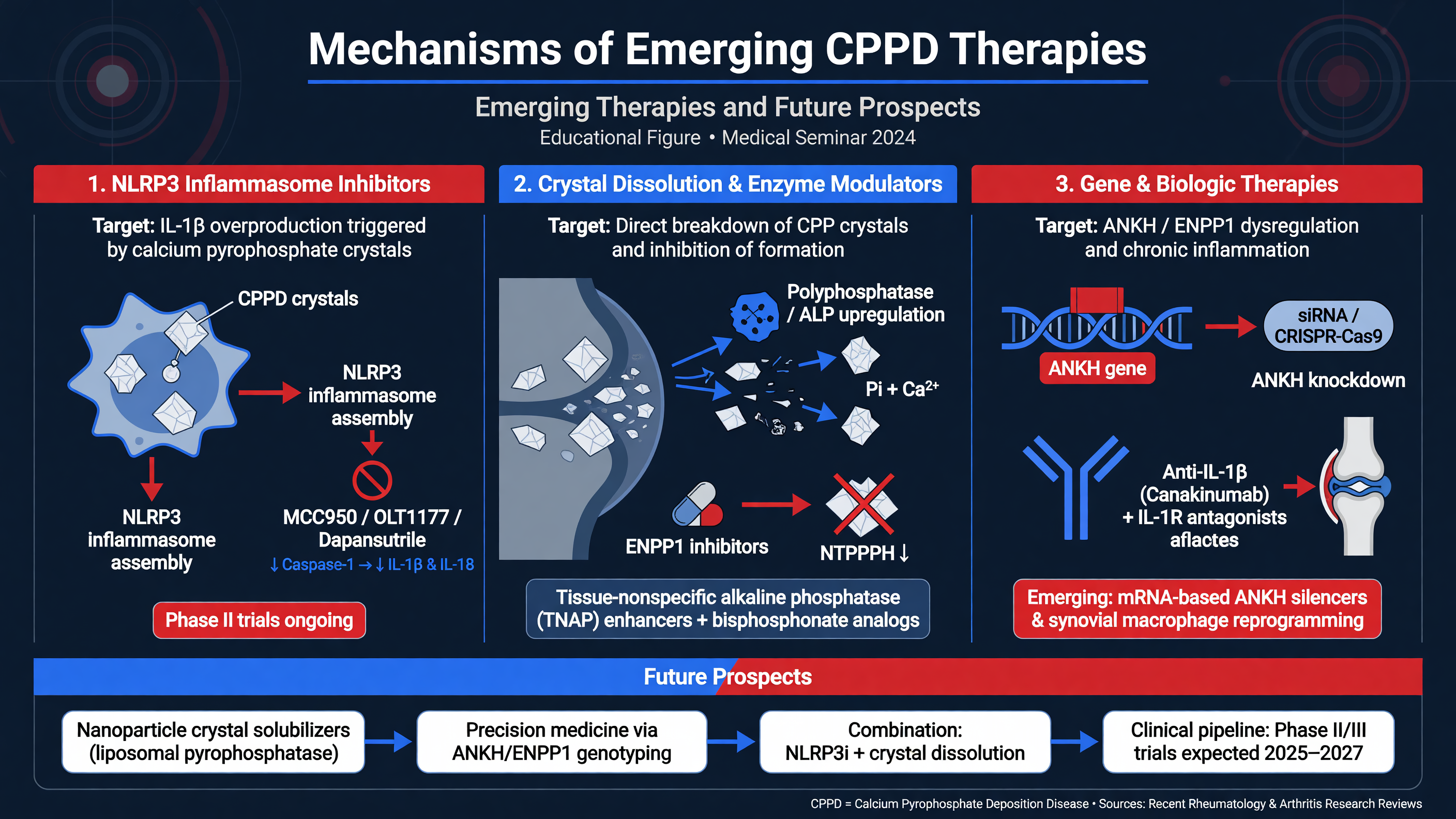
The inflammatory biology of CPPD offers plausible therapeutic targets, but biologic plausibility must not be confused with established effectiveness. No biologic is approved specifically to treat CPPD, and nearly all use remains off-label, specialist-directed, and supported by small trials, uncontrolled cohorts, or case series.
**Nuance:** Biologics suppress the host response to crystals; they do not remove the crystal burden. The indication should therefore be refractory inflammatory disease—not asymptomatic chondrocalcinosis or purely mechanical OA pain.
Anakinra blocks the IL-1 receptor and is the best-described biologic for acute refractory CPP arthritis or for patients in whom NSAIDs, colchicine, and glucocorticoids are unsafe. The usual reported regimen is 100 mg subcutaneously daily for three days. In severe renal impairment, every-other-day administration is commonly used because the drug is renally cleared. Active infection must be excluded, and neutrophil counts should be monitored.
A 2026 meta-analysis included six studies and only 84 patients, predominantly from observational cohorts. The pooled physician-assessed response between days 3 and 5 was 76%, with reductions in pain and CRP, but reported adverse events included infection, rash, injection-site reactions, and neutropenia. The estimate is encouraging rather than definitive (PMID: [41206733](https://pubmed.ncbi.nlm.nih.gov/41206733/)). Earlier systematic review data suggested stronger responses in acute than chronic CPPD (PMID: [32359034](https://pubmed.ncbi.nlm.nih.gov/32359034/)).
**Decision Point:** Anakinra is most defensible for a severe, crystal-supported acute flare that is refractory to conventional treatment or in which conventional treatment is contraindicated. It should not become a shortcut around an incomplete infection evaluation.
IL-6 blockade is an emerging strategy for persistent or frequently recurrent inflammatory CPPD. CPP crystals and IL-1β stimulate substantial IL-6 production by monocytes and synovial cells. In an open-label pilot study of 11 patients with severe refractory disease, intravenous tocilizumab—generally 8 mg/kg every four weeks—was associated with improvement in global disease activity at three months. There was no control group, and one patient developed a lung abscess (PMID: [32213498](https://pubmed.ncbi.nlm.nih.gov/32213498/)). A 2025 review identifies IL-6 as a promising target but explicitly emphasizes the need for randomized trials before broad adoption (PMID: [40550451](https://pubmed.ncbi.nlm.nih.gov/40550451/)).
If tocilizumab is considered, reported regimens include 8 mg/kg intravenously every four weeks or 162 mg subcutaneously weekly. Pre-treatment assessment should include infection and viral-hepatitis risk, tuberculosis screening as appropriate, vaccination review, CBC, liver enzymes, and lipids. Monitor for cytopenias, transaminitis, serious infection, and gastrointestinal perforation risk in diverticular disease. Because IL-6 blockade suppresses fever and CRP, a low CRP cannot safely exclude septic arthritis.
**MUST ACT:** Before escalating to anakinra or tocilizumab, reconfirm the phenotype. Persistent marginal erosions, psoriasis, inflammatory axial disease, high-titer anti-CCP antibodies, occult infection, or purely mechanical pain should redirect the diagnosis. TNF inhibitors are not evidence-based CPPD therapy, and empiric biologic cycling by analogy with RA can expose a misdiagnosed patient to harm.
Crystal-directed treatment remains the central unmet need. Correcting genuine hypomagnesemia is rational, but a small magnesium trial did not demonstrate radiographic crystal clearance. Probenecid can inhibit ANKH-related transport in experimental systems, yet early human investigation did not establish clinical benefit. Strategies involving ANKH, ENPP1, TNAP, nucleoside analogues, phosphocitrate, and direct NLRP3 inhibition remain preclinical or conceptual.
Manipulating pyrophosphate is biologically difficult: excess pyrophosphate promotes CPP formation in cartilage, but systemic pyrophosphate also restrains hydroxyapatite deposition in vessels and soft tissue. A successful therapy will likely need cartilage-selective delivery or highly localized pathway modulation to avoid exchanging articular CPP deposition for ectopic calcification elsewhere.
**Framework:** Future trials need crystal-confirmed, phenotype-specific cohorts; standardized flare definitions; validated pain and function outcomes; and separate endpoints for inflammation, crystal burden, and structural damage. The 2023 classification criteria and OMERACT imaging definitions provide the infrastructure that earlier studies lacked.
**Teaching Point:** The near-term innovation is better patient selection for existing anti-inflammatory agents. The long-term innovation is a safe therapy that prevents nucleation or removes CPP deposits.
**Audience Poll:** Which endpoint would be most convincing for a disease-modifying CPPD therapy: lower CRP, fewer flares, reduced ultrasound crystal burden, or slower structural progression?
---
## Clinical Cases and Differential Diagnosis
### Duration: 10 minutes
The inflammatory biology of CPPD offers plausible therapeutic targets, but biologic plausibility must not be confused with established effectiveness. No biologic is approved specifically to treat CPPD, and nearly all use remains off-label, specialist-directed, and supported by small trials, uncontrolled cohorts, or case series.
**Nuance:** Biologics suppress the host response to crystals; they do not remove the crystal burden. The indication should therefore be refractory inflammatory disease—not asymptomatic chondrocalcinosis or purely mechanical OA pain.
Anakinra blocks the IL-1 receptor and is the best-described biologic for acute refractory CPP arthritis or for patients in whom NSAIDs, colchicine, and glucocorticoids are unsafe. The usual reported regimen is 100 mg subcutaneously daily for three days. In severe renal impairment, every-other-day administration is commonly used because the drug is renally cleared. Active infection must be excluded, and neutrophil counts should be monitored.
A 2026 meta-analysis included six studies and only 84 patients, predominantly from observational cohorts. The pooled physician-assessed response between days 3 and 5 was 76%, with reductions in pain and CRP, but reported adverse events included infection, rash, injection-site reactions, and neutropenia. The estimate is encouraging rather than definitive (PMID: [41206733](https://pubmed.ncbi.nlm.nih.gov/41206733/)). Earlier systematic review data suggested stronger responses in acute than chronic CPPD (PMID: [32359034](https://pubmed.ncbi.nlm.nih.gov/32359034/)).
**Decision Point:** Anakinra is most defensible for a severe, crystal-supported acute flare that is refractory to conventional treatment or in which conventional treatment is contraindicated. It should not become a shortcut around an incomplete infection evaluation.
IL-6 blockade is an emerging strategy for persistent or frequently recurrent inflammatory CPPD. CPP crystals and IL-1β stimulate substantial IL-6 production by monocytes and synovial cells. In an open-label pilot study of 11 patients with severe refractory disease, intravenous tocilizumab—generally 8 mg/kg every four weeks—was associated with improvement in global disease activity at three months. There was no control group, and one patient developed a lung abscess (PMID: [32213498](https://pubmed.ncbi.nlm.nih.gov/32213498/)). A 2025 review identifies IL-6 as a promising target but explicitly emphasizes the need for randomized trials before broad adoption (PMID: [40550451](https://pubmed.ncbi.nlm.nih.gov/40550451/)).
If tocilizumab is considered, reported regimens include 8 mg/kg intravenously every four weeks or 162 mg subcutaneously weekly. Pre-treatment assessment should include infection and viral-hepatitis risk, tuberculosis screening as appropriate, vaccination review, CBC, liver enzymes, and lipids. Monitor for cytopenias, transaminitis, serious infection, and gastrointestinal perforation risk in diverticular disease. Because IL-6 blockade suppresses fever and CRP, a low CRP cannot safely exclude septic arthritis.
**MUST ACT:** Before escalating to anakinra or tocilizumab, reconfirm the phenotype. Persistent marginal erosions, psoriasis, inflammatory axial disease, high-titer anti-CCP antibodies, occult infection, or purely mechanical pain should redirect the diagnosis. TNF inhibitors are not evidence-based CPPD therapy, and empiric biologic cycling by analogy with RA can expose a misdiagnosed patient to harm.
Crystal-directed treatment remains the central unmet need. Correcting genuine hypomagnesemia is rational, but a small magnesium trial did not demonstrate radiographic crystal clearance. Probenecid can inhibit ANKH-related transport in experimental systems, yet early human investigation did not establish clinical benefit. Strategies involving ANKH, ENPP1, TNAP, nucleoside analogues, phosphocitrate, and direct NLRP3 inhibition remain preclinical or conceptual.
Manipulating pyrophosphate is biologically difficult: excess pyrophosphate promotes CPP formation in cartilage, but systemic pyrophosphate also restrains hydroxyapatite deposition in vessels and soft tissue. A successful therapy will likely need cartilage-selective delivery or highly localized pathway modulation to avoid exchanging articular CPP deposition for ectopic calcification elsewhere.
**Framework:** Future trials need crystal-confirmed, phenotype-specific cohorts; standardized flare definitions; validated pain and function outcomes; and separate endpoints for inflammation, crystal burden, and structural damage. The 2023 classification criteria and OMERACT imaging definitions provide the infrastructure that earlier studies lacked.
**Teaching Point:** The near-term innovation is better patient selection for existing anti-inflammatory agents. The long-term innovation is a safe therapy that prevents nucleation or removes CPP deposits.
**Audience Poll:** Which endpoint would be most convincing for a disease-modifying CPPD therapy: lower CRP, fewer flares, reduced ultrasound crystal burden, or slower structural progression?
---
## Clinical Cases and Differential Diagnosis
 ### Duration: 15 minutes
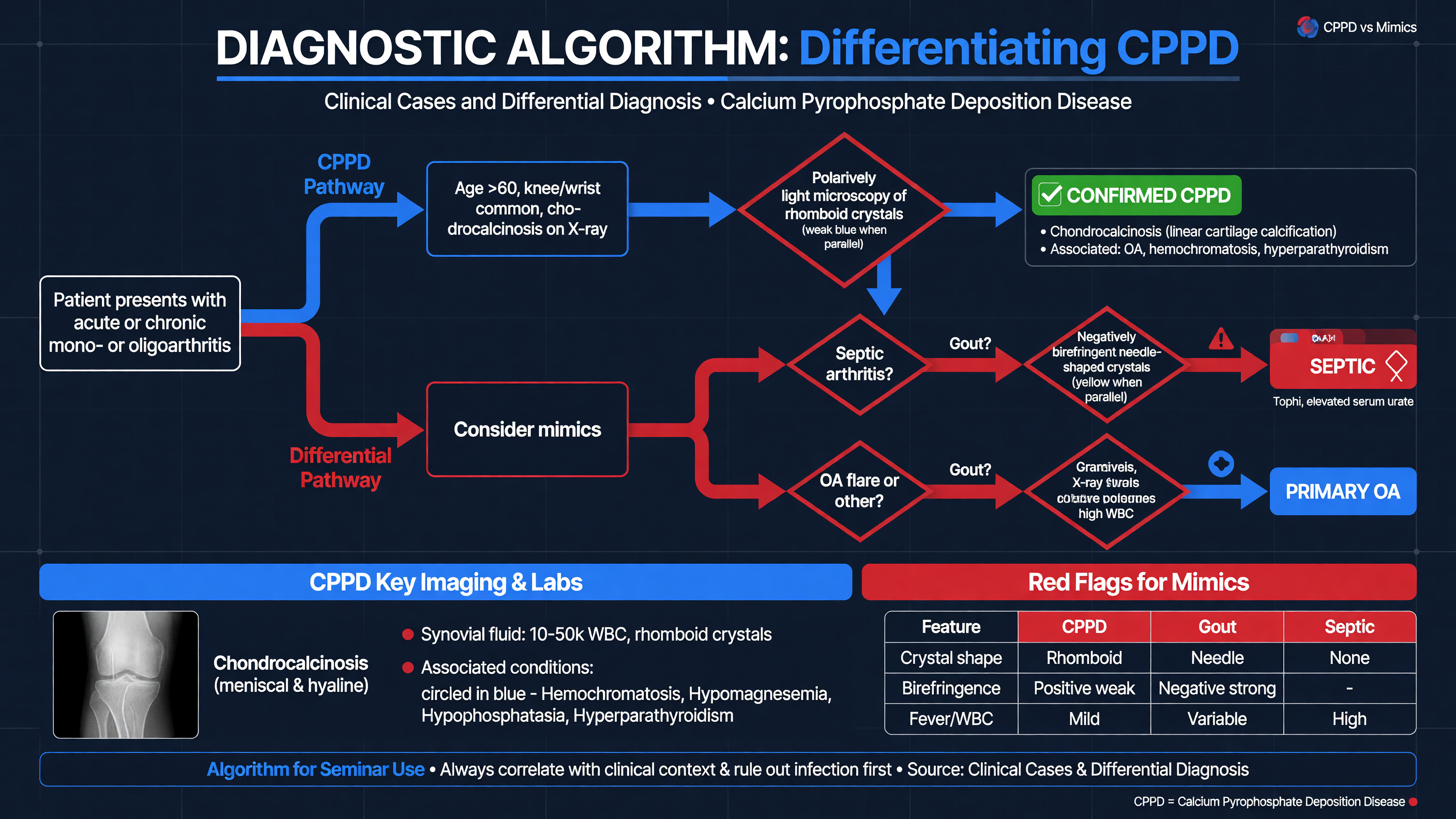
CPPD diagnosis requires pattern recognition without anchoring. An older patient with chondrocalcinosis may still have septic arthritis, gout, RA, fracture, or ordinary OA. Conversely, a negative radiograph or initial negative crystal examination does not eliminate CPPD.
**Framework:** Use a five-step diagnostic sequence:
1. Define the syndrome: acute hot joint, recurrent episodic arthritis, persistent inflammatory polyarthritis, mechanical OA-pattern pain, or axial disease.
2. Exclude immediate threats: septic arthritis, fracture, hemarthrosis, compartment-threatening effusion, or neurologic compression.
3. Obtain direct evidence where possible: aspirate for crystals and culture.
4. Image strategically: radiography or ultrasound for peripheral disease; CT for suspected axial CPPD.
5. Reconcile the entire pattern: age, distribution, metabolic associations, serology, erosions, response over time, and possible coexistence of disorders.
**MUST ACT:** In acute monoarthritis, neither chondrocalcinosis nor CPP crystals excludes infection. In a large retrospective series, approximately 5% of crystal-positive acute monoarthritis cases had concomitant infection, although the rate varies by clinical setting (PMID: [22133623](https://pubmed.ncbi.nlm.nih.gov/22133623/)). Obtain blood cultures when systemically ill and begin empiric antibiotics after appropriate sampling when the clinical probability of sepsis is substantial.
CPPD and gout may be clinically indistinguishable. CPPD favors later onset, knee, and wrist involvement; gout more often begins at the first MTP, ankle, or midfoot. These are tendencies, not rules. CPP crystals are short rods or rhomboids with weak positive birefringence; monosodium urate crystals are needle-shaped with strong negative birefringence. Serum urate may be normal during a gout flare, and both crystal types can coexist. On imaging, CPP deposits are usually intracartilaginous, whereas gout produces surface double contour, tophi, and—later—punched-out erosions with overhanging edges.
Chronic CPP inflammatory arthritis versus RA is a common diagnostic trap. CPPD is supported by onset after 60, episodic attacks, knee or wrist disease, cartilage deposits, MCP2–3 or radiocarpal OA, hook osteophytes, and limited marginal erosion. RA is supported by high-titer anti-CCP antibodies, persistent symmetric small-joint synovitis, marginal erosions, periarticular osteopenia, and extra-articular disease. Rheumatoid factor alone is weak evidence in an older adult. CPPD and RA may coexist; demonstrating crystals should not automatically terminate effective RA treatment.
**Teaching Point:** When a “seronegative RA” patient has unusual wrist or MCP structural disease, recurrent abrupt flares, and inadequate response to sequential RA biologics, revisit the diagnosis before escalating again.
CPPD-associated OA must also be distinguished from incidental chondrocalcinosis in primary OA. Mechanical pain, short-lived morning stiffness, and load-related symptoms should be treated as OA unless there is convincing active inflammation. Atypical distribution—wrist, MCP, elbow, ankle, lateral-compartment knee, or severe isolated patellofemoral disease—raises CPPD probability but does not prove that crystals are the current pain generator.
Shoulder and hip-girdle CPPD can mimic PMR. Peripheral wrist or knee synovitis, prior acute attacks, AC-joint deposits, or crowned-dens symptoms favor CPPD. Subacromial-subdeltoid bursitis and biceps tenosynovitis favor PMR but are not entirely specific. Steroid response is nondiagnostic. Continue to assess for giant-cell arteritis when headache, jaw claudication, scalp tenderness, visual symptoms, or vascular findings are present.
Basic calcium phosphate deposition is another calcium-crystal mimic. It more often causes amorphous periarticular calcification, calcific tendinitis, or destructive Milwaukee-shoulder arthropathy. Basic calcium phosphate crystals are not identified by routine polarized microscopy. CPPD more typically produces linear or punctate cartilage deposits.
Crowned dens syndrome should be considered in acute febrile neck pain with severe rotational restriction. Thin-section CT centered at C1–C2 is the diagnostic imaging study; MRI is added for neurologic deficits or concern for epidural infection, discitis, tumor, or cord compression. Meningitis, giant-cell arteritis, RA-related atlantoaxial disease, fracture, and malignancy remain competing diagnoses. Crowned dens syndrome is a radioclinical diagnosis—not merely an incidental calcified transverse ligament (PMID: [23806762](https://pubmed.ncbi.nlm.nih.gov/23806762/)).
**Decision Point:** If one finding does not explain the entire presentation, allow dual diagnoses. CPPD commonly coexists with OA and may coexist with gout, RA, or infection.
**Audience Poll:** A patient with CPP crystals, synovial WBC 70,000/µL, fever, and recent bacteremia has which diagnosis: CPPD alone, septic arthritis alone, both until cultures prove otherwise, or neither?
---
## Clinical Case Discussions
### Case 1: Chronic CPPD Mimicking Seronegative Rheumatoid Arthritis
A 72-year-old woman presents with eight months of bilateral wrist and MCP pain, two hours of morning stiffness, and intermittent knee swelling. RF and anti-CCP antibodies are negative. Prednisone repeatedly improves symptoms, but synovitis returns during tapering. She has received a provisional diagnosis of seronegative RA and is being considered for a TNF inhibitor.
Hand radiographs show joint-space narrowing at MCP2–3 and the radiocarpal joints, hook-like metacarpal-head osteophytes, and no convincing marginal erosions. Ultrasound demonstrates synovitis plus hyperechoic deposits within both triangular fibrocartilage complexes and knee menisci. Knee aspiration identifies weakly positively birefringent CPP crystals; Gram stain and culture are negative. Calcium, magnesium, phosphate, alkaline phosphatase, PTH, ferritin, and transferrin saturation are obtained because of the MCP pattern. Iron studies are normal.
**Decision Point:** The evidence supports chronic CPP crystal inflammatory arthritis with CPPD-associated structural disease. It does not prove that RA is impossible, but the absence of erosions and anti-CCP antibodies, episodic knee attacks, and characteristic deposits make empiric TNF inhibition inappropriate before CPPD-directed management.
She begins colchicine 0.6 mg daily after renal and interaction review, receives ultrasound-guided glucocorticoid treatment of the most symptomatic wrist, and starts hand therapy and lower-extremity strengthening. Flare frequency, swollen-joint burden, function, and toxicity are reassessed over three months. Hydroxychloroquine or specialist-directed biologic therapy would be considered only if objectively inflammatory disease persisted.
**Teaching Point:** CPPD should be reconsidered whenever late-onset “seronegative RA” has atypical OA-pattern damage, abrupt superimposed flares, or poor response to RA-directed biologics.
### Case 2: Crowned Dens Syndrome—A Pain in the Neck
A 57-year-old man presents with abrupt occipital and upper-cervical pain, temperature 38.2°C, CRP 146 mg/L, and near-complete inability to rotate his neck. He has no focal neurologic deficit, photophobia, rash, visual symptoms, or jaw claudication. Blood cultures are obtained. Thin-section noncontrast CT shows curvilinear calcification around the odontoid within the transverse and alar ligament region, with no fracture or destructive lesion.
The syndrome is compatible with crowned dens CPPD, but infection remains actively assessed because fever and CRP are nonspecific. MRI would be added for neurologic findings, persistent bacteremia, or concern for epidural infection or discitis. Because onset before 60 is atypical, calcium, magnesium, phosphate, alkaline phosphatase, PTH, ferritin, and transferrin saturation are checked. Magnesium is low in the setting of chronic proton-pump inhibitor and thiazide exposure.
After infection is reasonably excluded, he receives a short oral glucocorticoid course and magnesium replacement with medication review. Pain and rotation improve substantially over 72 hours.
**MUST ACT:** Do not perform a lumbar puncture, start prolonged antibiotics, or diagnose giant-cell arteritis solely from fever, neck pain, and inflammatory markers—but do not allow periodontoid calcification to suppress those diagnoses when their clinical features remain present.
**Teaching Point:** CT identifies the deposit; the clinical syndrome determines whether the deposit is relevant.
---
## Tonight on Shift
- Aspirate a first, atypical, or febrile hot joint; send crystals, cell count, Gram stain, and culture.
- Remember that CPP crystals and septic arthritis can coexist.
- Use radiography or ultrasound for peripheral CPPD and thin-section CT for suspected crowned dens syndrome.
- Choose intra-articular steroid, colchicine, NSAID, or systemic steroid according to joint number and comorbidity—not habit.
- Screen for magnesium, calcium/PTH, iron overload, low alkaline phosphatase, and familial disease when onset is early or unusually extensive.
- Reconsider the diagnosis before escalating chronic immunosuppression or an off-label biologic.
---
## References
- Abhishek A, Tedeschi SK, Pascart T, et al. The 2023 ACR/EULAR classification criteria for calcium pyrophosphate deposition disease. 2023. PMID: [37495237](https://pubmed.ncbi.nlm.nih.gov/37495237/).
- Andrés M, Sivera F, Pascual E. Progresses in the imaging of calcium pyrophosphate crystal disease. 2020. PMID: [31860551](https://pubmed.ncbi.nlm.nih.gov/31860551/).
- Cipolletta E, Filippou G, Scirè CA, et al. Diagnostic value of conventional radiography and musculoskeletal ultrasonography in CPPD. 2021. PMID: [33577959](https://pubmed.ncbi.nlm.nih.gov/33577959/).
- Cipolletta E, Di Matteo A, Scanu A, et al. Biologics in the treatment of CPPD: a systematic literature review. 2020. PMID: [32359034](https://pubmed.ncbi.nlm.nih.gov/32359034/).
- Filippou G, Sirotti S. How can ultrasonography help in the management of CPPD? 2023. PMID: [36943699](https://pubmed.ncbi.nlm.nih.gov/36943699/).
- Finckh A, McCarthy GM, Madigan A, et al. Methotrexate in chronic-recurrent CPPD: no significant effect in a randomized crossover trial. 2014. PMID: [25315665](https://pubmed.ncbi.nlm.nih.gov/25315665/).
- Issa W, Mercer R, Yinh J, et al. Imaging and management of calcium pyrophosphate deposition disease. 2025. PMID: [39775910](https://pubmed.ncbi.nlm.nih.gov/39775910/).
- Latourte A, Ea HK, Frazier A, et al. Tocilizumab in symptomatic calcium pyrophosphate deposition disease: a pilot study. 2020. PMID: [32213498](https://pubmed.ncbi.nlm.nih.gov/32213498/).
- Latourte A, Pascart T, Richette P. IL-6: a new target in crystal-induced arthritides. 2025. PMID: [40550451](https://pubmed.ncbi.nlm.nih.gov/40550451/).
- Mandl P, D’Agostino MA, Navarro-Compán V, et al. 2023 EULAR recommendations on imaging in crystal-induced arthropathies. 2024. PMID: [38320811](https://pubmed.ncbi.nlm.nih.gov/38320811/).
- Manigold T. Calcium pyrophosphate deposition disease—more than just pseudogout. 2024. PMID: [39508814](https://pubmed.ncbi.nlm.nih.gov/39508814/).
- Nakaphan P, Phutinart S, Pajareya P, et al. Efficacy and safety of anakinra for acute CPPD flare: a systematic review and meta-analysis. 2026. PMID: [41206733](https://pubmed.ncbi.nlm.nih.gov/41206733/).
- Pascart T, Filippou G, Lioté F, et al. Calcium pyrophosphate deposition disease. 2024. PMID: [39089298](https://pubmed.ncbi.nlm.nih.gov/39089298/).
- Pascart T, Robinet P, Ottaviani S, et al. COLCHICORT: colchicine versus prednisone in older patients with acute CPP crystal arthritis. 2023. PMID: [38251496](https://pubmed.ncbi.nlm.nih.gov/38251496/).
- Rosenthal AK, Ryan LM. Calcium pyrophosphate deposition disease. 2016. PMID: [27355536](https://pubmed.ncbi.nlm.nih.gov/27355536/).
- Tedeschi SK, Becce F, Pascart T, et al. Imaging features of CPPD: international consensus definitions. 2023. PMID: [35439343](https://pubmed.ncbi.nlm.nih.gov/35439343/).
- Thiele RG. New developments in imaging in crystalline arthritis. 2024. PMID: [39415374](https://pubmed.ncbi.nlm.nih.gov/39415374/).
- Zhang W, Doherty M, Pascual E, et al. EULAR recommendations for CPPD, Part II: management. 2011. PMID: [21257614](https://pubmed.ncbi.nlm.nih.gov/21257614/).
### Duration: 15 minutes
CPPD diagnosis requires pattern recognition without anchoring. An older patient with chondrocalcinosis may still have septic arthritis, gout, RA, fracture, or ordinary OA. Conversely, a negative radiograph or initial negative crystal examination does not eliminate CPPD.
**Framework:** Use a five-step diagnostic sequence:
1. Define the syndrome: acute hot joint, recurrent episodic arthritis, persistent inflammatory polyarthritis, mechanical OA-pattern pain, or axial disease.
2. Exclude immediate threats: septic arthritis, fracture, hemarthrosis, compartment-threatening effusion, or neurologic compression.
3. Obtain direct evidence where possible: aspirate for crystals and culture.
4. Image strategically: radiography or ultrasound for peripheral disease; CT for suspected axial CPPD.
5. Reconcile the entire pattern: age, distribution, metabolic associations, serology, erosions, response over time, and possible coexistence of disorders.
**MUST ACT:** In acute monoarthritis, neither chondrocalcinosis nor CPP crystals excludes infection. In a large retrospective series, approximately 5% of crystal-positive acute monoarthritis cases had concomitant infection, although the rate varies by clinical setting (PMID: [22133623](https://pubmed.ncbi.nlm.nih.gov/22133623/)). Obtain blood cultures when systemically ill and begin empiric antibiotics after appropriate sampling when the clinical probability of sepsis is substantial.
CPPD and gout may be clinically indistinguishable. CPPD favors later onset, knee, and wrist involvement; gout more often begins at the first MTP, ankle, or midfoot. These are tendencies, not rules. CPP crystals are short rods or rhomboids with weak positive birefringence; monosodium urate crystals are needle-shaped with strong negative birefringence. Serum urate may be normal during a gout flare, and both crystal types can coexist. On imaging, CPP deposits are usually intracartilaginous, whereas gout produces surface double contour, tophi, and—later—punched-out erosions with overhanging edges.
Chronic CPP inflammatory arthritis versus RA is a common diagnostic trap. CPPD is supported by onset after 60, episodic attacks, knee or wrist disease, cartilage deposits, MCP2–3 or radiocarpal OA, hook osteophytes, and limited marginal erosion. RA is supported by high-titer anti-CCP antibodies, persistent symmetric small-joint synovitis, marginal erosions, periarticular osteopenia, and extra-articular disease. Rheumatoid factor alone is weak evidence in an older adult. CPPD and RA may coexist; demonstrating crystals should not automatically terminate effective RA treatment.
**Teaching Point:** When a “seronegative RA” patient has unusual wrist or MCP structural disease, recurrent abrupt flares, and inadequate response to sequential RA biologics, revisit the diagnosis before escalating again.
CPPD-associated OA must also be distinguished from incidental chondrocalcinosis in primary OA. Mechanical pain, short-lived morning stiffness, and load-related symptoms should be treated as OA unless there is convincing active inflammation. Atypical distribution—wrist, MCP, elbow, ankle, lateral-compartment knee, or severe isolated patellofemoral disease—raises CPPD probability but does not prove that crystals are the current pain generator.
Shoulder and hip-girdle CPPD can mimic PMR. Peripheral wrist or knee synovitis, prior acute attacks, AC-joint deposits, or crowned-dens symptoms favor CPPD. Subacromial-subdeltoid bursitis and biceps tenosynovitis favor PMR but are not entirely specific. Steroid response is nondiagnostic. Continue to assess for giant-cell arteritis when headache, jaw claudication, scalp tenderness, visual symptoms, or vascular findings are present.
Basic calcium phosphate deposition is another calcium-crystal mimic. It more often causes amorphous periarticular calcification, calcific tendinitis, or destructive Milwaukee-shoulder arthropathy. Basic calcium phosphate crystals are not identified by routine polarized microscopy. CPPD more typically produces linear or punctate cartilage deposits.
Crowned dens syndrome should be considered in acute febrile neck pain with severe rotational restriction. Thin-section CT centered at C1–C2 is the diagnostic imaging study; MRI is added for neurologic deficits or concern for epidural infection, discitis, tumor, or cord compression. Meningitis, giant-cell arteritis, RA-related atlantoaxial disease, fracture, and malignancy remain competing diagnoses. Crowned dens syndrome is a radioclinical diagnosis—not merely an incidental calcified transverse ligament (PMID: [23806762](https://pubmed.ncbi.nlm.nih.gov/23806762/)).
**Decision Point:** If one finding does not explain the entire presentation, allow dual diagnoses. CPPD commonly coexists with OA and may coexist with gout, RA, or infection.
**Audience Poll:** A patient with CPP crystals, synovial WBC 70,000/µL, fever, and recent bacteremia has which diagnosis: CPPD alone, septic arthritis alone, both until cultures prove otherwise, or neither?
---
## Clinical Case Discussions
### Case 1: Chronic CPPD Mimicking Seronegative Rheumatoid Arthritis
A 72-year-old woman presents with eight months of bilateral wrist and MCP pain, two hours of morning stiffness, and intermittent knee swelling. RF and anti-CCP antibodies are negative. Prednisone repeatedly improves symptoms, but synovitis returns during tapering. She has received a provisional diagnosis of seronegative RA and is being considered for a TNF inhibitor.
Hand radiographs show joint-space narrowing at MCP2–3 and the radiocarpal joints, hook-like metacarpal-head osteophytes, and no convincing marginal erosions. Ultrasound demonstrates synovitis plus hyperechoic deposits within both triangular fibrocartilage complexes and knee menisci. Knee aspiration identifies weakly positively birefringent CPP crystals; Gram stain and culture are negative. Calcium, magnesium, phosphate, alkaline phosphatase, PTH, ferritin, and transferrin saturation are obtained because of the MCP pattern. Iron studies are normal.
**Decision Point:** The evidence supports chronic CPP crystal inflammatory arthritis with CPPD-associated structural disease. It does not prove that RA is impossible, but the absence of erosions and anti-CCP antibodies, episodic knee attacks, and characteristic deposits make empiric TNF inhibition inappropriate before CPPD-directed management.
She begins colchicine 0.6 mg daily after renal and interaction review, receives ultrasound-guided glucocorticoid treatment of the most symptomatic wrist, and starts hand therapy and lower-extremity strengthening. Flare frequency, swollen-joint burden, function, and toxicity are reassessed over three months. Hydroxychloroquine or specialist-directed biologic therapy would be considered only if objectively inflammatory disease persisted.
**Teaching Point:** CPPD should be reconsidered whenever late-onset “seronegative RA” has atypical OA-pattern damage, abrupt superimposed flares, or poor response to RA-directed biologics.
### Case 2: Crowned Dens Syndrome—A Pain in the Neck
A 57-year-old man presents with abrupt occipital and upper-cervical pain, temperature 38.2°C, CRP 146 mg/L, and near-complete inability to rotate his neck. He has no focal neurologic deficit, photophobia, rash, visual symptoms, or jaw claudication. Blood cultures are obtained. Thin-section noncontrast CT shows curvilinear calcification around the odontoid within the transverse and alar ligament region, with no fracture or destructive lesion.
The syndrome is compatible with crowned dens CPPD, but infection remains actively assessed because fever and CRP are nonspecific. MRI would be added for neurologic findings, persistent bacteremia, or concern for epidural infection or discitis. Because onset before 60 is atypical, calcium, magnesium, phosphate, alkaline phosphatase, PTH, ferritin, and transferrin saturation are checked. Magnesium is low in the setting of chronic proton-pump inhibitor and thiazide exposure.
After infection is reasonably excluded, he receives a short oral glucocorticoid course and magnesium replacement with medication review. Pain and rotation improve substantially over 72 hours.
**MUST ACT:** Do not perform a lumbar puncture, start prolonged antibiotics, or diagnose giant-cell arteritis solely from fever, neck pain, and inflammatory markers—but do not allow periodontoid calcification to suppress those diagnoses when their clinical features remain present.
**Teaching Point:** CT identifies the deposit; the clinical syndrome determines whether the deposit is relevant.
---
## Tonight on Shift
- Aspirate a first, atypical, or febrile hot joint; send crystals, cell count, Gram stain, and culture.
- Remember that CPP crystals and septic arthritis can coexist.
- Use radiography or ultrasound for peripheral CPPD and thin-section CT for suspected crowned dens syndrome.
- Choose intra-articular steroid, colchicine, NSAID, or systemic steroid according to joint number and comorbidity—not habit.
- Screen for magnesium, calcium/PTH, iron overload, low alkaline phosphatase, and familial disease when onset is early or unusually extensive.
- Reconsider the diagnosis before escalating chronic immunosuppression or an off-label biologic.
---
## References
- Abhishek A, Tedeschi SK, Pascart T, et al. The 2023 ACR/EULAR classification criteria for calcium pyrophosphate deposition disease. 2023. PMID: [37495237](https://pubmed.ncbi.nlm.nih.gov/37495237/).
- Andrés M, Sivera F, Pascual E. Progresses in the imaging of calcium pyrophosphate crystal disease. 2020. PMID: [31860551](https://pubmed.ncbi.nlm.nih.gov/31860551/).
- Cipolletta E, Filippou G, Scirè CA, et al. Diagnostic value of conventional radiography and musculoskeletal ultrasonography in CPPD. 2021. PMID: [33577959](https://pubmed.ncbi.nlm.nih.gov/33577959/).
- Cipolletta E, Di Matteo A, Scanu A, et al. Biologics in the treatment of CPPD: a systematic literature review. 2020. PMID: [32359034](https://pubmed.ncbi.nlm.nih.gov/32359034/).
- Filippou G, Sirotti S. How can ultrasonography help in the management of CPPD? 2023. PMID: [36943699](https://pubmed.ncbi.nlm.nih.gov/36943699/).
- Finckh A, McCarthy GM, Madigan A, et al. Methotrexate in chronic-recurrent CPPD: no significant effect in a randomized crossover trial. 2014. PMID: [25315665](https://pubmed.ncbi.nlm.nih.gov/25315665/).
- Issa W, Mercer R, Yinh J, et al. Imaging and management of calcium pyrophosphate deposition disease. 2025. PMID: [39775910](https://pubmed.ncbi.nlm.nih.gov/39775910/).
- Latourte A, Ea HK, Frazier A, et al. Tocilizumab in symptomatic calcium pyrophosphate deposition disease: a pilot study. 2020. PMID: [32213498](https://pubmed.ncbi.nlm.nih.gov/32213498/).
- Latourte A, Pascart T, Richette P. IL-6: a new target in crystal-induced arthritides. 2025. PMID: [40550451](https://pubmed.ncbi.nlm.nih.gov/40550451/).
- Mandl P, D’Agostino MA, Navarro-Compán V, et al. 2023 EULAR recommendations on imaging in crystal-induced arthropathies. 2024. PMID: [38320811](https://pubmed.ncbi.nlm.nih.gov/38320811/).
- Manigold T. Calcium pyrophosphate deposition disease—more than just pseudogout. 2024. PMID: [39508814](https://pubmed.ncbi.nlm.nih.gov/39508814/).
- Nakaphan P, Phutinart S, Pajareya P, et al. Efficacy and safety of anakinra for acute CPPD flare: a systematic review and meta-analysis. 2026. PMID: [41206733](https://pubmed.ncbi.nlm.nih.gov/41206733/).
- Pascart T, Filippou G, Lioté F, et al. Calcium pyrophosphate deposition disease. 2024. PMID: [39089298](https://pubmed.ncbi.nlm.nih.gov/39089298/).
- Pascart T, Robinet P, Ottaviani S, et al. COLCHICORT: colchicine versus prednisone in older patients with acute CPP crystal arthritis. 2023. PMID: [38251496](https://pubmed.ncbi.nlm.nih.gov/38251496/).
- Rosenthal AK, Ryan LM. Calcium pyrophosphate deposition disease. 2016. PMID: [27355536](https://pubmed.ncbi.nlm.nih.gov/27355536/).
- Tedeschi SK, Becce F, Pascart T, et al. Imaging features of CPPD: international consensus definitions. 2023. PMID: [35439343](https://pubmed.ncbi.nlm.nih.gov/35439343/).
- Thiele RG. New developments in imaging in crystalline arthritis. 2024. PMID: [39415374](https://pubmed.ncbi.nlm.nih.gov/39415374/).
- Zhang W, Doherty M, Pascual E, et al. EULAR recommendations for CPPD, Part II: management. 2011. PMID: [21257614](https://pubmed.ncbi.nlm.nih.gov/21257614/).