Advanced Prostate Cancer Imaging: Lutetium-177 PSMA SPECT CT and Beyond
Lutetium-177 PSMA SPECT CT and Beyond
Radiology · Seminar week 28 · released August 31, 2026 · includes a discussion video
The seminar will explore the predictive value of Lutetium-177 PSMA SPECT CT on overall survival in metastatic castration-resistant prostate cancer, as detailed in a recent…
Learning Objectives
By the end of this seminar, learners will be able to:
- Differentiate pretreatment PSMA PET/CT, lutetium-177–PSMA radioligand therapy, and post-therapy lutetium-177 SPECT/CT.
- Explain how PSMA targeting and lutetium-177 emissions produce tumor irradiation while enabling quantitative imaging.
- Critically appraise the design, adaptive-treatment strategy, efficacy, toxicity, and limitations of ENZA-p.
- Distinguish prognostic from predictive biomarkers and integrate tumor volume, uptake, laboratory data, symptoms, and disease distribution into treatment decisions.
- Apply labeled dosing, toxicity modification, response assessment, and radiation-safety principles to practical patient management.
- Evaluate emerging response-adapted and dosimetry-guided strategies without overextending research thresholds into routine care.
- Anticipate biological, technical, operational, and equity challenges as PSMA theranostics moves earlier in prostate cancer.
Introduction to Theranostics in Cancer Imaging
Duration: 10–12 Minutes
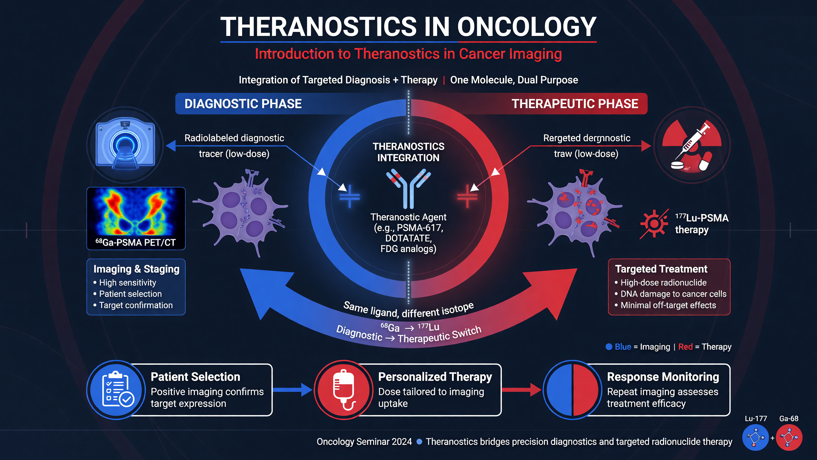
Teaching Point: Theranostics is not simply “imaging plus treatment.” It is a closed clinical loop in which a molecular target is demonstrated, the same target is irradiated, and subsequent biologic and imaging data are used to judge whether the target remains therapeutically relevant. The classic examples are radioiodine in differentiated thyroid cancer and somatostatin-receptor imaging paired with lutetium-177 dotatate in neuroendocrine tumors. In prostate cancer, PSMA PET/CT identifies target-expressing disease; lutetium-177–PSMA-617, also called lutetium Lu 177 vipivotide tetraxetan, delivers beta radiation; and its gamma emissions permit post-treatment planar imaging or SPECT/CT.
That distinction matters because the examinations answer different questions. Pretreatment PSMA PET asks, “Is enough of the clinically important tumor PSMA-positive to make target-directed therapy plausible?” Post-therapy lutetium-177 SPECT/CT asks, “Where did the administered treatment actually go, how heterogeneous was delivery, and how did tumor burden or uptake change across cycles?” Diagnostic CT, bone imaging, symptoms, and laboratory trends still ask whether cancer is anatomically or clinically progressing. No single modality replaces the others.
Framework: Think of prostate theranostics as four linked decisions: target qualification, treatment delivery, response surveillance, and adaptation. Qualification requires more than an attractive maximum-intensity projection: every sizable lesion must be reviewed against CT, and suspicious PSMA-low disease may require fluorodeoxyglucose (FDG) PET or biopsy. Delivery requires adequate marrow and renal reserve. Surveillance integrates symptoms, PSA, CBC, renal function, anatomic imaging, and post-therapy distribution. Adaptation may mean continuing, holding, reducing, stopping, or changing treatment—but only when the supporting evidence is sufficiently mature.
Randomized trials establish clinical value. In VISION, 831 patients with PSMA-positive metastatic castration-resistant prostate cancer (mCRPC), previously treated with an androgen-receptor pathway inhibitor (ARPI) and taxane chemotherapy, were randomized to lutetium-177–PSMA-617 plus protocol-permitted standard care or standard care alone. Imaging progression-free survival was 8.7 versus 3.4 months and overall survival 15.3 versus 11.3 months (hazard ratios 0.40 and 0.62, respectively; PMID: 34161051). TheraP, a randomized phase 2 comparison after docetaxel, produced a PSA decline of at least 50% in 66% with lutetium-177–PSMA-617 versus 37% with cabazitaxel, with fewer grade 3–4 adverse events, although mature overall survival was similar (PMID: 33581798; PMID: 38043558). PSMAfore then moved therapy before taxane in selected patients progressing after one ARPI: radiographic progression-free survival was 9.3 versus 5.6 months compared with an ARPI switch, while intention-to-treat overall survival was not significantly different in the setting of approximately 60% crossover (PMID: 39293462; PMID: 40680993).
Decision Point: In the United States, the 2025 indication includes adults with PSMA-positive mCRPC previously treated with an ARPI who are either appropriate to delay taxane chemotherapy or have already received a taxane. Selection uses an approved PSMA PET agent, not lutetium SPECT. Continue androgen deprivation, compare reasonable alternatives—including taxane therapy, genomically selected PARP inhibition, palliation, or a trial—and incorporate patient goals. “PSMA-positive” is necessary, but it is not the entire treatment decision.
Nuance: PSMA means prostate-specific membrane antigen, but the target is neither completely prostate-specific nor uniformly expressed. Physiologic activity occurs in lacrimal and salivary glands, kidneys, small bowel, and sympathetic ganglia; benign bone remodeling, inflammation, and neovasculature in other tumors can also be avid. Conversely, lineage-plastic or neuroendocrine prostate cancer may lose PSMA while remaining aggressive. Theranostics is therefore a strategy for managing heterogeneity, not a guarantee that one ligand sees every lethal clone.
MUST ACT: Before debating molecular eligibility, address oncologic emergencies: suspected spinal cord or cauda equina compression, unstable pathologic fracture, obstructive uropathy, severe marrow failure, hypercalcemia, uncontrolled pain, or rapidly progressive visceral disease. Targeted radiopharmaceutical therapy does not substitute for urgent MRI, decompression, steroids when indicated, drainage, stabilization, transfusion support, or palliative external-beam radiation.
Audience Poll: A patient has strongly PSMA-avid bone and nodal disease but a rapidly enlarging liver lesion with uptake below liver background. Is he “PSMA-positive,” or does the discordant lesion require a different diagnostic and therapeutic plan?
Mechanism of Lutetium-177 PSMA SPECT CT
Duration: 10–12 Minutes
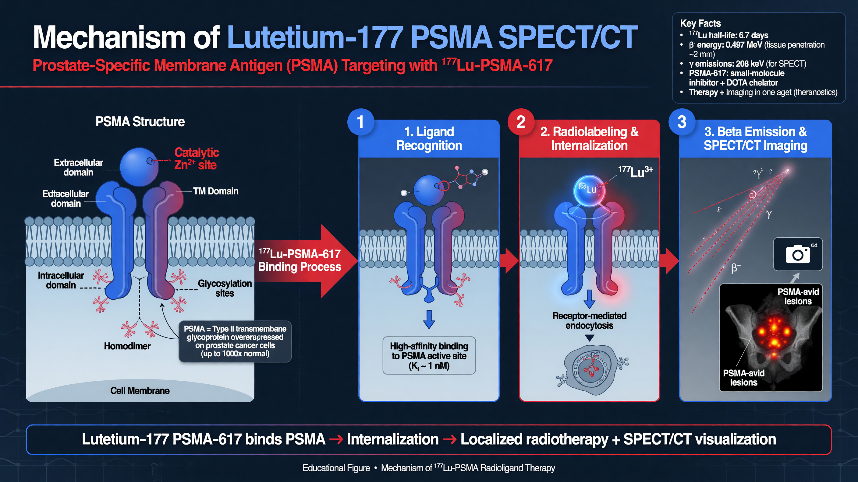
PSMA is a type II transmembrane glutamate carboxypeptidase encoded by FOLH1. Its extracellular domain is highly expressed on most clinically significant prostate adenocarcinomas, often increasing with disease progression and androgen-receptor blockade. PSMA-617 is a small-molecule ligand joined to a DOTA chelator that securely coordinates lutetium-177. After intravenous administration, ligand binding and receptor-mediated internalization retain radioactivity near the tumor cell. The beta particles injure DNA directly and through reactive oxygen species; their short but nonzero range creates a cross-fire effect that can irradiate neighboring cells with lower PSMA density. Cross-fire helps with microscopic heterogeneity but cannot sterilize a macroscopic PSMA-null clone.
Lutetium-177 has a physical half-life of 6.647 days. It emits beta-minus particles with a maximum energy of approximately 498 keV, a mean tissue range near 0.67 mm, and a maximum range of roughly 2 mm. It also emits 113-keV and 208-keV photons at low abundance, allowing gamma-camera imaging. This dual emission is the physical foundation of the theranostic workflow: the beta component treats, while the photon component reveals delivered biodistribution. The therapy is the radioligand; SPECT/CT is the post-treatment measurement tool.
Teaching Point: A post-therapy study is not a conventional diagnostic PSMA scan. At approximately 24 hours, SPECT shows retained lutetium-177–PSMA activity in tumors and physiologic organs after a therapeutic administration. CT supplies attenuation correction and anatomic localization and may disclose findings that uptake alone misses: a PSMA-low liver metastasis, hydronephrosis, fracture, pleural effusion, or extensive marrow replacement. ENZA-p obtained SPECT/CT 24 hours after every treatment. Other dosimetry protocols acquire images at multiple points—such as 4, 24, 48–72, and occasionally 168 hours—to model clearance and calculate time-integrated activity.
Framework: Quantitative SPECT requires a calibrated chain: camera sensitivity measurement; consistent acquisition timing; attenuation, scatter, and collimator-response correction; iterative reconstruction; segmentation of tumors and organs; conversion from counts to activity concentration; time-activity curves; and absorbed-dose calculation in gray. Partial-volume loss can markedly underestimate activity in small lesions. A single 24-hour SUV is therefore not synonymous with absorbed dose. Formal dosimetry is strongest with several time points, although validated single-time-point methods may reduce patient and departmental burden (quantitative lutetium-177 SPECT guidance, PMID: 26471692).
The current labeled regimen is 7.4 GBq (200 mCi) intravenously every six weeks for six doses, or until progression or unacceptable toxicity. Hydration and frequent voiding reduce urinary residence time. Before and during treatment, assess CBC and renal function; extensive skeletal disease, previous taxanes, wide-field radiotherapy, and baseline cytopenias increase marrow risk. Common toxicities include fatigue, nausea, xerostomia, anemia, thrombocytopenia, leukopenia, and less commonly clinically important renal injury. The joint EANM/SNMMI procedure guideline provides selection, administration, follow-up, and relative-contraindication guidance (PMID: 37246997).
Decision Point: Use post-therapy SPECT first to verify plausible target engagement and distribution. If previously avid disease shows little delivered activity, check timing, calibration, injection/extravasation, interval therapy, and biologic loss of PSMA. If a new CT lesion has minimal uptake while other lesions respond, assume interlesional heterogeneity until proven otherwise; FDG PET or biopsy may be more consequential than averaging the whole patient into one SUV.
Interpretive pitfalls are predictable. Salivary, lacrimal, renal, bowel, and ganglionic uptake is physiologic. Healing fractures and degenerative lesions can be PSMA-avid. Urinary activity can obscure pelvic disease. Skeletal tumor burden can make marrow dosimetry uncertain because activity in adjacent metastases contributes cross-dose. Early after enzalutamide, increased PSMA expression may represent pharmacologic upregulation rather than tumor growth; in ENZA-p, mean uptake increased by day 15 in 68% of evaluable patients (PMID: 41986500). Volume, new lesions, CT morphology, PSA, and symptoms must arbitrate apparent molecular “flare.”
MUST ACT: Do not attribute a falling blood count automatically to radioligand toxicity. The differential includes diffuse marrow progression, bleeding, infection, nutritional deficiency, renal disease, another drug, and therapy-related suppression. Review the post-therapy skeletal pattern, CT, reticulocytes, smear, and clinical tempo; hold or modify treatment according to severity while investigating reversible and malignant causes.
Nuance: Cohort dosimetry suggests kidneys, salivary/lacrimal glands, and marrow are principal organs at risk, but external-beam dose limits cannot be transferred uncritically to protracted, heterogeneous radiopharmaceutical exposure. Tumor absorbed dose correlates with response in prospective studies, yet no randomized trial has shown that escalating activity to a personalized tumor-dose target improves survival (PMID: 30291192). Dosimetry is biologically persuasive and operationally promising, but it is not yet a universal dose-prescription standard.
Audience Poll: Which is most dangerous: modestly falling whole-body SPECT SUV, a new PSMA-low liver lesion, or stable uptake with worsening pancytopenia? The answer depends on recognizing that intensity, clonal escape, and organ reserve describe different failure modes.
ENZA-p Trial: Design and Key Findings
Duration: 12 Minutes
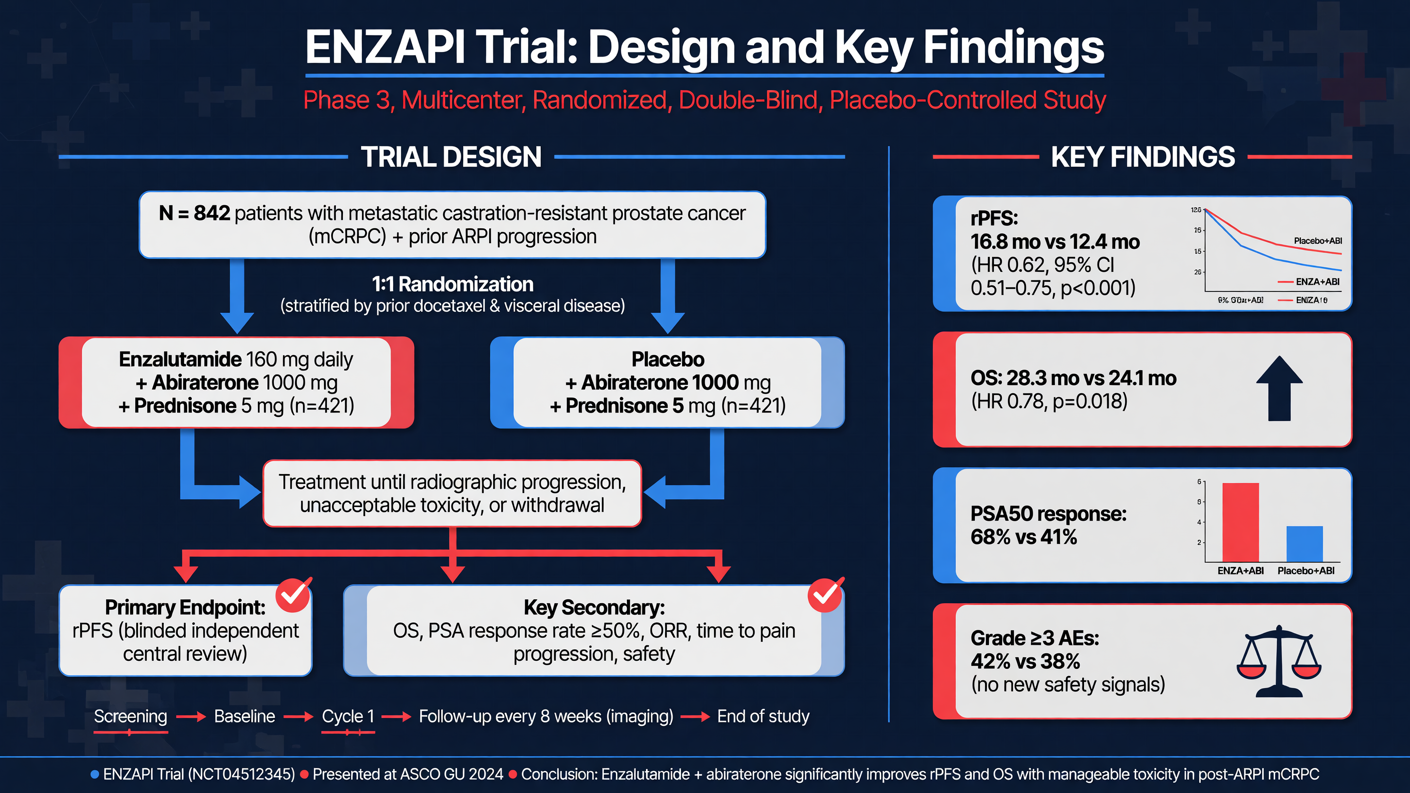
Teaching Point: The correct trial name is ENZA-p—ANZUP 1901, NCT04419402—not “ENZAPI.” ENZA-p did not test whether lutetium SPECT detects more metastases. It tested whether adding adaptively scheduled lutetium-177–PSMA-617 to enzalutamide improves outcomes in poor-risk, PSMA-positive mCRPC, while embedding serial PET and post-treatment SPECT as biomarkers.
The biologic premise was complementary targeting. Androgen-receptor blockade treats AR-dependent clones and can upregulate FOLH1/PSMA in some resistant subpopulations; lutetium-177–PSMA-617 irradiates sufficiently PSMA-expressing clones. ENZA-p was an investigator-initiated, open-label, randomized phase 2 trial at 15 Australian hospitals. It enrolled 162 men—83 assigned to combination and 79 to enzalutamide alone—with progressive mCRPC, PSA above 5 ng/mL, ECOG performance status 0–2, adequate organ function, and no previous docetaxel or ARPI for the castration-resistant state. Prior docetaxel or abiraterone for hormone-sensitive metastatic disease was allowed.
Participants required at least two features associated with early enzalutamide failure: LDH or alkaline phosphatase at or above the institutional upper limit of normal, albumin below 35 g/L, metastatic disease at initial diagnosis, less than three years from diagnosis, more than five bone metastases, visceral metastases, PSA doubling time under 84 days, opioid-requiring pain for more than 14 days, or prior abiraterone in hormone-sensitive disease. Significant small-cell/neuroendocrine, sarcomatoid, or spindle-cell components were excluded. PET eligibility required at least one lesion with SUVmax at least 15 and SUVmax at least 10 in every larger assessable lesion. Of 220 screened patients, 40—18%—were excluded for insufficient PSMA expression.
Framework: Both groups received enzalutamide 160 mg orally each day. The combination group received 7.5 GBq lutetium-177–PSMA-617 at weeks 2 and 8. Centrally reviewed PSMA PET/CT at week 12 then determined cycle number: patients with persistent tumor uptake above blood pool received two additional 7.5-GBq doses at weeks 16 and 24; those without residual PSMA-positive disease stopped after two. This was response-adapted two-versus-four-cycle treatment, not SPECT-derived activity prescription. Post-therapy SPECT/CT was acquired 24 hours after each dose. Nine of 83 participants stopped after one or two doses because no residual target was visible, five received three doses because of progression, and 67 received all four.
The primary endpoint was PSA progression-free survival, defined from randomization to confirmed PSA progression, nonprotocol anticancer therapy, or death. At a median follow-up of 20 months, median PSA progression-free survival was 13.0 months with combination versus 7.8 months with enzalutamide alone (HR 0.43, 95% CI 0.29–0.63; p<0.0001). Clinical progression-free survival was 14.0 versus 9.4 months (HR 0.47). Radiographic progression-free survival was 16.0 versus 12.0 months (HR 0.68, 95% CI 0.45–1.03), a secondary comparison whose confidence interval crossed 1. PSA50 responses occurred in 93% versus 68%, PSA90 responses in 78% versus 37%, and pain improved in 61% versus 27% of evaluable symptomatic patients (PMID: 38621400).
With median follow-up of 34 months, median overall survival was 34 versus 26 months (HR 0.55, 95% CI 0.36–0.84; p=0.0053). Physical-function deterioration-free survival was 10.64 versus 3.42 months, and global health/quality-of-life deterioration-free survival was 8.71 versus 3.32 months. Importantly, 38% of the control group later received lutetium-PSMA off protocol, complicating—and plausibly diluting—the treatment contrast (PMID: 39956124).
Nuance: Safety was not cost-free, but severe-event rates were similar. At mature follow-up, grade 3–5 adverse events occurred in 46% with combination and 44% with enzalutamide. In the combination group, common all-grade events included fatigue 77%, nausea 48%, and dry mouth 41%; grade 3 anemia occurred in 4%. Patient-reported xerostomia reached 74% versus 57%, illustrating how clinician-coded toxicity can underestimate symptom burden. No death was attributed to study treatment.
Decision Point: ENZA-p is compelling phase 2 evidence for early treatment intensification in a specifically selected, high-risk, largely ARPI-naïve mCRPC population. It does not establish routine enzalutamide-plus-lutetium therapy for every patient, does not validate four cycles as superior to the labeled six-cycle monotherapy course, and does not prove biological synergy. Modern generalizability is also limited because many patients now receive an ARPI during hormone-sensitive disease. Phase 3 confirmation and sequencing data remain necessary.
Audience Poll: Which feature most limits immediate adoption: phase 2 size, selected ARPI-naïve biology, PET thresholds, an active but narrower comparator, or the fact that the adaptive gate used PET rather than SPECT?
Survival Prediction and Treatment Decision-Making
Duration: 12 Minutes
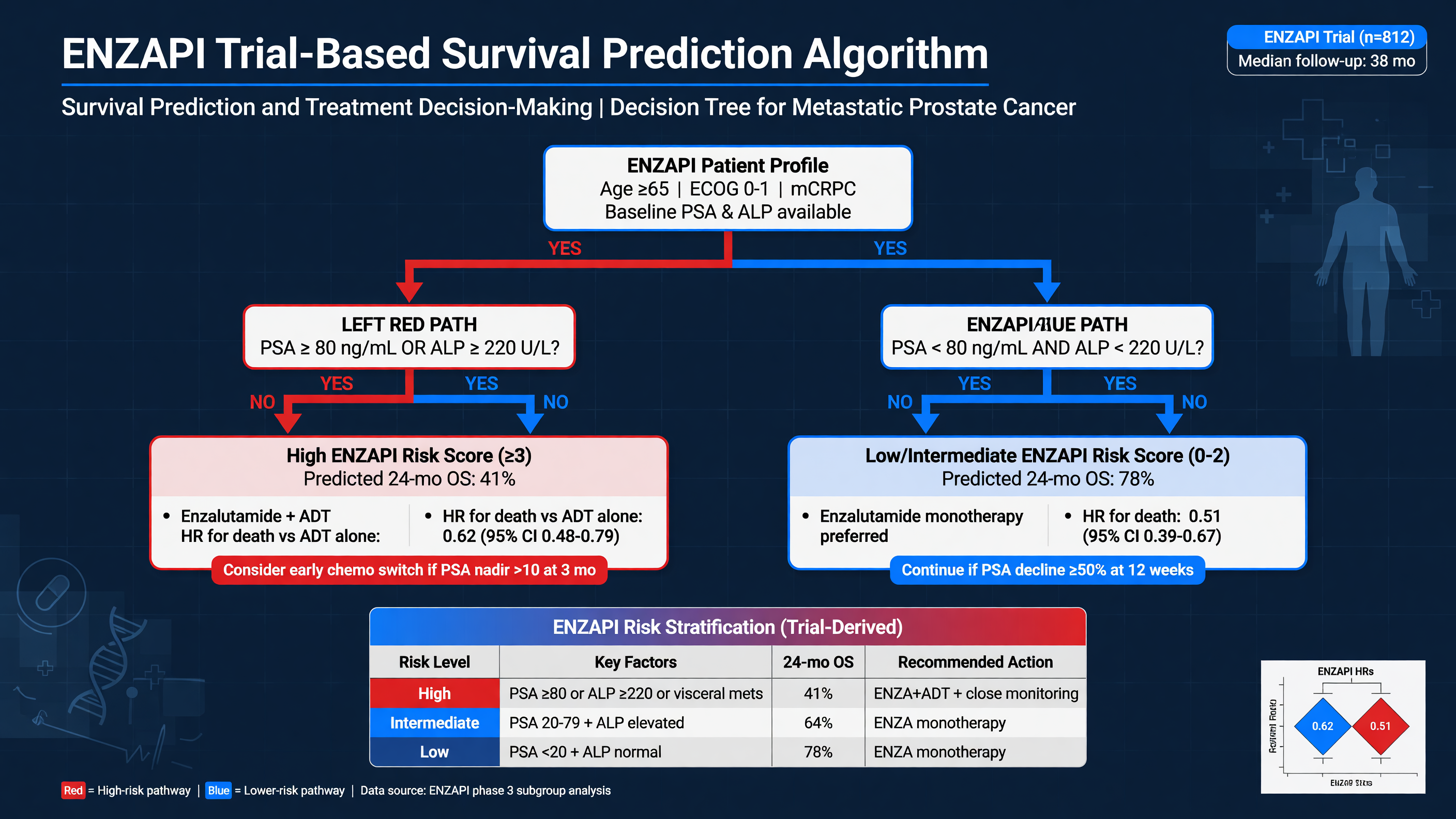
Framework: A prognostic marker identifies outcome regardless of assigned treatment; a predictive marker identifies differential benefit from one treatment compared with another. This distinction prevents two common errors: denying an effective therapy because a patient has poor prognosis, and assuming that a marker associated with survival proves that changing therapy according to that marker will improve survival.
ECOG status, progression tempo, opioid-requiring pain, liver metastases, diffuse osseous or marrow disease, and short time from diagnosis convey aggressiveness. Hemoglobin and platelet count reflect reserve and marrow involvement; alkaline phosphatase tracks skeletal burden; LDH can signal high-volume or dedifferentiated disease; albumin reflects systemic illness. PSA can underrepresent AR-independent or neuroendocrine transformation. A rapidly growing visceral lesion, lytic disease, rising LDH, and relatively modest PSA should trigger review of histology and consideration of FDG PET or biopsy.
The VISION post hoc multivariable overall-survival model combined whole-body PSMA SUVmax, time since diagnosis, opioid use, AST, hemoglobin, lymphocytes, nodal disease, LDH, alkaline phosphatase, and neutrophils, achieving a C-index of 0.73. The radiographic-progression model also included liver metastases. Performance remained similar when SUVmax was removed, reinforcing that outcome reflects whole-patient and whole-tumor state, not one image number (PMID: 39430616). These models support counseling and trial stratification; they are not validated rationing instruments.
ENZA-p sharpened the imaging question. In a prespecified baseline PET substudy, median PSMA-positive total tumor volume (TTV) was 234 mL. With enzalutamide alone, median survival was 39 months below versus 20 months above that median. With combination treatment, it was 35 versus 28 months, and the treatment-by-volume interaction was significant (p=0.0078). Baseline TTV was therefore strongly prognostic and potentially predictive of greater incremental benefit from adding lutetium in high-burden disease. Baseline SUVmean, however, was neither prognostic nor predictive in that combination setting (PMID: 40752515). The 234-mL median is an internal research cutpoint, not a clinical eligibility threshold.
At three months, molecular response carried independent information. Among 152 ENZA-p participants, any increase rather than decrease in PET-derived TTV was associated with shorter survival (HR 2.52) and two-year survival of 30% versus 67%. Residual TTV above versus below the study median of 103 mL was associated with two-year survival of 34% versus 76% (HR 3.76), independent of treatment arm and PSA response (PMID: 41956861). This makes interim tumor volume a credible prognostic biomarker, but it does not prove that switching treatment at 103 mL improves outcome.
The 2026 post-therapy SPECT analysis is especially relevant to this seminar. Seventy-four ENZA-p combination patients underwent quantitative SPECT/CT 24 hours after dose 1 and again after dose 2 at six weeks. Median SPECT TTV fell from 236 to 65 mL, a median reduction of 57%. Twelve patients met the study definition of SPECT complete response—residual TTV below 1 mL. Their two-year survival was 83% versus 67% without SPECT complete response (HR 0.26). Two-year survival was 76% in patients with PSA90 response but no SPECT complete response and 54% in those with neither marker (Ayati et al., Radiology 2026; PMID: 42048586). The cohort and complete-response subgroup were small; the threshold is promising, not a standard stop rule.
Decision Point: Use a layered algorithm. First confirm the indication and rule out emergencies. Second review every sizable lesion on PSMA PET plus diagnostic CT; add FDG PET or biopsy when aggressive discordance is plausible. Third assess organ reserve, prior therapy, symptoms, and alternatives. Fourth establish baseline PSA, CBC, renal function, disease burden, and patient goals. During therapy, integrate clinical benefit, serial laboratories, conventional restaging, and post-therapy target engagement. A new nonavid lesion or clinical deterioration should outweigh a reassuring average SUV.
MUST ACT: Do not continue or stop treatment solely because of one early PSA value, a single SUV, the ENZA-p 234-mL or 103-mL medians, or the SPECT <1-mL research definition. If imaging and clinical data disagree, resolve the discordance: confirm scan comparability, look for new lesions, review CT morphology, consider FDG PET, and biopsy a result that would redirect therapy.
Nuance: More PSMA uptake can mean more deliverable target, but high tumor volume simultaneously portends worse prognosis and can create a “tumor-sink” distribution. Conversely, a dramatic fall in uptake may reflect response or loss of target expression. Volume change and new lesions generally outperform isolated SUV change because they better capture whole-body disease evolution. A 2026 meta-analysis of seven early post-treatment SPECT studies found that new lesions and TTV increase predicted inferior survival, while changes in SUVmean or SUVmax did not reliably do so (PMID: 42110431).
Audience Poll: Would you change therapy for a patient with PSA90 response but a new low-uptake liver lesion on cycle-2 SPECT? The safest answer is not “continue because PSA fell”; it is “characterize the discordant clone before it dictates the patient’s prognosis.”
Adaptive Dosing Strategies in Precision Oncology
Duration: 10–12 Minutes
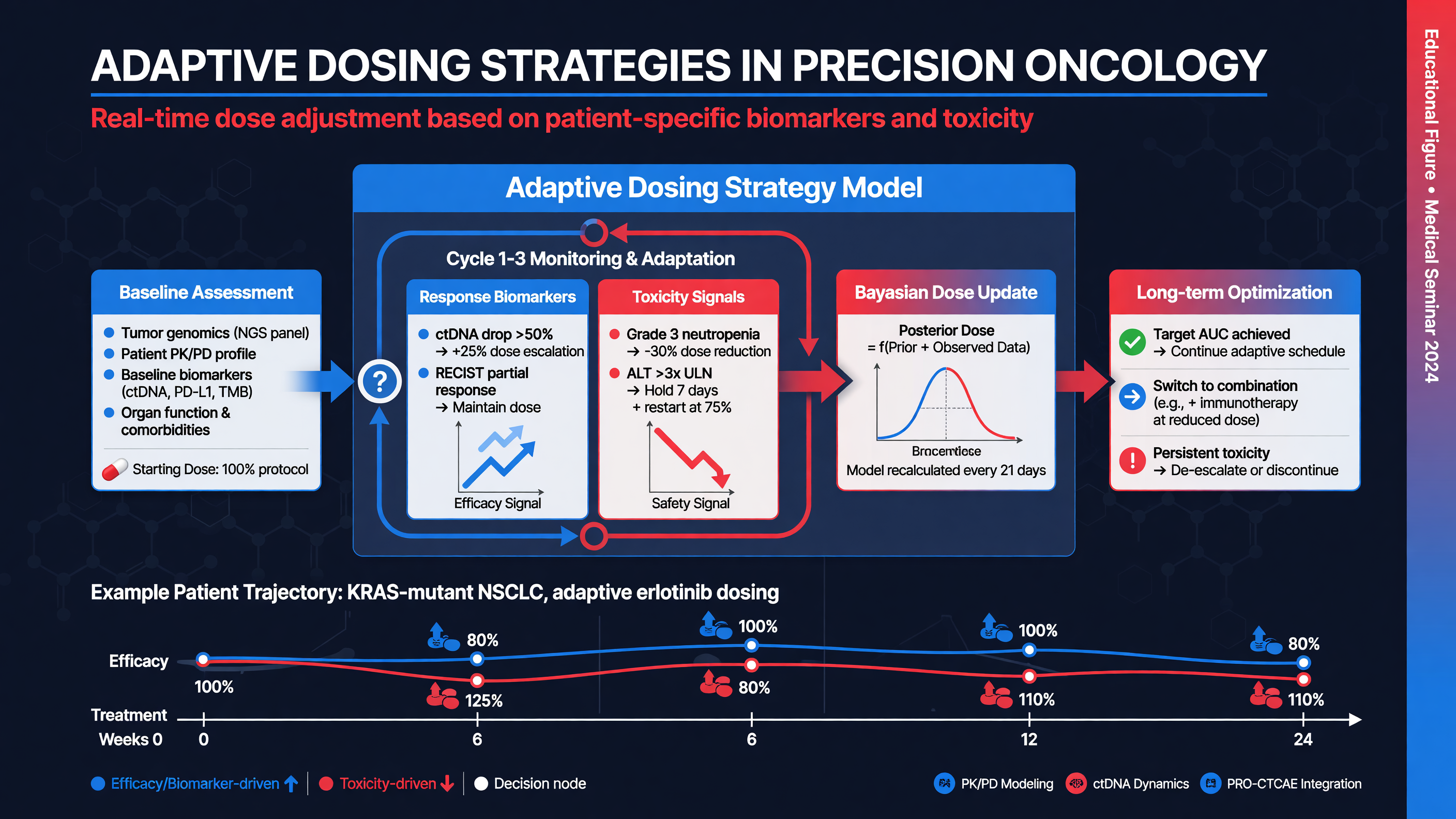
“Adaptive dosing” is often used imprecisely. At least four distinct strategies sit under that label: toxicity-based holding or reduction; response-adapted cycle number; individualized administered activity based on dosimetry; and retreatment after a prior response. Only the first is routine in the current product label. ENZA-p tested the second. The third and fourth remain center-dependent or investigational.
Framework: Start from the labeled reference regimen: 7.4 GBq intravenously every six weeks for up to six doses, while continuing castration, until progression or unacceptable toxicity. Before each cycle, reassess symptoms, performance status, CBC, creatinine and calculated clearance, hydration, obstruction, and interval anticancer treatment. A patient with a deep response still needs toxicity review; a patient with a poor PSA response still needs confirmation that target-positive disease persists and that another immediately effective option exists.
The U.S. label permits one 20% reduction to 5.9 GBq without re-escalation. Clinically significant myelosuppression or renal toxicity generally requires withholding treatment until recovery, then resuming or reducing according to severity; a second required reduction or recurrent severe toxicity leads to discontinuation. Persistent toxicity-related delay beyond four weeks should prompt discontinuation consideration. These are safety adaptations, not evidence that lower activity preserves equal cancer control. Hydration and frequent voiding reduce urinary exposure, while cardiac or renal comorbidity may require individualized fluid planning.
Teaching Point: ENZA-p’s adaptation was deliberately simple. Everyone in the combination arm received 7.5 GBq at weeks 2 and 8. Patients with persistent week-12 PET uptake above blood pool received doses 3 and 4; those without visible target stopped. The administered activity was not calculated from kidney, marrow, or tumor absorbed dose, and SPECT did not control the original decision. This design demonstrated that biomarker-gated cycle number is feasible, but only 11% stopped at two cycles, and the trial was not designed to prove that stopping spared toxicity without sacrificing survival.
Post-therapy SPECT offers a more scalable feedback signal because the therapeutic injection itself supplies the tracer. Quantitative imaging can show delivered tumor burden, emerging new lesions, and interlesional heterogeneity without another PET injection. In three prospective trial cohorts, new metastases on cycle-2 SPECT remained independently associated with worse overall survival after accounting for PSA change (PMID: 40610228). In the ENZA-p SPECT substudy, TTV complete response at dose 2 identified a favorable group (PMID: 42048586). These studies justify prospective adaptive trials; they do not yet justify automatic cessation, escalation, or denial in routine care.
Nuance: Dosimetry-guided treatment aims to prescribe absorbed dose rather than a fixed activity. Serial calibrated SPECT estimates time-integrated activity in tumors, kidneys, salivary glands, and marrow. In principle, a patient with low tumor dose and generous organ reserve could receive more activity, whereas a patient with high renal or marrow exposure could receive less, wait longer, or stop earlier. In practice, kinetics vary by cycle; marrow dose is difficult to estimate in diffuse bone disease; small-lesion recovery is imperfect; acquisition and reconstruction differ across cameras; and accepted organ thresholds largely derive from other radiation contexts. Prospective dose-response data are encouraging, but randomized survival evidence for dosimetry-guided escalation is absent.
Decision Point: Adapt for three reasons, in this order. First, protect the patient from clinically important marrow, renal, salivary, or systemic toxicity. Second, identify unequivocal treatment failure—new lesions, lost target, anatomic progression, or clinical decline—and redirect therapy. Third, within a protocol or validated program, optimize cumulative tumor dose or cycle number. Do not confuse a technically measurable parameter with a clinically validated action threshold.
Rechallenge is another form of adaptation. A patient who achieved a meaningful, durable response, recovered organ function, and retains concordant PSMA-positive disease may respond to later lutetium retreatment. Selection bias is substantial in existing series, cumulative marrow exposure matters, and alternative active treatments must be considered. Prospective trials should define minimum prior response, treatment-free interval, acceptable cumulative dose, and the role of post-treatment SPECT in selecting rechallenge.
MUST ACT: When cytopenia, renal decline, severe xerostomia, or worsening performance status appears, pause and establish causality before the next radioactive dose. When a new low-uptake lesion appears, do not “dose through” presumed target-negative progression without multidisciplinary review. Adaptive oncology is safest when adaptation is explicit, evidence-linked, and reversible.
Audience Poll: If cycle-2 SPECT shows an 80% TTV reduction but platelets are falling and a new nonavid liver lesion is enlarging, should the next action be another full dose, a reduced dose, a delay, or a diagnostic pivot? The case contains three competing signals, and average response cannot erase clonal escape or organ risk.
Challenges and Future Directions in Theranostic Imaging
Duration: 10–12 Minutes
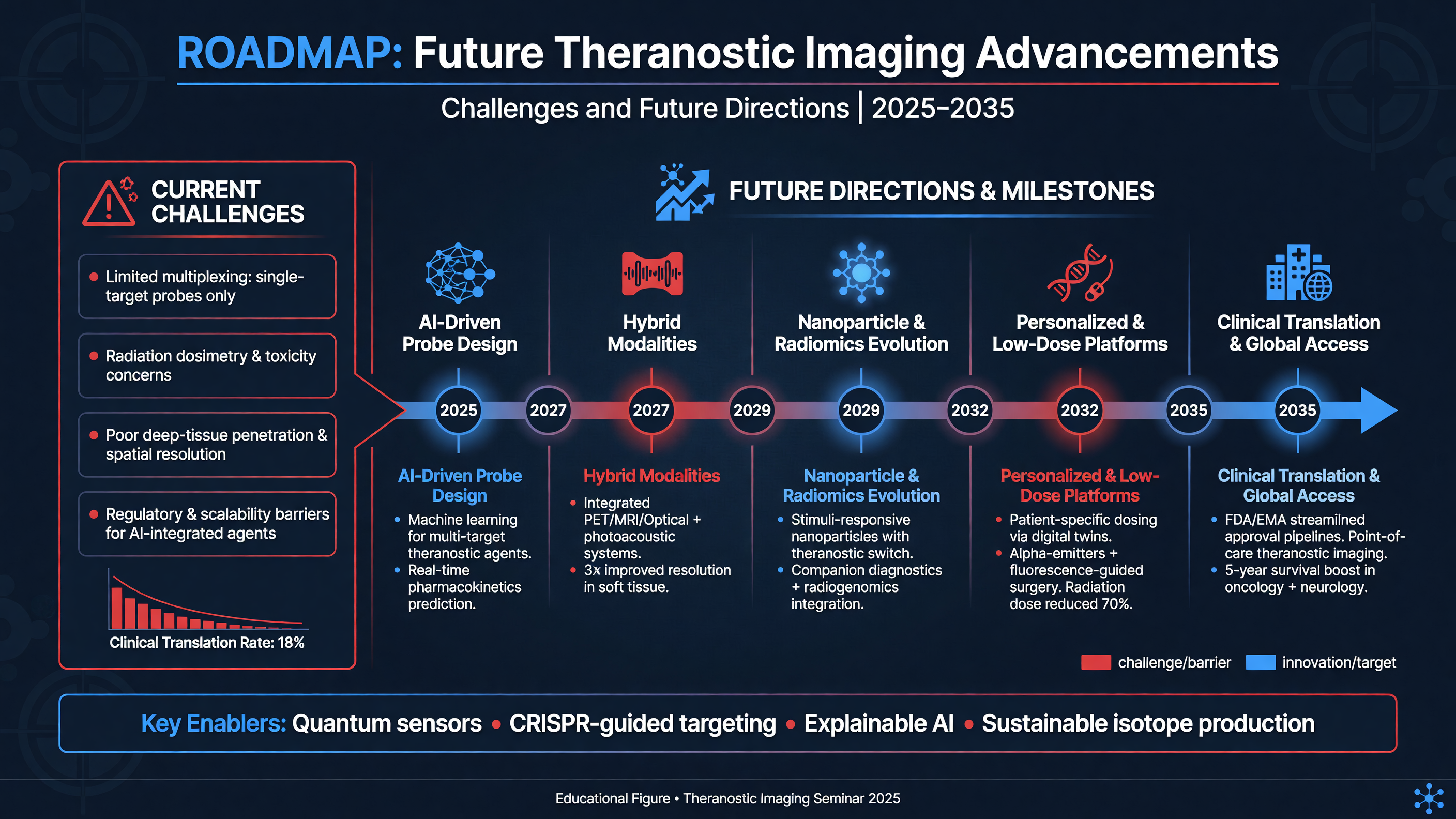
The central biological challenge is heterogeneity across space and time. PSMA expression varies between patients, between lesions, and within the same lesion. Androgen-receptor blockade can transiently raise expression; therapy can select PSMA-low clones; neuroendocrine or other lineage-plastic disease may become FDG-avid and PSMA-poor. Short-range beta cross-fire cannot compensate for a large target-negative population. Future selection must therefore move beyond a binary “positive/negative” PET label toward joint measures of target intensity, total burden, discordant volume, anatomic risk, and tumor genomics.
MUST ACT: Suspect aggressive variant transformation when clinical behavior is out of proportion to PSA, particularly with rapid visceral progression, lytic lesions, rising LDH, hypercalcemia, or low PSMA uptake. Obtain FDG PET and/or tissue when the result can change treatment. A target-negative liver clone may require taxane or platinum-based therapy rather than additional PSMA-directed radiation.
Technical standardization is the second barrier. Quantitative SPECT is more available than PET and can be embedded after each dose, but it has lower spatial resolution and is sensitive to acquisition time, camera calibration, collimator choice, scatter correction, reconstruction, segmentation, and partial-volume error. Total tumor volume can change simply because thresholds or software change. Multicenter harmonization, phantom qualification, reference datasets, and uncertainty reporting are prerequisites before an SUV or absorbed-dose threshold becomes portable. RECIP-style response systems that combine tumor-volume change and new lesions offer a useful research language, but they still require prospective treatment-switch validation.
Framework: Artificial intelligence can remove major workflow bottlenecks by segmenting whole-body disease, excluding physiologic organs, matching lesions longitudinally, computing TTV and uptake metrics, and supporting voxel-level dosimetry. The most valuable output is not an autonomous “continue/stop” command; it is a reproducible map of burden, heterogeneity, delivered dose, and uncertainty for physician review. Domain shift across tracers, scanners, reconstruction methods, and patient populations can silently corrupt models. Ganglia, fractures, degenerative disease, urinary activity, and low-dose CT artifacts remain classic failure modes. External validation and human correction are mandatory.
The therapeutic horizon includes higher-linear-energy-transfer radionuclides and rational combinations. Actinium-225–PSMA produces short-range alpha tracks with clustered DNA damage and has shown activity after lutetium failure, but current evidence is dominated by early-phase and nonrandomized cohorts. Xerostomia, lacrimal injury, marrow and renal exposure, daughter redistribution, isotope scarcity, and difficult imaging prevent routine substitution (phase 1 ^225Ac-J591, PMID: 37922438). Terbium-161 emits beta particles plus short-range conversion and Auger electrons; the first-in-human VIOLET phase 1/2 study supports further evaluation, not standard use (PMID: 40617237). Lead-212, astatine-211, copper isotopes, antibodies, and albumin-binding ligands similarly remain investigational.
Combination therapy aims to overcome resistance: ARPIs may increase PSMA and suppress complementary clones; PARP or DNA-damage-response inhibitors may radiosensitize; immunotherapy could exploit radiation-induced immune signaling; and external-beam radiation can control oligoprogressive escape. ENZA-p supports the combination concept, while UpFrontPSMA and LUNAR show that radioligand therapy is being studied in hormone-sensitive and oligorecurrent disease (PMID: 39293461; PMID: 41223345). Earlier use creates a higher bar for long-term safety, fertility counseling, renal surveillance, marrow recovery, and late myeloid neoplasms.
Nuance: As treatment moves earlier, the comparator becomes stronger and the opportunity cost larger. A radiographic progression-free survival advantage over an ARPI switch does not prove superiority to docetaxel for every taxane-fit patient. Likewise, a favorable response biomarker does not establish the optimal sequence with cabazitaxel, PARP inhibition, radium-223, metastasis-directed therapy, or emerging agents. Trials must measure overall survival, symptoms, time to deterioration, subsequent-treatment feasibility, financial toxicity, and quality-adjusted survival—not merely PSA response.
Implementation is itself a clinical technology. A safe program requires authorized nuclear-medicine physicians, medical oncology and urology integration, technologists, nurses, physicists, radiation-safety expertise, calibrated cameras, radiopharmacy and waste processes, reliable isotope supply, patient-release instructions, and rapid access for urgent complications. Rural distance, reimbursement, manufacturing bottlenecks, and limited PET capacity can turn an effective therapy into an inequitable one. ENZA-p did not collect ethnicity data, and pivotal trials underrepresented some populations; prognostic algorithms trained on such cohorts can reproduce access bias.
Decision Point: The next generation of trials should test explicit adaptive actions: continue versus stop after deep molecular response; switch versus continue after new SPECT lesions; fixed activity versus dosimetry-guided escalation; and lutetium alone versus biologically rational combinations. Imaging must be analytically validated, the decision rule specified in advance, and patient-centered outcomes measured. A prognostic association is only the first half of precision oncology; the second half is proving that acting on it helps.
Audience Poll: Which bottleneck most limits your center today—patient selection, discordant disease, isotope supply, treatment chairs, quantitative SPECT calibration, dosimetry staffing, reimbursement, or coordination across specialties?
Case Discussion: Advanced Prostate Cancer Management
Presentation
A 72-year-old man with de novo metastatic prostate adenocarcinoma received androgen deprivation, docetaxel, and later abiraterone during hormone-sensitive disease. Twenty months later, despite castrate testosterone, his PSA rises from 18 to 46 ng/mL over eight weeks. He has new thoracic back pain requiring opioids but no weakness, sensory level, or sphincter symptoms. CT shows progressive sclerotic skeletal metastases and retroperitoneal nodes; MRI excludes epidural disease. Hemoglobin is 10.2 g/dL, platelets 138 × 10^9/L, alkaline phosphatase is 2.1 times the upper limit of normal, LDH is mildly elevated, albumin is 34 g/L, and creatinine clearance is 58 mL/min. Tumor sequencing shows no actionable homologous-recombination repair alteration and no MSI-high phenotype.
MUST ACT: The immediate back-pain question is spinal instability or cord compression, not radioligand eligibility. Because urgent MRI is negative and pain is controlled, the team can proceed to systemic planning while considering focal external-beam radiation for a mechanically or symptomatically dominant lesion.
PSMA PET/CT demonstrates uptake above liver in extensive nodal and osseous disease. A 1.4-cm liver lesion, however, is only faintly avid. The group reviews prior CT and obtains FDG PET, which shows intense FDG uptake in the liver lesion but no other discordant site. Biopsy confirms prostate adenocarcinoma without small-cell morphology, but with low PSMA expression. This lesion is not an incidental footnote: it represents a clone likely to receive inadequate radiation.
Decision Point: Reasonable strategies include taxane chemotherapy, local ablation or stereotactic radiation to the isolated discordant liver focus followed by lutetium-177–PSMA-617 for the dominant concordant burden, or a clinical trial. The choice depends on taxane fitness, pace of liver progression, symptoms, patient preference, and multidisciplinary confidence that the discordant site can be controlled. PSMA positivity in most disease should not erase a biologically important exception.
After discussion, the patient prefers to defer another taxane and undergoes stereotactic treatment of the isolated liver lesion, then starts labeled lutetium-177–PSMA-617 at 7.4 GBq every six weeks while continuing androgen deprivation. He receives hydration instructions and individualized radiation-safety counseling. Baseline CBC, creatinine clearance, symptoms, PSA, and imaging are documented. Twenty-four-hour post-therapy SPECT/CT confirms high delivery to the known skeletal and nodal disease without unexpected organ distribution.
At cycle 2, PSA has fallen 72%, pain and opioid use have decreased, hemoglobin is 9.4 g/dL, and platelets are 96 × 10^9/L. Quantitative SPECT shows a large TTV reduction and no new avid lesions. CT shows stable treated liver disease. The response is encouraging, but the cytopenias require a differential: treatment effect, extensive skeletal tumor, delayed chemotherapy injury, bleeding, renal contribution, or nutritional deficiency.
Framework: The team reviews the blood-count trajectory, reticulocyte count, smear, iron/B12/folate status, renal function, marrow distribution on imaging, and concomitant drugs. They delay the next cycle until recovery rather than treating the SPECT response as permission to ignore marrow risk. If recovered adequately, treatment may resume according to the label; if toxicity meets reduction criteria, use the single permitted reduction to 5.9 GBq without re-escalation. Recurrent severe myelosuppression would end lutetium therapy.
Teaching Point: This case demonstrates the complete theranostic loop. PET qualified most—but not all—disease. FDG PET and biopsy exposed a target-negative clone. Local therapy controlled the exception. Post-therapy SPECT confirmed delivered treatment and quantified early response. Laboratory monitoring prevented a favorable image from overriding organ reserve. At every step, the actionable unit was the integrated patient, not the brightest lesion, the lowest PSA, or one tumor-volume threshold.
Nuance: If cycle-2 SPECT had shown a new lesion despite falling PSA, that would be an adverse prognostic signal and a reason for urgent anatomic correlation, not an automatic conclusion that all therapy had failed. If SPECT uptake had disappeared everywhere with stable or enlarging CT lesions, loss of PSMA rather than complete response would need consideration. Molecular silence can mean eradication—or escape.
Tonight on Shift
- Separate the tools: use approved PSMA PET/CT to establish target expression; use post-therapy lutetium-177 SPECT/CT to confirm delivery, assess heterogeneity, and support response or dosimetry.
- Find the dangerous exception: correlate every sizable lesion with CT; pursue FDG PET or biopsy for rapidly progressive, visceral, lytic, low-PSA, or PSMA-low discordant disease.
- Protect organ reserve: before every cycle review CBC, creatinine clearance, symptoms, hydration, obstruction, marrow burden, and prior treatment; investigate cytopenias rather than assuming one cause.
- Know the reference regimen: 7.4 GBq every six weeks for up to six doses; use label-directed holds and the single 20% reduction to 5.9 GBq when toxicity requires it.
- Treat biomarkers as evidence, not commands: ENZA-p PET TTV and six-week SPECT TTV are prognostic and hypothesis-generating; no research median, SUV, PSA response, or <1-mL threshold is a stand-alone stop/switch rule.
- Escalate discordance, not averages: worsening symptoms, new lesions, an enlarging nonavid clone, or organ toxicity should trigger multidisciplinary reassessment even when PSA or whole-body uptake appears favorable.
Read this seminar as Markdown · All seminars · Lecture library · Question bank