# Advanced Management of Pediatric Vaso-Occlusive Episodes in Sickle Cell Disease
## Learning Objectives
1. Explain the molecular, vascular, inflammatory, and neurologic mechanisms that produce vaso-occlusive pain.
2. Distinguish an uncomplicated vaso-occlusive episode from acute chest syndrome, infection, sequestration, aplasia, stroke, and other emergencies.
3. Construct an individualized, time-sensitive, multimodal analgesic plan for children presenting with severe pain.
4. Apply hydration and respiratory-prevention protocols without causing fluid overload or opioid-related hypoventilation.
5. Select simple transfusion or red-cell exchange according to the clinical indication, baseline hemoglobin, and hyperviscosity risk.
6. Anticipate alloimmunization, delayed hemolytic transfusion reactions, hyperhemolysis, and iron overload.
7. Integrate family expertise, disease-modifying therapy, discharge education, and equitable communication into acute care.
---
## Introduction to Sickle Cell Disease and Vaso-Occlusive Episodes
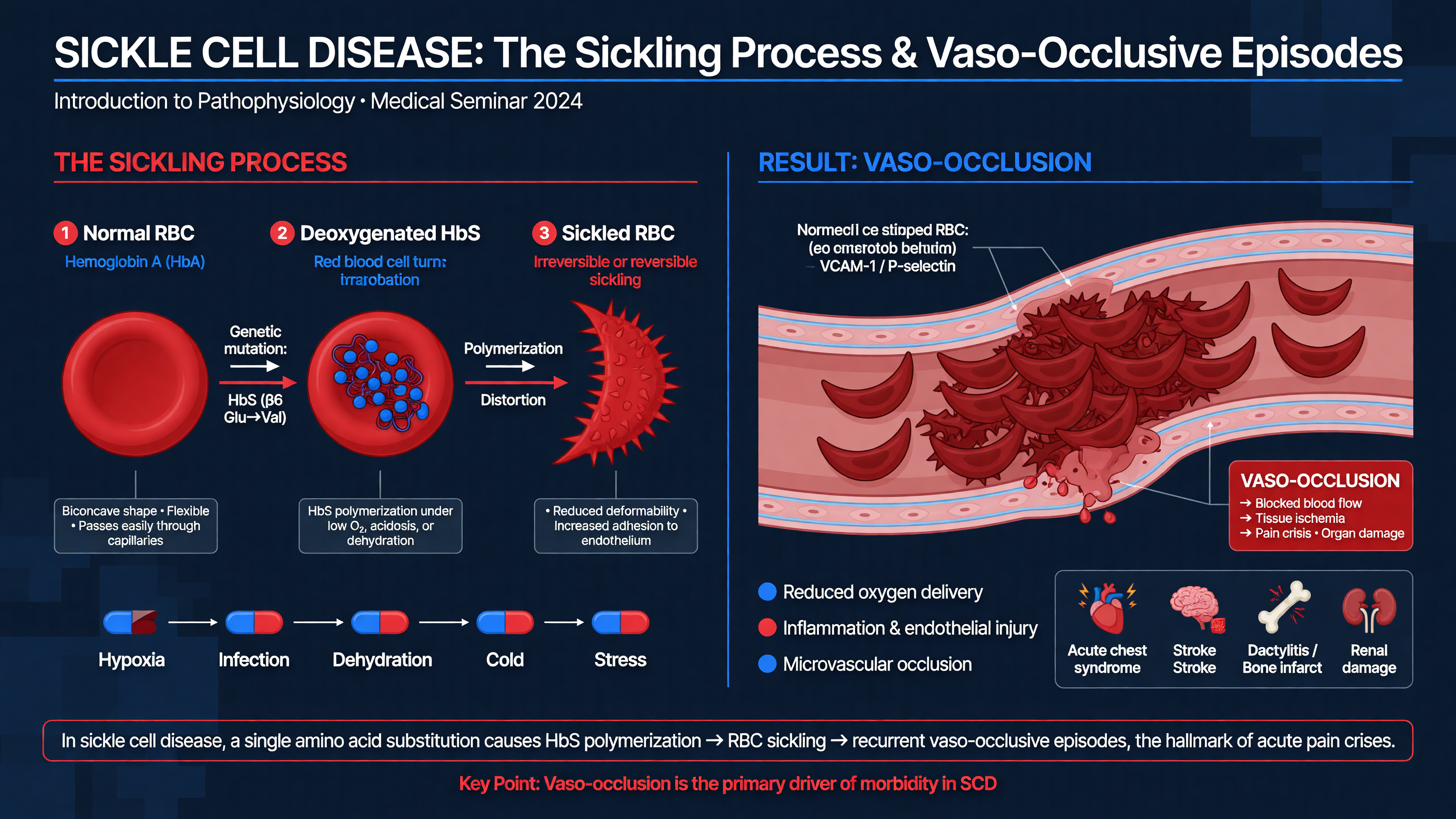 Sickle cell disease (SCD) comprises several inherited hemoglobin disorders in which hemoglobin S is present. The pathogenic HBB variant substitutes valine for glutamic acid at position 6 of the β-globin chain. In HbSS and HbSβ⁰-thalassemia, little or no normal adult hemoglobin is produced; HbSC and HbSβ⁺-thalassemia usually have higher baseline hemoglobin concentrations but can still cause severe vaso-occlusion, acute chest syndrome, retinopathy, avascular necrosis, and other organ injury. Genotype influences population-level risk, but it does not reliably predict the severity of an individual child’s pain.
When oxygen tension falls, deoxygenated HbS molecules polymerize into long intracellular fibers. Polymerization is exquisitely sensitive to intracellular HbS concentration: erythrocyte dehydration, acidosis, and prolonged microvascular transit shorten the delay before polymer formation. Repeated cycles of sickling and unsickling damage the membrane, expose adhesion molecules, alter ion transport, and produce dense, poorly deformable erythrocytes. Fetal hemoglobin interrupts HbS polymer formation, explaining much of the protective effect of hydroxyurea.
**Framework:** Follow the path from molecule to symptom: HbS polymerization causes erythrocyte distortion; membrane injury and dehydration reduce deformability; erythrocytes, reticulocytes, neutrophils, and platelets adhere to activated endothelium; intermittent obstruction produces ischemia; reperfusion releases oxidants and inflammatory mediators; repeated nociceptive input produces peripheral and central sensitization. VOE is therefore a dynamic multicellular process, not simply a collection of rigid cells plugging capillaries. Selectins, neutrophil extracellular traps, complement, free heme, and endothelial activation all amplify obstruction and inflammation (PMID: [29542687](https://pubmed.ncbi.nlm.nih.gov/29542687/); PMID: [30332562](https://pubmed.ncbi.nlm.nih.gov/30332562/)).
Hemolysis contributes a parallel vascular injury phenotype. Cell-free hemoglobin consumes nitric oxide, while heme promotes oxidative stress and innate immune activation. The result is vasoconstriction, endothelial dysfunction, platelet activation, and increased vascular adhesiveness. These pathways overlap with, but are not identical to, the mechanisms driving acute pain.
**Teaching Point:** A vaso-occlusive episode is a clinical diagnosis. There is no laboratory value, radiograph, or physical sign that confirms uncomplicated VOE or quantifies its severity. Severe pain may coexist with normal vital signs, a reassuring appearance, and hemoglobin near baseline.
Cold exposure, dehydration, infection, hypoxemia, acidosis, sleep-disordered breathing, intense exertion, and emotional stress can precipitate an episode, but many events have no identifiable trigger. Asking “what caused this?” can uncover treatable infection or poor access to medication, yet it should never become an exercise in blaming the child or family.
Newborn screening permits early penicillin prophylaxis, immunization, spleen education, transcranial Doppler surveillance, hydroxyurea counseling, and connection with comprehensive SCD care. These interventions have transformed childhood survival. Nevertheless, VOEs remain the most frequent acute complication and the leading reason for emergency visits and hospitalization. Healthcare utilization captures only part of the burden because families treat many episodes at home.
**MUST ACT:** Treat reported pain while evaluating for dangerous alternatives. Do not require laboratory confirmation, visible distress, tachycardia, or imaging before initiating analgesia.
**Nuance:** “Vaso-occlusive episode” is preferred to “crisis” because it describes the pathophysiology without suggesting chaos, culpability, or a single uniform presentation. It also emphasizes that an acute encounter occurs within a chronic disease marked by cumulative vascular injury and recurrent pain.
**Audience Poll:** Which intervention changes the immediate experience of a child with severe VOE most reliably: an additional diagnostic test, rapid individualized analgesia, routine oxygen, or an empiric blood transfusion?
---
## Clinical Presentation and Pathophysiology of VOEs
Sickle cell disease (SCD) comprises several inherited hemoglobin disorders in which hemoglobin S is present. The pathogenic HBB variant substitutes valine for glutamic acid at position 6 of the β-globin chain. In HbSS and HbSβ⁰-thalassemia, little or no normal adult hemoglobin is produced; HbSC and HbSβ⁺-thalassemia usually have higher baseline hemoglobin concentrations but can still cause severe vaso-occlusion, acute chest syndrome, retinopathy, avascular necrosis, and other organ injury. Genotype influences population-level risk, but it does not reliably predict the severity of an individual child’s pain.
When oxygen tension falls, deoxygenated HbS molecules polymerize into long intracellular fibers. Polymerization is exquisitely sensitive to intracellular HbS concentration: erythrocyte dehydration, acidosis, and prolonged microvascular transit shorten the delay before polymer formation. Repeated cycles of sickling and unsickling damage the membrane, expose adhesion molecules, alter ion transport, and produce dense, poorly deformable erythrocytes. Fetal hemoglobin interrupts HbS polymer formation, explaining much of the protective effect of hydroxyurea.
**Framework:** Follow the path from molecule to symptom: HbS polymerization causes erythrocyte distortion; membrane injury and dehydration reduce deformability; erythrocytes, reticulocytes, neutrophils, and platelets adhere to activated endothelium; intermittent obstruction produces ischemia; reperfusion releases oxidants and inflammatory mediators; repeated nociceptive input produces peripheral and central sensitization. VOE is therefore a dynamic multicellular process, not simply a collection of rigid cells plugging capillaries. Selectins, neutrophil extracellular traps, complement, free heme, and endothelial activation all amplify obstruction and inflammation (PMID: [29542687](https://pubmed.ncbi.nlm.nih.gov/29542687/); PMID: [30332562](https://pubmed.ncbi.nlm.nih.gov/30332562/)).
Hemolysis contributes a parallel vascular injury phenotype. Cell-free hemoglobin consumes nitric oxide, while heme promotes oxidative stress and innate immune activation. The result is vasoconstriction, endothelial dysfunction, platelet activation, and increased vascular adhesiveness. These pathways overlap with, but are not identical to, the mechanisms driving acute pain.
**Teaching Point:** A vaso-occlusive episode is a clinical diagnosis. There is no laboratory value, radiograph, or physical sign that confirms uncomplicated VOE or quantifies its severity. Severe pain may coexist with normal vital signs, a reassuring appearance, and hemoglobin near baseline.
Cold exposure, dehydration, infection, hypoxemia, acidosis, sleep-disordered breathing, intense exertion, and emotional stress can precipitate an episode, but many events have no identifiable trigger. Asking “what caused this?” can uncover treatable infection or poor access to medication, yet it should never become an exercise in blaming the child or family.
Newborn screening permits early penicillin prophylaxis, immunization, spleen education, transcranial Doppler surveillance, hydroxyurea counseling, and connection with comprehensive SCD care. These interventions have transformed childhood survival. Nevertheless, VOEs remain the most frequent acute complication and the leading reason for emergency visits and hospitalization. Healthcare utilization captures only part of the burden because families treat many episodes at home.
**MUST ACT:** Treat reported pain while evaluating for dangerous alternatives. Do not require laboratory confirmation, visible distress, tachycardia, or imaging before initiating analgesia.
**Nuance:** “Vaso-occlusive episode” is preferred to “crisis” because it describes the pathophysiology without suggesting chaos, culpability, or a single uniform presentation. It also emphasizes that an acute encounter occurs within a chronic disease marked by cumulative vascular injury and recurrent pain.
**Audience Poll:** Which intervention changes the immediate experience of a child with severe VOE most reliably: an additional diagnostic test, rapid individualized analgesia, routine oxygen, or an empiric blood transfusion?
---
## Clinical Presentation and Pathophysiology of VOEs
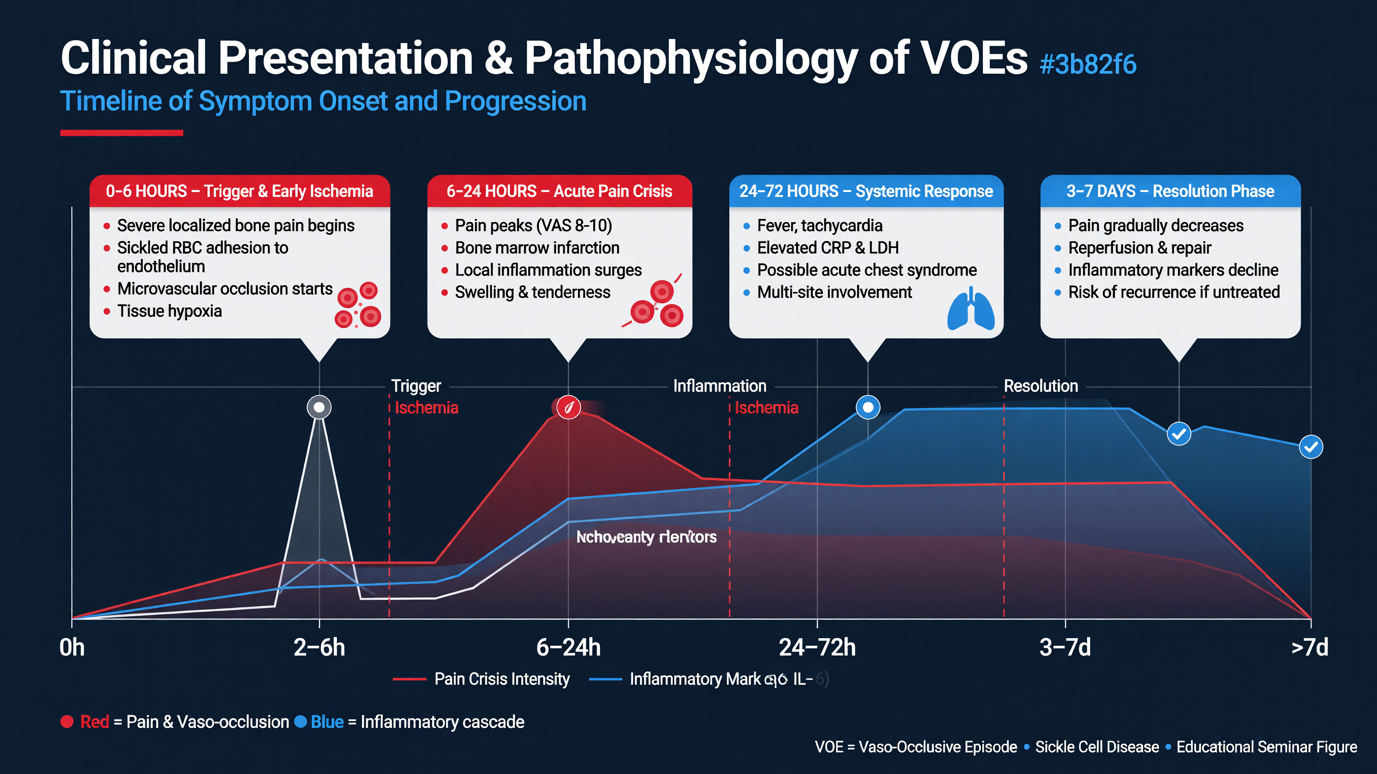 VOE pain may begin abruptly or intensify over several hours. It is commonly deep, throbbing, or aching pain in the long bones, back, ribs, sternum, or abdomen; multiple sites are frequent. Infants may present with dactylitis, irritability, refusal to crawl, or decreased use of an extremity. Older children may have focal or generalized pain, guarding, impaired walking, shallow breathing, nausea, or sleep disruption. The child’s description and caregiver’s observation are primary data.
Initial assessment should establish genotype, baseline hemoglobin and reticulocyte count, usual oxygen saturation, prior acute chest syndrome or stroke, spleen status, asthma, renal disease, current hydroxyurea and analgesic exposure, prior effective opioid doses, recent transfusions, and known antibodies. Ask whether the pain is typical in location and quality. Record temperature, respiratory rate, work of breathing, oxygen saturation relative to baseline, perfusion, neurologic status, hydration, and an age-appropriate pain score. Examine the chest, abdomen and spleen, joints, bones, skin, and neurologic system without repeatedly manipulating painful areas.
**MUST ACT:** Before labeling an encounter “uncomplicated VOE,” explicitly screen for fever, hypoxemia, cough, chest pain, increased work of breathing, focal neurologic findings, altered mental status, priapism, rapidly enlarging spleen, pallor, syncope, focal joint swelling, and peritoneal signs.
The differential diagnosis should be driven by anatomy and trajectory. Focal bone pain with persistent fever, swelling, or bacteremia raises concern for osteomyelitis; both *Staphylococcus aureus* and *Salmonella* species are important. A hot, immobile joint requires urgent evaluation for septic arthritis. Persistent hip or shoulder pain with restricted motion suggests avascular necrosis. Chest or upper-back pain may represent rib infarction, pneumonia, asthma, pulmonary embolism, or evolving acute chest syndrome. Abdominal pain may arise from constipation, opioid ileus, cholelithiasis, cholecystitis, appendicitis, pancreatitis, pyelonephritis, or hepatic or splenic sequestration.
Abrupt anemia with splenomegaly, thrombocytopenia, and reticulocytosis suggests splenic sequestration. Severe anemia with reticulocytopenia suggests transient aplasia, often from parvovirus B19. Pain, jaundice, dark urine, and worsening anemia within 21 days of transfusion should trigger evaluation for delayed hemolytic transfusion reaction or hyperhemolysis. Headache, weakness, aphasia, visual change, seizure, or new gait abnormality is stroke until proven otherwise.
**Framework:** Separate the encounter into three concurrent questions: Is the pain being treated? Is there a precipitating illness? Is a time-sensitive SCD complication developing? These questions should be revisited after every meaningful clinical change.
For ED or hospital presentations, obtain a CBC and reticulocyte count and compare them with the child’s steady state. A hemoglobin decrease of at least 2 g/dL requires evaluation for sequestration, aplasia, infection, acute chest syndrome, blood loss, or delayed hemolysis. Renal function and electrolytes inform NSAID and fluid decisions. Obtain blood cultures promptly for fever of at least 38.5°C and a type and screen when transfusion may become necessary. Imaging should answer a clinical question: chest radiography for respiratory symptoms, fever with thoracic pain, hypoxemia, or abnormal lung findings; ultrasonography for biliary or splenic disease; and targeted MRI when osteomyelitis or avascular necrosis remains concerning.
**Decision Point:** Typical pain with stable oxygenation and no red flags requires limited testing and rapid treatment. Atypical location, focal examination findings, systemic illness, or deviation from baseline justifies broader evaluation—but diagnostic work must proceed in parallel with analgesia.
Acute chest syndrome (ACS) may emerge one to three days after pain begins. Thoracic infarction causes splinting; opioids can compound hypoventilation; atelectasis produces regional hypoxemia; infection, pulmonary vaso-occlusion, and marrow fat embolism intensify lung injury. The National Acute Chest Syndrome Study demonstrated the heterogeneous infectious and noninfectious causes of ACS (PMID: [10861320](https://pubmed.ncbi.nlm.nih.gov/10861320/)). An initially normal chest radiograph does not exclude evolving disease.
**Nuance:** Leukocytosis, elevated inflammatory markers, fever, and marrow edema can occur in both infarction and infection. Trends, cultures, anatomic localization, and clinical evolution are more informative than a single result.
**Audience Poll:** A child reports 9/10 bilateral leg pain but is quietly watching a tablet with normal vital signs. Which finding best determines whether analgesia is indicated? The child’s self-report remains the gold standard.
---
## Pharmacologic Management of Pain in VOEs
VOE pain may begin abruptly or intensify over several hours. It is commonly deep, throbbing, or aching pain in the long bones, back, ribs, sternum, or abdomen; multiple sites are frequent. Infants may present with dactylitis, irritability, refusal to crawl, or decreased use of an extremity. Older children may have focal or generalized pain, guarding, impaired walking, shallow breathing, nausea, or sleep disruption. The child’s description and caregiver’s observation are primary data.
Initial assessment should establish genotype, baseline hemoglobin and reticulocyte count, usual oxygen saturation, prior acute chest syndrome or stroke, spleen status, asthma, renal disease, current hydroxyurea and analgesic exposure, prior effective opioid doses, recent transfusions, and known antibodies. Ask whether the pain is typical in location and quality. Record temperature, respiratory rate, work of breathing, oxygen saturation relative to baseline, perfusion, neurologic status, hydration, and an age-appropriate pain score. Examine the chest, abdomen and spleen, joints, bones, skin, and neurologic system without repeatedly manipulating painful areas.
**MUST ACT:** Before labeling an encounter “uncomplicated VOE,” explicitly screen for fever, hypoxemia, cough, chest pain, increased work of breathing, focal neurologic findings, altered mental status, priapism, rapidly enlarging spleen, pallor, syncope, focal joint swelling, and peritoneal signs.
The differential diagnosis should be driven by anatomy and trajectory. Focal bone pain with persistent fever, swelling, or bacteremia raises concern for osteomyelitis; both *Staphylococcus aureus* and *Salmonella* species are important. A hot, immobile joint requires urgent evaluation for septic arthritis. Persistent hip or shoulder pain with restricted motion suggests avascular necrosis. Chest or upper-back pain may represent rib infarction, pneumonia, asthma, pulmonary embolism, or evolving acute chest syndrome. Abdominal pain may arise from constipation, opioid ileus, cholelithiasis, cholecystitis, appendicitis, pancreatitis, pyelonephritis, or hepatic or splenic sequestration.
Abrupt anemia with splenomegaly, thrombocytopenia, and reticulocytosis suggests splenic sequestration. Severe anemia with reticulocytopenia suggests transient aplasia, often from parvovirus B19. Pain, jaundice, dark urine, and worsening anemia within 21 days of transfusion should trigger evaluation for delayed hemolytic transfusion reaction or hyperhemolysis. Headache, weakness, aphasia, visual change, seizure, or new gait abnormality is stroke until proven otherwise.
**Framework:** Separate the encounter into three concurrent questions: Is the pain being treated? Is there a precipitating illness? Is a time-sensitive SCD complication developing? These questions should be revisited after every meaningful clinical change.
For ED or hospital presentations, obtain a CBC and reticulocyte count and compare them with the child’s steady state. A hemoglobin decrease of at least 2 g/dL requires evaluation for sequestration, aplasia, infection, acute chest syndrome, blood loss, or delayed hemolysis. Renal function and electrolytes inform NSAID and fluid decisions. Obtain blood cultures promptly for fever of at least 38.5°C and a type and screen when transfusion may become necessary. Imaging should answer a clinical question: chest radiography for respiratory symptoms, fever with thoracic pain, hypoxemia, or abnormal lung findings; ultrasonography for biliary or splenic disease; and targeted MRI when osteomyelitis or avascular necrosis remains concerning.
**Decision Point:** Typical pain with stable oxygenation and no red flags requires limited testing and rapid treatment. Atypical location, focal examination findings, systemic illness, or deviation from baseline justifies broader evaluation—but diagnostic work must proceed in parallel with analgesia.
Acute chest syndrome (ACS) may emerge one to three days after pain begins. Thoracic infarction causes splinting; opioids can compound hypoventilation; atelectasis produces regional hypoxemia; infection, pulmonary vaso-occlusion, and marrow fat embolism intensify lung injury. The National Acute Chest Syndrome Study demonstrated the heterogeneous infectious and noninfectious causes of ACS (PMID: [10861320](https://pubmed.ncbi.nlm.nih.gov/10861320/)). An initially normal chest radiograph does not exclude evolving disease.
**Nuance:** Leukocytosis, elevated inflammatory markers, fever, and marrow edema can occur in both infarction and infection. Trends, cultures, anatomic localization, and clinical evolution are more informative than a single result.
**Audience Poll:** A child reports 9/10 bilateral leg pain but is quietly watching a tablet with normal vital signs. Which finding best determines whether analgesia is indicated? The child’s self-report remains the gold standard.
---
## Pharmacologic Management of Pain in VOEs
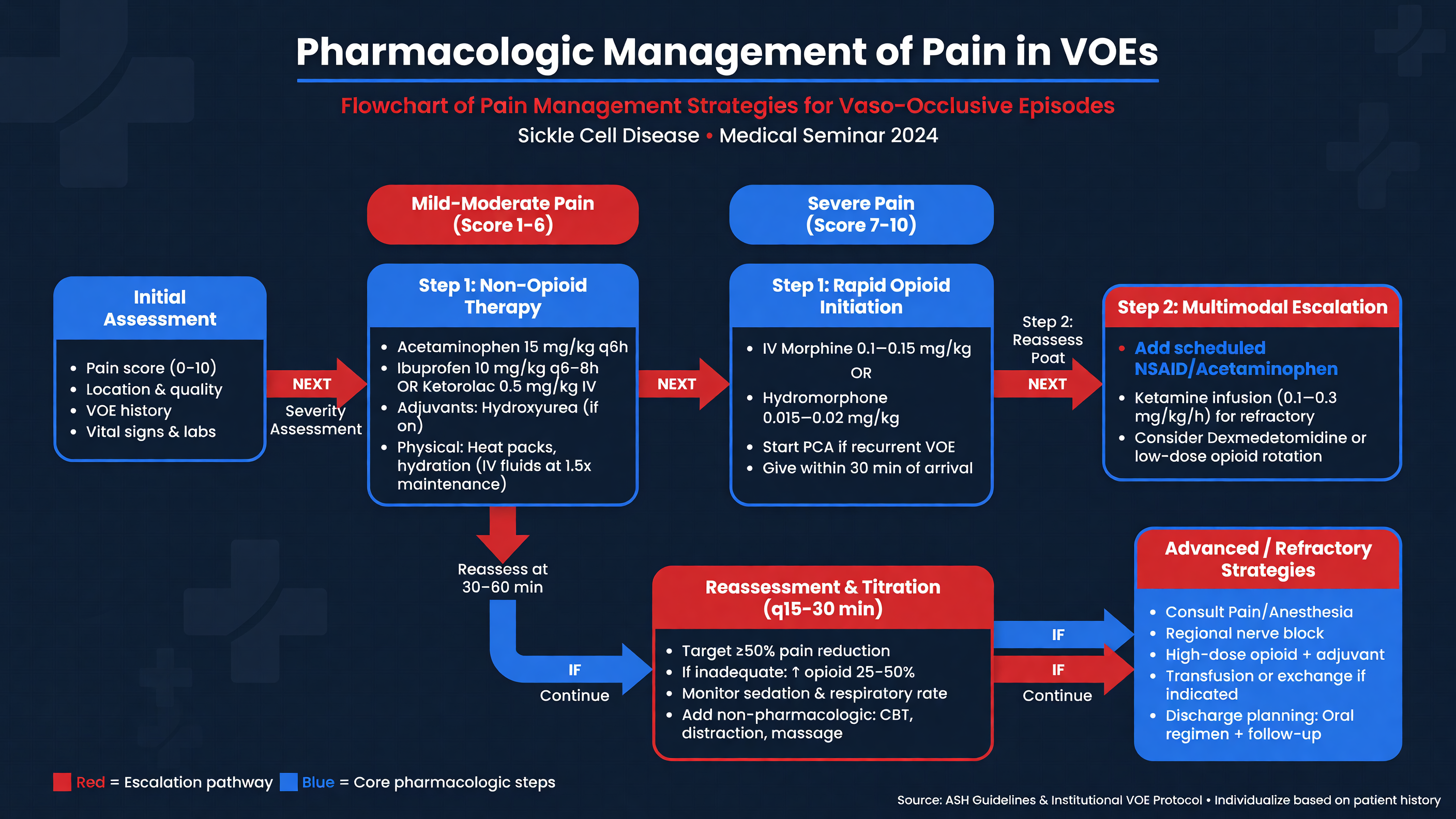 Pain treatment is a time-critical intervention. NHLBI recommends initiating analgesia within 30 minutes of triage or 60 minutes of registration; ASH recommends treatment within one hour of arrival with reassessment every 30–60 minutes. For persistent severe pain, reassessment and redosing every 15–30 minutes may be required during initial titration ([NHLBI Expert Panel Report](https://www.nhlbi.nih.gov/resources/evidence-based-management-sickle-cell-disease-expert-panel-report-2014); ASH pain guideline, PMID: [32559294](https://pubmed.ncbi.nlm.nih.gov/32559294/)). Recent multicenter pediatric data associate opioid delivery within 60 minutes—and a timely second dose when needed—with lower hospitalization odds, although the observational design cannot prove causation (PMID: [40892426](https://pubmed.ncbi.nlm.nih.gov/40892426/)).
**MUST ACT:** Locate the individualized pain plan immediately. Use the child’s prior effective drug, dose, and route when safe. Do not restart an opioid-tolerant adolescent at an opioid-naïve dose solely because a generic pathway is easier to access.
**Framework:** Acute treatment has four components: immediate opioid titration for severe pain, scheduled nonopioid analgesia when safe, prevention and monitoring of adverse effects, and repeated assessment of pain, function, and evolving complications.
If no individualized plan exists, a contemporary pediatric starting example for severe pain is morphine 0.1–0.15 mg/kg IV or hydromorphone 0.01–0.015 mg/kg IV, adjusted for prior opioid exposure and organ dysfunction. Intranasal fentanyl 2 micrograms/kg, usually capped at 100 micrograms, can provide rapid bridging analgesia while IV access is obtained. Reassess 15–30 minutes after parenteral medication; if pain remains severe without excessive sedation, respiratory depression, or hypotension, repeat or increase the opioid according to the institutional protocol. A common pathway gives approximately 50% of the initial dose for early repeat doses, while NHLBI permits cautious escalation of roughly 25% until control is achieved. Pain relief, not somnolence, is the endpoint ([CHOP pediatric pain pathway](https://www.chop.edu/clinical-pathway/sickle-cell-disease-with-pain-clinical-pathway)).
For mild or improving pain, oral morphine or oxycodone may be appropriate when absorption is reliable. Avoid intramuscular injections because they are painful, have unpredictable absorption, and create another potentially confusing pain site. Meperidine should generally be avoided because accumulation of normeperidine can cause neurotoxicity and seizures.
Use one NSAID when renal function, hydration, platelets, gastrointestinal risk, and bleeding risk permit. Examples include ibuprofen 10 mg/kg orally every 6–8 hours or ketorolac 0.5 mg/kg IV every 6–8 hours; age-specific maximum doses and duration must follow the local formulary. Do not combine ketorolac with ibuprofen or another NSAID. Acetaminophen 10–15 mg/kg every 4–6 hours can be scheduled, with total exposure limited to the lower of 75 mg/kg/day or 4 g/day and reduced further for hepatic disease or malnutrition. Evidence for opioid-sparing benefit from NSAIDs and IV acetaminophen is modest, but multimodal therapy can improve comfort and function in selected patients (Canadian Paediatric Society guidance, PMID: [35273671](https://pubmed.ncbi.nlm.nih.gov/35273671/)).
When repeated boluses are required, transition to scheduled dosing or patient-controlled analgesia rather than PRN-only treatment. PCA settings should be derived from recent opioid consumption and prior successful admissions. ASH found insufficient evidence to favor routine basal-plus-demand dosing over demand-only PCA; a basal infusion may benefit an opioid-tolerant patient but can increase sedation in an opioid-naïve child. Only the patient should activate a patient-controlled dose. Use an objective sedation scale, continuous oxygen monitoring when indicated, and capnography for high-risk patients. Prescribe constipation prevention, nausea treatment, and oral—not repeatedly intravenous—antihistamine when pruritus requires therapy.
**Decision Point:** If pain remains severe despite appropriately titrated opioids, first reassess the diagnosis, IV delivery, dose history, anxiety, neuropathic features, and complications. “Refractory pain” should not be declared after one conservative dose.
Ketamine is a specialist adjunct, not a reflexive replacement for opioid titration. ASH conditionally suggests an inpatient subanesthetic infusion beginning at 0.1–0.3 mg/kg/hour, up to 1 mg/kg/hour, for pain inadequately controlled with opioids in centers with appropriate expertise and monitoring. Lubega and colleagues randomized 240 children aged 7–18 years to ketamine 1 mg/kg or morphine 0.1 mg/kg infused over 10 minutes. Ketamine produced noninferior maximal pain reduction and acted faster, but its effect was brief and transient nystagmus, dysphoria, salivation, and other adverse effects were substantially more frequent (PMID: [29794277](https://pubmed.ncbi.nlm.nih.gov/29794277/)).
**Nuance:** The Lubega single-infusion regimen is not the same intervention as ASH’s continuous adjunctive infusion. The trial establishes analgesic activity; it does not justify routine substitution of a 1 mg/kg ketamine infusion for an individualized opioid pathway.
Local heat, repositioning, massage, distraction, breathing exercises, virtual reality, child-life support, and caregiver presence should begin early. Routine oxygen does not treat normoxic VOE. Systemic corticosteroids should not be used solely for VOE pain because rebound pain and rehospitalization may outweigh shorter initial symptoms; reserve them for a separate compelling indication such as significant asthma, with hematology input.
**Teaching Point:** Numeric pain reduction matters, but so do the abilities to breathe deeply, walk, drink, sleep, and engage. A child need not reach zero pain before discharge, but pain must be manageable with a clear oral plan and no evolving complication.
**Audience Poll:** After two appropriately titrated opioid doses, a child remains at 8/10 pain but is alert and breathing comfortably. Is the next step to withhold medication, repeat treatment with reassessment, or conclude that the child is drug-seeking?
---
## Hydration Protocols and Prevention of Acute Chest Syndrome
Pain treatment is a time-critical intervention. NHLBI recommends initiating analgesia within 30 minutes of triage or 60 minutes of registration; ASH recommends treatment within one hour of arrival with reassessment every 30–60 minutes. For persistent severe pain, reassessment and redosing every 15–30 minutes may be required during initial titration ([NHLBI Expert Panel Report](https://www.nhlbi.nih.gov/resources/evidence-based-management-sickle-cell-disease-expert-panel-report-2014); ASH pain guideline, PMID: [32559294](https://pubmed.ncbi.nlm.nih.gov/32559294/)). Recent multicenter pediatric data associate opioid delivery within 60 minutes—and a timely second dose when needed—with lower hospitalization odds, although the observational design cannot prove causation (PMID: [40892426](https://pubmed.ncbi.nlm.nih.gov/40892426/)).
**MUST ACT:** Locate the individualized pain plan immediately. Use the child’s prior effective drug, dose, and route when safe. Do not restart an opioid-tolerant adolescent at an opioid-naïve dose solely because a generic pathway is easier to access.
**Framework:** Acute treatment has four components: immediate opioid titration for severe pain, scheduled nonopioid analgesia when safe, prevention and monitoring of adverse effects, and repeated assessment of pain, function, and evolving complications.
If no individualized plan exists, a contemporary pediatric starting example for severe pain is morphine 0.1–0.15 mg/kg IV or hydromorphone 0.01–0.015 mg/kg IV, adjusted for prior opioid exposure and organ dysfunction. Intranasal fentanyl 2 micrograms/kg, usually capped at 100 micrograms, can provide rapid bridging analgesia while IV access is obtained. Reassess 15–30 minutes after parenteral medication; if pain remains severe without excessive sedation, respiratory depression, or hypotension, repeat or increase the opioid according to the institutional protocol. A common pathway gives approximately 50% of the initial dose for early repeat doses, while NHLBI permits cautious escalation of roughly 25% until control is achieved. Pain relief, not somnolence, is the endpoint ([CHOP pediatric pain pathway](https://www.chop.edu/clinical-pathway/sickle-cell-disease-with-pain-clinical-pathway)).
For mild or improving pain, oral morphine or oxycodone may be appropriate when absorption is reliable. Avoid intramuscular injections because they are painful, have unpredictable absorption, and create another potentially confusing pain site. Meperidine should generally be avoided because accumulation of normeperidine can cause neurotoxicity and seizures.
Use one NSAID when renal function, hydration, platelets, gastrointestinal risk, and bleeding risk permit. Examples include ibuprofen 10 mg/kg orally every 6–8 hours or ketorolac 0.5 mg/kg IV every 6–8 hours; age-specific maximum doses and duration must follow the local formulary. Do not combine ketorolac with ibuprofen or another NSAID. Acetaminophen 10–15 mg/kg every 4–6 hours can be scheduled, with total exposure limited to the lower of 75 mg/kg/day or 4 g/day and reduced further for hepatic disease or malnutrition. Evidence for opioid-sparing benefit from NSAIDs and IV acetaminophen is modest, but multimodal therapy can improve comfort and function in selected patients (Canadian Paediatric Society guidance, PMID: [35273671](https://pubmed.ncbi.nlm.nih.gov/35273671/)).
When repeated boluses are required, transition to scheduled dosing or patient-controlled analgesia rather than PRN-only treatment. PCA settings should be derived from recent opioid consumption and prior successful admissions. ASH found insufficient evidence to favor routine basal-plus-demand dosing over demand-only PCA; a basal infusion may benefit an opioid-tolerant patient but can increase sedation in an opioid-naïve child. Only the patient should activate a patient-controlled dose. Use an objective sedation scale, continuous oxygen monitoring when indicated, and capnography for high-risk patients. Prescribe constipation prevention, nausea treatment, and oral—not repeatedly intravenous—antihistamine when pruritus requires therapy.
**Decision Point:** If pain remains severe despite appropriately titrated opioids, first reassess the diagnosis, IV delivery, dose history, anxiety, neuropathic features, and complications. “Refractory pain” should not be declared after one conservative dose.
Ketamine is a specialist adjunct, not a reflexive replacement for opioid titration. ASH conditionally suggests an inpatient subanesthetic infusion beginning at 0.1–0.3 mg/kg/hour, up to 1 mg/kg/hour, for pain inadequately controlled with opioids in centers with appropriate expertise and monitoring. Lubega and colleagues randomized 240 children aged 7–18 years to ketamine 1 mg/kg or morphine 0.1 mg/kg infused over 10 minutes. Ketamine produced noninferior maximal pain reduction and acted faster, but its effect was brief and transient nystagmus, dysphoria, salivation, and other adverse effects were substantially more frequent (PMID: [29794277](https://pubmed.ncbi.nlm.nih.gov/29794277/)).
**Nuance:** The Lubega single-infusion regimen is not the same intervention as ASH’s continuous adjunctive infusion. The trial establishes analgesic activity; it does not justify routine substitution of a 1 mg/kg ketamine infusion for an individualized opioid pathway.
Local heat, repositioning, massage, distraction, breathing exercises, virtual reality, child-life support, and caregiver presence should begin early. Routine oxygen does not treat normoxic VOE. Systemic corticosteroids should not be used solely for VOE pain because rebound pain and rehospitalization may outweigh shorter initial symptoms; reserve them for a separate compelling indication such as significant asthma, with hematology input.
**Teaching Point:** Numeric pain reduction matters, but so do the abilities to breathe deeply, walk, drink, sleep, and engage. A child need not reach zero pain before discharge, but pain must be manageable with a clear oral plan and no evolving complication.
**Audience Poll:** After two appropriately titrated opioid doses, a child remains at 8/10 pain but is alert and breathing comfortably. Is the next step to withhold medication, repeat treatment with reassessment, or conclude that the child is drug-seeking?
---
## Hydration Protocols and Prevention of Acute Chest Syndrome
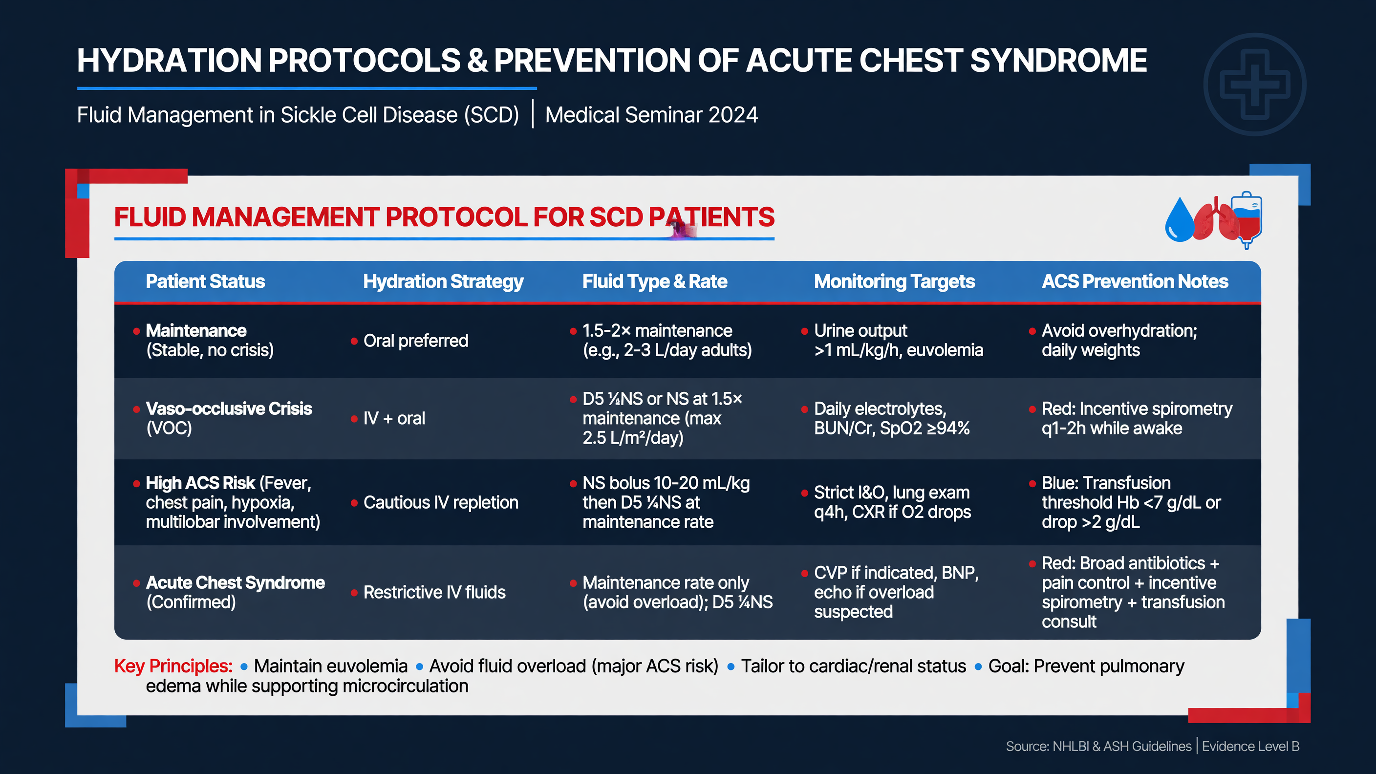 Hydration corrects a real fluid deficit; it does not mechanically “flush” an occluded microcirculation. Hyposthenuria makes many children vulnerable to dehydration, but indiscriminate IV fluid can cause pulmonary edema, worsen gas exchange, and contribute to ACS. ASH found insufficient direct evidence to recommend routine IV fluid as an analgesic treatment, while NHLBI advises no more than maintenance-rate IV hydration for a euvolemic patient who cannot drink.
**Framework:** Assign the child to one of three fluid states. A euvolemic child drinking adequately needs oral fluids, not an IV bolus. A euvolemic child unable to drink may receive IV fluid at no more than maintenance, with all oral intake and medication carriers counted. A child with objective hypovolemia or shock needs isotonic crystalloid in reassessed pediatric resuscitation aliquots. Severe pain alone is not evidence of hypovolemia.
Use an isotonic maintenance solution with dextrose and potassium added when clinically appropriate, guided by age, urine output, electrolytes, and institutional policy. Balanced crystalloid is reasonable when volume replacement is required, although pediatric comparative evidence in SCD remains limited. Record strict intake and output, daily weight, respiratory findings, oxygen requirement, creatinine, and electrolytes. Reduce IV fluid as oral intake improves and account for transfused blood in the total fluid balance. New weight gain, edema, crackles, tachypnea, or oxygen need requires immediate reassessment for both fluid overload and ACS.
**MUST ACT:** Do not order an automatic 20 mL/kg bolus or 1.5-times-maintenance infusion for every VOE. In a euvolemic child, more fluid is not better. If ACS is suspected, many pediatric pathways limit total oral-plus-IV intake to approximately two-thirds to three-quarters maintenance, individualized to perfusion and renal function.
ACS is defined by a new pulmonary infiltrate involving at least one lung segment together with fever, chest pain, cough, tachypnea, wheeze, increased work of breathing, or hypoxemia relative to baseline. Pneumonia and ACS cannot be reliably separated at presentation. Infection, pulmonary infarction, fat embolism, atelectasis, and hypoventilation may coexist, producing a feedback loop in which hypoxemia accelerates HbS polymerization and further pulmonary vaso-occlusion.
Prevention begins on admission. Provide effective analgesia without oversedation, position the child upright, encourage ambulation, and prescribe coached incentive spirometry—typically 10 sustained maximal inspirations every two hours while awake. Younger children may use bubbles, pinwheels, or positive expiratory pressure devices. In the randomized Bellet trial of patients hospitalized with chest or back pain, pulmonary complications occurred during 1 of 19 spirometry admissions versus 8 of 19 control admissions (PMID: [7637747](https://pubmed.ncbi.nlm.nih.gov/7637747/)). The trial was small, but the intervention is low risk and remains standard practice.
**Teaching Point:** Adequate analgesia prevents splinting; excessive sedation promotes hypoventilation. The safest plan combines active pain treatment, objective sedation monitoring, incentive spirometry, mobilization, and surveillance for respiratory change.
Use continuous pulse oximetry for chest or back pain, opioid escalation, sleep-related desaturation, or respiratory symptoms. Give supplemental oxygen when saturation is below 95% on room air or meaningfully below the child’s baseline; do not use routine oxygen as a treatment for normoxic limb pain. A normal initial radiograph does not exclude evolving ACS. Repeat imaging promptly if fever, cough, tachypnea, hypoxemia, or examination findings develop.
**Nuance:** In young children, fever and cough may dominate while pain and auscultatory findings are minimal. In adolescents, severe chest pain, dyspnea, multilobar disease, and fat embolism may produce rapid deterioration.
When ACS is suspected, obtain a chest radiograph, CBC and reticulocytes compared with baseline, metabolic panel, type and screen, blood culture if febrile, and respiratory viral testing. Blood gas assessment is appropriate for severe disease or discordance between appearance and pulse oximetry. Follow hemoglobin, platelets, oxygen requirement, work of breathing, and radiographic progression.
Treat all ACS in the hospital. Begin antibiotics covering typical and atypical respiratory organisms—for example, ceftriaxone 50 mg/kg IV every 24 hours, maximum 2 g, plus azithromycin 10 mg/kg on day 1, maximum 500 mg, followed by 5 mg/kg daily on days 2–5, maximum 250 mg. Adapt this example to allergy, prior ceftriaxone reactions, local resistance, culture results, renal function, and sepsis or MRSA risk. Provide bronchodilator therapy for wheeze or established asthma, not routinely. Escalate from low-flow oxygen to high-flow nasal cannula, noninvasive ventilation, or invasive support according to work of breathing and gas exchange.
**Decision Point:** Falling saturation, increasing oxygen need, multilobar infiltrates, progressive anemia, altered mental status, or respiratory fatigue should trigger PICU and hematology involvement and an urgent transfusion decision—not another fluid bolus.
**Audience Poll:** A euvolemic child receiving maintenance IV fluid develops a new oxygen requirement and crackles. Which two diagnoses must be considered simultaneously? Fluid overload and evolving acute chest syndrome.
---
## Blood Transfusion: Indications and Complications
Hydration corrects a real fluid deficit; it does not mechanically “flush” an occluded microcirculation. Hyposthenuria makes many children vulnerable to dehydration, but indiscriminate IV fluid can cause pulmonary edema, worsen gas exchange, and contribute to ACS. ASH found insufficient direct evidence to recommend routine IV fluid as an analgesic treatment, while NHLBI advises no more than maintenance-rate IV hydration for a euvolemic patient who cannot drink.
**Framework:** Assign the child to one of three fluid states. A euvolemic child drinking adequately needs oral fluids, not an IV bolus. A euvolemic child unable to drink may receive IV fluid at no more than maintenance, with all oral intake and medication carriers counted. A child with objective hypovolemia or shock needs isotonic crystalloid in reassessed pediatric resuscitation aliquots. Severe pain alone is not evidence of hypovolemia.
Use an isotonic maintenance solution with dextrose and potassium added when clinically appropriate, guided by age, urine output, electrolytes, and institutional policy. Balanced crystalloid is reasonable when volume replacement is required, although pediatric comparative evidence in SCD remains limited. Record strict intake and output, daily weight, respiratory findings, oxygen requirement, creatinine, and electrolytes. Reduce IV fluid as oral intake improves and account for transfused blood in the total fluid balance. New weight gain, edema, crackles, tachypnea, or oxygen need requires immediate reassessment for both fluid overload and ACS.
**MUST ACT:** Do not order an automatic 20 mL/kg bolus or 1.5-times-maintenance infusion for every VOE. In a euvolemic child, more fluid is not better. If ACS is suspected, many pediatric pathways limit total oral-plus-IV intake to approximately two-thirds to three-quarters maintenance, individualized to perfusion and renal function.
ACS is defined by a new pulmonary infiltrate involving at least one lung segment together with fever, chest pain, cough, tachypnea, wheeze, increased work of breathing, or hypoxemia relative to baseline. Pneumonia and ACS cannot be reliably separated at presentation. Infection, pulmonary infarction, fat embolism, atelectasis, and hypoventilation may coexist, producing a feedback loop in which hypoxemia accelerates HbS polymerization and further pulmonary vaso-occlusion.
Prevention begins on admission. Provide effective analgesia without oversedation, position the child upright, encourage ambulation, and prescribe coached incentive spirometry—typically 10 sustained maximal inspirations every two hours while awake. Younger children may use bubbles, pinwheels, or positive expiratory pressure devices. In the randomized Bellet trial of patients hospitalized with chest or back pain, pulmonary complications occurred during 1 of 19 spirometry admissions versus 8 of 19 control admissions (PMID: [7637747](https://pubmed.ncbi.nlm.nih.gov/7637747/)). The trial was small, but the intervention is low risk and remains standard practice.
**Teaching Point:** Adequate analgesia prevents splinting; excessive sedation promotes hypoventilation. The safest plan combines active pain treatment, objective sedation monitoring, incentive spirometry, mobilization, and surveillance for respiratory change.
Use continuous pulse oximetry for chest or back pain, opioid escalation, sleep-related desaturation, or respiratory symptoms. Give supplemental oxygen when saturation is below 95% on room air or meaningfully below the child’s baseline; do not use routine oxygen as a treatment for normoxic limb pain. A normal initial radiograph does not exclude evolving ACS. Repeat imaging promptly if fever, cough, tachypnea, hypoxemia, or examination findings develop.
**Nuance:** In young children, fever and cough may dominate while pain and auscultatory findings are minimal. In adolescents, severe chest pain, dyspnea, multilobar disease, and fat embolism may produce rapid deterioration.
When ACS is suspected, obtain a chest radiograph, CBC and reticulocytes compared with baseline, metabolic panel, type and screen, blood culture if febrile, and respiratory viral testing. Blood gas assessment is appropriate for severe disease or discordance between appearance and pulse oximetry. Follow hemoglobin, platelets, oxygen requirement, work of breathing, and radiographic progression.
Treat all ACS in the hospital. Begin antibiotics covering typical and atypical respiratory organisms—for example, ceftriaxone 50 mg/kg IV every 24 hours, maximum 2 g, plus azithromycin 10 mg/kg on day 1, maximum 500 mg, followed by 5 mg/kg daily on days 2–5, maximum 250 mg. Adapt this example to allergy, prior ceftriaxone reactions, local resistance, culture results, renal function, and sepsis or MRSA risk. Provide bronchodilator therapy for wheeze or established asthma, not routinely. Escalate from low-flow oxygen to high-flow nasal cannula, noninvasive ventilation, or invasive support according to work of breathing and gas exchange.
**Decision Point:** Falling saturation, increasing oxygen need, multilobar infiltrates, progressive anemia, altered mental status, or respiratory fatigue should trigger PICU and hematology involvement and an urgent transfusion decision—not another fluid bolus.
**Audience Poll:** A euvolemic child receiving maintenance IV fluid develops a new oxygen requirement and crackles. Which two diagnoses must be considered simultaneously? Fluid overload and evolving acute chest syndrome.
---
## Blood Transfusion: Indications and Complications
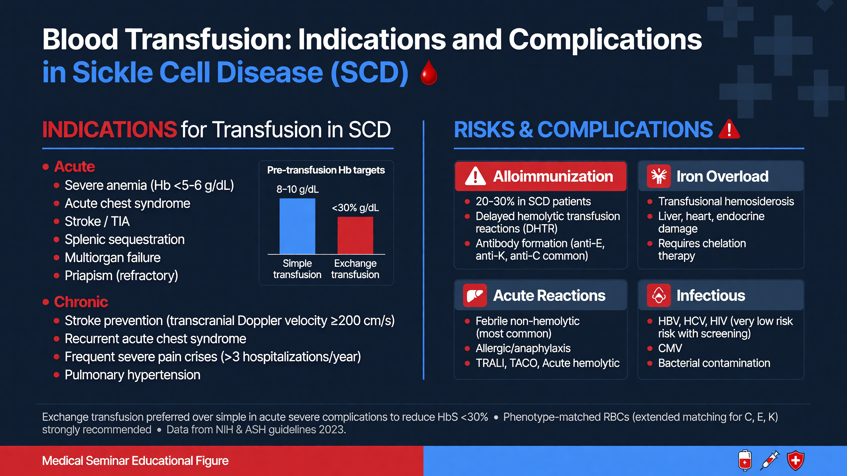 Transfusion improves oxygen-carrying capacity and, when performed as an exchange, lowers the fraction of circulating HbS. It also introduces risks: hyperviscosity, volume overload, alloimmunization, acute and delayed hemolysis, infection, vascular-access complications, and iron loading. The decision must therefore be tied to a specific indication and physiologic target.
**MUST ACT:** Do not transfuse an uncomplicated VOE solely because pain is severe. Transfusion has not been shown to terminate uncomplicated pain and exposes the child to immediate and lifelong risk.
Simple transfusion is appropriate for symptomatic acute anemia, transient aplastic crisis, severe splenic sequestration, acute blood loss, and many moderate ACS presentations when baseline hemoglobin leaves room for a safe increment. A hemoglobin decrease of at least 2 g/dL should trigger diagnostic evaluation, but transfusion depends on symptoms, mechanism, trajectory, and baseline. In splenic sequestration, use cautious aliquots—often 5 mL/kg followed by reassessment—because trapped erythrocytes may re-enter the circulation and produce an unexpectedly high hemoglobin.
For symptomatic ACS with hemoglobin more than 1 g/dL below baseline, a common simple-transfusion dose is 10 mL/kg packed red cells. In HbSS or HbSβ⁰-thalassemia, avoid raising hemoglobin much above 10 g/dL with simple transfusion because viscosity rises as hematocrit increases while substantial HbS remains. HbSC and HbSβ⁺ patients may begin near this threshold and therefore have little room for a top-up.
**Framework:** Ask whether the immediate objective is to raise hemoglobin, reduce HbS, or accomplish both. Simple transfusion primarily raises oxygen-carrying capacity. Exchange transfusion rapidly lowers HbS while limiting the rise in hematocrit and net iron.
Automated or manual red-cell exchange is favored for severe or rapidly progressive ACS, particularly with escalating hypoxemia, multilobar disease, increasing respiratory support, falling hemoglobin despite simple transfusion, or pretransfusion hemoglobin around 9 g/dL or higher. Common targets are hemoglobin near 10 g/dL and HbS below 30%, individualized with hematology and apheresis. Acute focal neurologic deficit is also a transfusion emergency: initiate transfusion urgently, preferably exchange, without waiting for MRI confirmation. If profound anemia makes immediate exchange unsafe or unavailable, simple transfusion may be used while exchange is arranged.
Other accepted indications include multisystem organ failure, severe hepatic sequestration or intrahepatic cholestasis, selected high-risk surgery, and chronic stroke prevention. The STOP trial established chronic transfusion for children with abnormal transcranial Doppler velocities; this preventive indication should not be confused with treatment of an isolated pain episode.
**Decision Point:** Moderate ACS with low hemoglobin generally permits simple transfusion. Severe or rapidly progressive ACS, high pretransfusion hemoglobin, or failure to improve after simple transfusion favors exchange. The ASH transfusion guideline emphasizes this severity-based approach (PMID: [31985807](https://pubmed.ncbi.nlm.nih.gov/31985807/)).
Before the first transfusion, obtain an extended antigen profile whenever possible. At minimum, this should include C/c, E/e, K, Jkᵃ/Jkᵇ, Fyᵃ/Fyᵇ, M/N, and S/s. Genotyping is preferred because recent transfusion can make serologic phenotyping inaccurate and because genotyping detects partial RH variants. Provide leukoreduced, HbS-negative units matched for Rh and Kell at minimum, plus antigen-negative blood for every current or historical clinically significant antibody. Historical antibodies matter even when they are no longer detectable.
**Nuance:** “Phenotype matched” is not a complete order. Specify which antigens are matched, review all prior blood-bank records, and involve transfusion medicine early when antibodies or RH variants are present.
Delayed hemolytic transfusion reaction may masquerade as another VOE. Suspect it within 21 days of transfusion when pain, dark urine, jaundice, fatigue, or worsening anemia develops. Hemoglobin may fall below the pretransfusion value; LDH and bilirubin rise; HbA falls while the HbS fraction rises; and the reticulocyte count may be either high or inappropriately low. The direct antiglobulin test and antibody screen can be negative.
Hyperhemolysis involves destruction of donor and autologous erythrocytes. Avoid additional transfusion unless anemia is life-threatening because further blood may accelerate hemolysis. Obtain urgent hematology and transfusion-medicine consultation. ASH-supported regimens include IV immunoglobulin 0.4–1 g/kg/day to a total of 2 g/kg plus methylprednisolone or prednisone 1–4 mg/kg/day; eculizumab may be considered for severe ongoing hyperhemolysis, while rituximab is used principally when future transfusion is anticipated.
Chronic simple transfusion inevitably causes iron accumulation. Ferritin is distorted by inflammation; ASH recommends liver iron measurement by validated MRI every one to two years for chronically transfused patients. Automated exchange can achieve neutral or negative iron balance but requires specialized staff, compatible units, reliable access, and monitoring for citrate-related hypocalcemia.
**Teaching Point:** Give families a transfusion card listing genotype, antigen profile, historical antibodies, matching requirements, and the home blood bank’s contact information.
**Audience Poll:** A child returns with back pain, jaundice, and hemoglobin below the pretransfusion value 10 days after receiving blood. Is the safest first assumption recurrent uncomplicated VOE or delayed hemolytic transfusion reaction?
---
## Family-Centered Care and Patient Education Strategies
Transfusion improves oxygen-carrying capacity and, when performed as an exchange, lowers the fraction of circulating HbS. It also introduces risks: hyperviscosity, volume overload, alloimmunization, acute and delayed hemolysis, infection, vascular-access complications, and iron loading. The decision must therefore be tied to a specific indication and physiologic target.
**MUST ACT:** Do not transfuse an uncomplicated VOE solely because pain is severe. Transfusion has not been shown to terminate uncomplicated pain and exposes the child to immediate and lifelong risk.
Simple transfusion is appropriate for symptomatic acute anemia, transient aplastic crisis, severe splenic sequestration, acute blood loss, and many moderate ACS presentations when baseline hemoglobin leaves room for a safe increment. A hemoglobin decrease of at least 2 g/dL should trigger diagnostic evaluation, but transfusion depends on symptoms, mechanism, trajectory, and baseline. In splenic sequestration, use cautious aliquots—often 5 mL/kg followed by reassessment—because trapped erythrocytes may re-enter the circulation and produce an unexpectedly high hemoglobin.
For symptomatic ACS with hemoglobin more than 1 g/dL below baseline, a common simple-transfusion dose is 10 mL/kg packed red cells. In HbSS or HbSβ⁰-thalassemia, avoid raising hemoglobin much above 10 g/dL with simple transfusion because viscosity rises as hematocrit increases while substantial HbS remains. HbSC and HbSβ⁺ patients may begin near this threshold and therefore have little room for a top-up.
**Framework:** Ask whether the immediate objective is to raise hemoglobin, reduce HbS, or accomplish both. Simple transfusion primarily raises oxygen-carrying capacity. Exchange transfusion rapidly lowers HbS while limiting the rise in hematocrit and net iron.
Automated or manual red-cell exchange is favored for severe or rapidly progressive ACS, particularly with escalating hypoxemia, multilobar disease, increasing respiratory support, falling hemoglobin despite simple transfusion, or pretransfusion hemoglobin around 9 g/dL or higher. Common targets are hemoglobin near 10 g/dL and HbS below 30%, individualized with hematology and apheresis. Acute focal neurologic deficit is also a transfusion emergency: initiate transfusion urgently, preferably exchange, without waiting for MRI confirmation. If profound anemia makes immediate exchange unsafe or unavailable, simple transfusion may be used while exchange is arranged.
Other accepted indications include multisystem organ failure, severe hepatic sequestration or intrahepatic cholestasis, selected high-risk surgery, and chronic stroke prevention. The STOP trial established chronic transfusion for children with abnormal transcranial Doppler velocities; this preventive indication should not be confused with treatment of an isolated pain episode.
**Decision Point:** Moderate ACS with low hemoglobin generally permits simple transfusion. Severe or rapidly progressive ACS, high pretransfusion hemoglobin, or failure to improve after simple transfusion favors exchange. The ASH transfusion guideline emphasizes this severity-based approach (PMID: [31985807](https://pubmed.ncbi.nlm.nih.gov/31985807/)).
Before the first transfusion, obtain an extended antigen profile whenever possible. At minimum, this should include C/c, E/e, K, Jkᵃ/Jkᵇ, Fyᵃ/Fyᵇ, M/N, and S/s. Genotyping is preferred because recent transfusion can make serologic phenotyping inaccurate and because genotyping detects partial RH variants. Provide leukoreduced, HbS-negative units matched for Rh and Kell at minimum, plus antigen-negative blood for every current or historical clinically significant antibody. Historical antibodies matter even when they are no longer detectable.
**Nuance:** “Phenotype matched” is not a complete order. Specify which antigens are matched, review all prior blood-bank records, and involve transfusion medicine early when antibodies or RH variants are present.
Delayed hemolytic transfusion reaction may masquerade as another VOE. Suspect it within 21 days of transfusion when pain, dark urine, jaundice, fatigue, or worsening anemia develops. Hemoglobin may fall below the pretransfusion value; LDH and bilirubin rise; HbA falls while the HbS fraction rises; and the reticulocyte count may be either high or inappropriately low. The direct antiglobulin test and antibody screen can be negative.
Hyperhemolysis involves destruction of donor and autologous erythrocytes. Avoid additional transfusion unless anemia is life-threatening because further blood may accelerate hemolysis. Obtain urgent hematology and transfusion-medicine consultation. ASH-supported regimens include IV immunoglobulin 0.4–1 g/kg/day to a total of 2 g/kg plus methylprednisolone or prednisone 1–4 mg/kg/day; eculizumab may be considered for severe ongoing hyperhemolysis, while rituximab is used principally when future transfusion is anticipated.
Chronic simple transfusion inevitably causes iron accumulation. Ferritin is distorted by inflammation; ASH recommends liver iron measurement by validated MRI every one to two years for chronically transfused patients. Automated exchange can achieve neutral or negative iron balance but requires specialized staff, compatible units, reliable access, and monitoring for citrate-related hypocalcemia.
**Teaching Point:** Give families a transfusion card listing genotype, antigen profile, historical antibodies, matching requirements, and the home blood bank’s contact information.
**Audience Poll:** A child returns with back pain, jaundice, and hemoglobin below the pretransfusion value 10 days after receiving blood. Is the safest first assumption recurrent uncomplicated VOE or delayed hemolytic transfusion reaction?
---
## Family-Centered Care and Patient Education Strategies
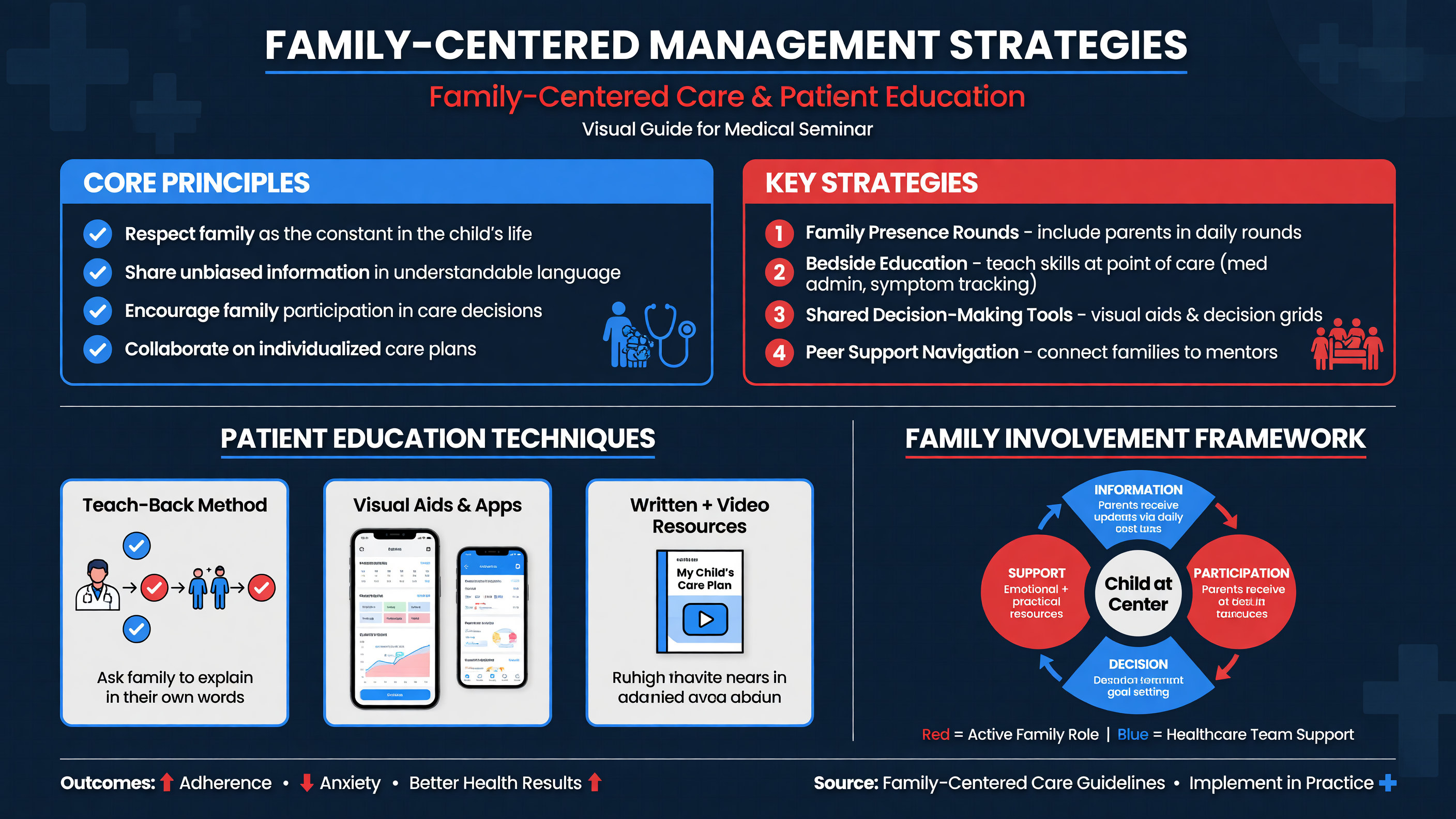 Children and caregivers are experts in the child’s baseline, prior treatment response, and early warning signs. Clinicians contribute physiologic assessment and access to acute interventions. High-quality care depends on combining both sources of expertise. A request for a specific opioid or dose often reflects prior success or tolerance; it is not evidence of drug-seeking.
**MUST ACT:** Use the child’s self-report, investigate dangerous alternatives, and treat pain without stigmatizing language. Recurrent attendance should prompt a better longitudinal plan—not progressively slower analgesia.
Create an individualized plan jointly with the family, hematology team, emergency clinicians, nursing, pharmacy, and pain specialists when needed. Place it in the EHR and give the family a portable copy. It should include baseline hemoglobin and oxygen saturation, prior effective medication and doses, NSAID contraindications, reassessment intervals, transfusion history, ACS-prevention measures, expected admission strategy, and discharge criteria. ASH endorses tailored opioid dosing based on baseline therapy and prior effective treatment (PMID: [32559294](https://pubmed.ncbi.nlm.nih.gov/32559294/)).
**Framework:** A usable home plan answers four questions: What should we do when pain begins? Which medicines may be repeated, at what interval, and to what maximum? Which symptoms require immediate hospital evaluation? Whom can we contact after hours?
Home management may include warmth, rest, normal oral hydration, distraction, scheduled acetaminophen or one NSAID when safe, and a prescribed short-acting opioid for breakthrough pain. Instructions must account for combination products to prevent duplicate acetaminophen dosing and should include opioid storage, constipation prevention, and naloxone education where applicable. Use teach-back: ask the caregiver or adolescent to explain the plan in their own words.
Immediate evaluation is required for fever of at least 38.5°C, chest pain with cough or dyspnea, falling oxygen saturation, new neurologic symptoms, severe headache, pallor or syncope, an enlarging spleen, inability to drink or urinate, uncontrolled pain, or priapism lasting four hours. Families should be encouraged to seek advice earlier during stuttering priapism rather than waiting for the four-hour threshold. Fever may be the only sign of invasive infection in a functionally asplenic child ([CDC fever guidance](https://www.cdc.gov/sickle-cell/complications/fever.html)).
**Teaching Point:** Discharge readiness is functional and contextual. The child should be able to drink, breathe deeply, move safely, and manage pain with the oral plan. The family must have medication in hand, transport, a working telephone, clear return precautions, and timely hematology follow-up.
Every acute visit is also an opportunity to optimize prevention. Hydroxyurea is first-line disease-modifying therapy for children with HbSS or HbSβ⁰-thalassemia and should be offered from 9 months of age regardless of prior symptom severity. It increases HbF, improves erythrocyte hydration and deformability, and reduces leukocyte- and reticulocyte-mediated adhesion. BABY HUG used 20 mg/kg/day in infants and reduced pain, dactylitis, ACS, hospitalization, and transfusion (PMID: [21571150](https://pubmed.ncbi.nlm.nih.gov/21571150/)).
A common pediatric protocol starts hydroxyurea at 20 mg/kg once daily, checks CBC, differential, reticulocytes, platelets, renal and hepatic function, MCV, and HbF, and monitors counts approximately every four weeks during titration. Increase by about 5 mg/kg/day every eight weeks toward mild myelosuppression, up to 35 mg/kg/day, then monitor every two to three months when stable. Benefit develops over months, not hours; hydroxyurea should not be stopped merely because a child presents with an uncomplicated VOE. Recurrent events despite optimized therapy should prompt assessment of adherence, dosing, sleep-disordered breathing, asthma, psychosocial stressors, and access before declaring treatment failure.
**Nuance:** Adherence questions should be curious rather than accusatory. Pharmacy shortages, formulation taste, caregiver schedules, unstable housing, transportation, insurance, and laboratory access may be more important than motivation.
Longitudinal care also includes penicillin prophylaxis where indicated, complete pneumococcal, meningococcal, *Haemophilus influenzae* type b, influenza, and routine immunization, annual transcranial Doppler screening from ages 2–16 for HbSS/HbSβ⁰, renal and ophthalmologic surveillance, reproductive and genetic counseling, and discussion of curative therapies for eligible patients. Schools need individualized accommodations for hydration, unrestricted bathroom use, temperature exposure, fatigue, pain medication, missed work, and emergency response.
Screen for anxiety, depression, sleep problems, school avoidance, cognitive difficulty, caregiver strain, and trauma from repeated healthcare encounters. Adolescents need confidential time, transition preparation, and explicit education about alcohol, smoking or vaping, sexual health, pregnancy, and opioid safety.
**Decision Point:** If the family cannot obtain medication, return for reassessment, or follow the plan safely, that is a clinical disposition problem—not a reason to document “noncompliance.”
**Audience Poll:** Does your discharge process confirm that prescriptions were filled and understood, or does it only confirm that they were electronically sent?
---
## Case Studies and Interactive Discussions
Children and caregivers are experts in the child’s baseline, prior treatment response, and early warning signs. Clinicians contribute physiologic assessment and access to acute interventions. High-quality care depends on combining both sources of expertise. A request for a specific opioid or dose often reflects prior success or tolerance; it is not evidence of drug-seeking.
**MUST ACT:** Use the child’s self-report, investigate dangerous alternatives, and treat pain without stigmatizing language. Recurrent attendance should prompt a better longitudinal plan—not progressively slower analgesia.
Create an individualized plan jointly with the family, hematology team, emergency clinicians, nursing, pharmacy, and pain specialists when needed. Place it in the EHR and give the family a portable copy. It should include baseline hemoglobin and oxygen saturation, prior effective medication and doses, NSAID contraindications, reassessment intervals, transfusion history, ACS-prevention measures, expected admission strategy, and discharge criteria. ASH endorses tailored opioid dosing based on baseline therapy and prior effective treatment (PMID: [32559294](https://pubmed.ncbi.nlm.nih.gov/32559294/)).
**Framework:** A usable home plan answers four questions: What should we do when pain begins? Which medicines may be repeated, at what interval, and to what maximum? Which symptoms require immediate hospital evaluation? Whom can we contact after hours?
Home management may include warmth, rest, normal oral hydration, distraction, scheduled acetaminophen or one NSAID when safe, and a prescribed short-acting opioid for breakthrough pain. Instructions must account for combination products to prevent duplicate acetaminophen dosing and should include opioid storage, constipation prevention, and naloxone education where applicable. Use teach-back: ask the caregiver or adolescent to explain the plan in their own words.
Immediate evaluation is required for fever of at least 38.5°C, chest pain with cough or dyspnea, falling oxygen saturation, new neurologic symptoms, severe headache, pallor or syncope, an enlarging spleen, inability to drink or urinate, uncontrolled pain, or priapism lasting four hours. Families should be encouraged to seek advice earlier during stuttering priapism rather than waiting for the four-hour threshold. Fever may be the only sign of invasive infection in a functionally asplenic child ([CDC fever guidance](https://www.cdc.gov/sickle-cell/complications/fever.html)).
**Teaching Point:** Discharge readiness is functional and contextual. The child should be able to drink, breathe deeply, move safely, and manage pain with the oral plan. The family must have medication in hand, transport, a working telephone, clear return precautions, and timely hematology follow-up.
Every acute visit is also an opportunity to optimize prevention. Hydroxyurea is first-line disease-modifying therapy for children with HbSS or HbSβ⁰-thalassemia and should be offered from 9 months of age regardless of prior symptom severity. It increases HbF, improves erythrocyte hydration and deformability, and reduces leukocyte- and reticulocyte-mediated adhesion. BABY HUG used 20 mg/kg/day in infants and reduced pain, dactylitis, ACS, hospitalization, and transfusion (PMID: [21571150](https://pubmed.ncbi.nlm.nih.gov/21571150/)).
A common pediatric protocol starts hydroxyurea at 20 mg/kg once daily, checks CBC, differential, reticulocytes, platelets, renal and hepatic function, MCV, and HbF, and monitors counts approximately every four weeks during titration. Increase by about 5 mg/kg/day every eight weeks toward mild myelosuppression, up to 35 mg/kg/day, then monitor every two to three months when stable. Benefit develops over months, not hours; hydroxyurea should not be stopped merely because a child presents with an uncomplicated VOE. Recurrent events despite optimized therapy should prompt assessment of adherence, dosing, sleep-disordered breathing, asthma, psychosocial stressors, and access before declaring treatment failure.
**Nuance:** Adherence questions should be curious rather than accusatory. Pharmacy shortages, formulation taste, caregiver schedules, unstable housing, transportation, insurance, and laboratory access may be more important than motivation.
Longitudinal care also includes penicillin prophylaxis where indicated, complete pneumococcal, meningococcal, *Haemophilus influenzae* type b, influenza, and routine immunization, annual transcranial Doppler screening from ages 2–16 for HbSS/HbSβ⁰, renal and ophthalmologic surveillance, reproductive and genetic counseling, and discussion of curative therapies for eligible patients. Schools need individualized accommodations for hydration, unrestricted bathroom use, temperature exposure, fatigue, pain medication, missed work, and emergency response.
Screen for anxiety, depression, sleep problems, school avoidance, cognitive difficulty, caregiver strain, and trauma from repeated healthcare encounters. Adolescents need confidential time, transition preparation, and explicit education about alcohol, smoking or vaping, sexual health, pregnancy, and opioid safety.
**Decision Point:** If the family cannot obtain medication, return for reassessment, or follow the plan safely, that is a clinical disposition problem—not a reason to document “noncompliance.”
**Audience Poll:** Does your discharge process confirm that prescriptions were filled and understood, or does it only confirm that they were electronically sent?
---
## Case Studies and Interactive Discussions
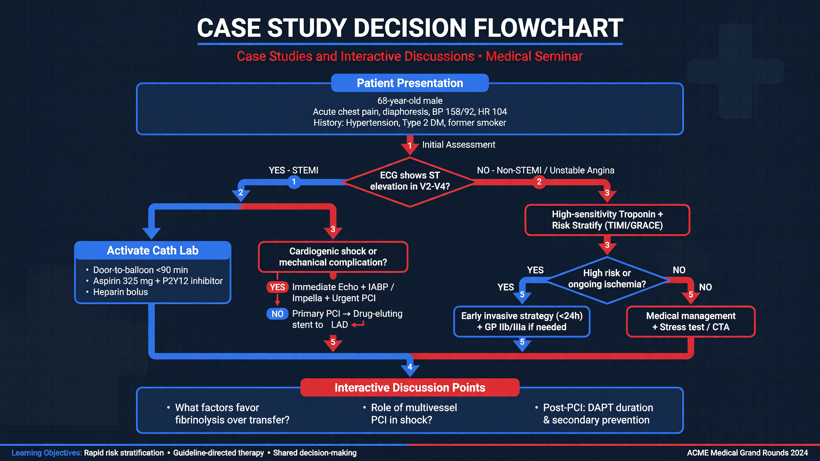 ### Progressive Case: Pain Evolving Into Acute Chest Syndrome
A 10-year-old child weighing 32 kg with HbSS presents with 10 hours of severe chest and upper-back pain. Baseline hemoglobin is 8.3 g/dL and baseline oxygen saturation is 98%. The child takes hydroxyurea but has missed several doses because the refill was delayed. There is a history of asthma and one previous ACS admission. Temperature is 37.9°C, heart rate 122/min, respiratory rate 26/min, blood pressure 112/68 mm Hg, and room-air saturation 94%. Pain is 9/10. The child is splinting but has no wheeze or crackles, no focal neurologic deficit, and no palpable spleen.
**Audience Poll:** Should the team obtain a chest radiograph before giving analgesia? The correct approach is parallel action: begin treatment immediately while evaluating the relative hypoxemia and thoracic pain.
The individualized plan is unavailable. IV access is requested, but intranasal fentanyl 2 micrograms/kg can be given if access will delay analgesia. Once access is established, a reasonable initial regimen is morphine 0.1 mg/kg IV—3.2 mg—plus ketorolac 0.5 mg/kg IV, capped according to the pediatric formulary, and acetaminophen 15 mg/kg. Apply continuous pulse oximetry, begin incentive spirometry, and reassess pain, sedation, respiratory rate, and oxygenation within 20–30 minutes. Do not give a routine large crystalloid bolus: the child is euvolemic. Because nausea limits drinking, begin IV fluid at no more than maintenance and count all medication carriers.
CBC, reticulocytes, metabolic panel, type and screen, and chest radiography are obtained. Hemoglobin is 7.9 g/dL with appropriate reticulocytosis; renal function is normal. The initial radiograph shows no infiltrate.
**Decision Point:** Does the normal radiograph make discharge safe? No. Persistent severe thoracic pain, saturation four points below baseline, prior ACS, and need for repeated IV opioid support admission and close respiratory reassessment.
Pain remains 8/10 after the first dose, but the child is alert with stable ventilation. A protocolized repeat opioid dose is appropriate; withholding analgesia would worsen splinting. Scheduled nonopioid therapy, incentive spirometry every two hours while awake, upright positioning, and ambulation as tolerated are continued. A demand-only or individualized PCA can replace serial boluses if repeated dosing is required. Ketamine infusion could be considered later for opioid-refractory pain with appropriate expertise, but a single conservative opioid dose does not establish refractoriness.
Twelve hours later, temperature rises to 38.7°C, respiratory rate to 38/min, and saturation falls to 88% on room air. Crackles are now audible. Repeat radiography shows a new right middle- and lower-lobe infiltrate; hemoglobin is 6.9 g/dL.
**MUST ACT:** This is ACS until proven otherwise. Obtain blood culture without delaying antibiotics, provide oxygen, repeat CBC and reticulocytes, notify hematology and the PICU, and reassess fluid balance and respiratory support.
Begin a third-generation cephalosporin plus macrolide according to local policy—for example, ceftriaxone 50 mg/kg IV daily plus azithromycin 10 mg/kg on day 1. Continue careful analgesia, incentive spirometry, mobilization when safe, and bronchodilator treatment if wheeze develops. Reduce total fluid toward approximately two-thirds to three-quarters maintenance because ACS is present, while ensuring adequate perfusion.
The hemoglobin has fallen 1.4 g/dL below baseline, there is clinically significant hypoxemia, and pretransfusion hemoglobin leaves room for a safe increment. After reviewing the antibody history and extended genotype, give approximately 10 mL/kg of appropriately matched packed red cells, targeting hemoglobin around 9–10 g/dL rather than normalizing it.
**Audience Poll:** What finding would move the plan from simple transfusion to urgent exchange? Rapidly increasing oxygen or ventilatory support, multilobar progression, worsening anemia after simple transfusion, or high pretransfusion hemoglobin would favor exchange, generally targeting HbS below 30%.
The child improves after simple transfusion, oxygen, antibiotics, spirometry, and controlled analgesia. Had hypoxemia progressed despite oxygen or noninvasive support, exchange transfusion and intensive care escalation would have been required. Pneumonia remains part of the ACS differential and is treated empirically; these diagnoses are not mutually exclusive. Pulmonary embolism would deserve additional evaluation if hypoxemia or pain were disproportionate, particularly with a central line or thrombosis history.
Before discharge, convert parenteral opioids deliberately to a time-limited oral regimen, confirm stable oxygenation awake and asleep, demonstrate spirometry, and verify oral intake and ambulation. Reconcile hydroxyurea, solve the refill barrier, provide fever and respiratory return precautions, and arrange prompt hematology follow-up. Review the transfusion card and counsel the family to report jaundice, dark urine, fatigue, or recurrent pain during the next three weeks because delayed hemolysis may initially resemble another VOE.
**Teaching Point:** The first diagnosis—VOE—was correct but incomplete over time. Safe SCD care depends on serial reassessment rather than treating the admission label as permanent.
**Framework:** In every evolving case, repeat five questions: Is pain controlled? Is ventilation safe? Is oxygenation changing? Is hemoglobin changing relative to baseline? Has a new indication for antibiotics, transfusion, imaging, or intensive care appeared?
---
## Tonight on Shift
- Give the first individualized analgesic within 30 minutes of triage or 60 minutes of registration; reassess and retitrate severe pain every 15–30 minutes.
- Screen every VOE for fever, respiratory change, neurologic deficit, sequestration, aplasia, priapism, focal infection, and recent transfusion.
- Prefer oral hydration when possible; if euvolemic and unable to drink, keep IV fluid at no more than maintenance and count every fluid source.
- Prevent ACS with effective analgesia, objective sedation monitoring, incentive spirometry every two hours while awake, mobilization, and pulse-oximetry surveillance.
- Do not transfuse uncomplicated pain; use simple transfusion for selected anemia or moderate ACS and exchange for stroke or severe, progressive ACS.
- Before discharge, confirm a workable oral plan, medication access, fever and ACS return precautions, transfusion history, hydroxyurea follow-up, and family teach-back.
### Progressive Case: Pain Evolving Into Acute Chest Syndrome
A 10-year-old child weighing 32 kg with HbSS presents with 10 hours of severe chest and upper-back pain. Baseline hemoglobin is 8.3 g/dL and baseline oxygen saturation is 98%. The child takes hydroxyurea but has missed several doses because the refill was delayed. There is a history of asthma and one previous ACS admission. Temperature is 37.9°C, heart rate 122/min, respiratory rate 26/min, blood pressure 112/68 mm Hg, and room-air saturation 94%. Pain is 9/10. The child is splinting but has no wheeze or crackles, no focal neurologic deficit, and no palpable spleen.
**Audience Poll:** Should the team obtain a chest radiograph before giving analgesia? The correct approach is parallel action: begin treatment immediately while evaluating the relative hypoxemia and thoracic pain.
The individualized plan is unavailable. IV access is requested, but intranasal fentanyl 2 micrograms/kg can be given if access will delay analgesia. Once access is established, a reasonable initial regimen is morphine 0.1 mg/kg IV—3.2 mg—plus ketorolac 0.5 mg/kg IV, capped according to the pediatric formulary, and acetaminophen 15 mg/kg. Apply continuous pulse oximetry, begin incentive spirometry, and reassess pain, sedation, respiratory rate, and oxygenation within 20–30 minutes. Do not give a routine large crystalloid bolus: the child is euvolemic. Because nausea limits drinking, begin IV fluid at no more than maintenance and count all medication carriers.
CBC, reticulocytes, metabolic panel, type and screen, and chest radiography are obtained. Hemoglobin is 7.9 g/dL with appropriate reticulocytosis; renal function is normal. The initial radiograph shows no infiltrate.
**Decision Point:** Does the normal radiograph make discharge safe? No. Persistent severe thoracic pain, saturation four points below baseline, prior ACS, and need for repeated IV opioid support admission and close respiratory reassessment.
Pain remains 8/10 after the first dose, but the child is alert with stable ventilation. A protocolized repeat opioid dose is appropriate; withholding analgesia would worsen splinting. Scheduled nonopioid therapy, incentive spirometry every two hours while awake, upright positioning, and ambulation as tolerated are continued. A demand-only or individualized PCA can replace serial boluses if repeated dosing is required. Ketamine infusion could be considered later for opioid-refractory pain with appropriate expertise, but a single conservative opioid dose does not establish refractoriness.
Twelve hours later, temperature rises to 38.7°C, respiratory rate to 38/min, and saturation falls to 88% on room air. Crackles are now audible. Repeat radiography shows a new right middle- and lower-lobe infiltrate; hemoglobin is 6.9 g/dL.
**MUST ACT:** This is ACS until proven otherwise. Obtain blood culture without delaying antibiotics, provide oxygen, repeat CBC and reticulocytes, notify hematology and the PICU, and reassess fluid balance and respiratory support.
Begin a third-generation cephalosporin plus macrolide according to local policy—for example, ceftriaxone 50 mg/kg IV daily plus azithromycin 10 mg/kg on day 1. Continue careful analgesia, incentive spirometry, mobilization when safe, and bronchodilator treatment if wheeze develops. Reduce total fluid toward approximately two-thirds to three-quarters maintenance because ACS is present, while ensuring adequate perfusion.
The hemoglobin has fallen 1.4 g/dL below baseline, there is clinically significant hypoxemia, and pretransfusion hemoglobin leaves room for a safe increment. After reviewing the antibody history and extended genotype, give approximately 10 mL/kg of appropriately matched packed red cells, targeting hemoglobin around 9–10 g/dL rather than normalizing it.
**Audience Poll:** What finding would move the plan from simple transfusion to urgent exchange? Rapidly increasing oxygen or ventilatory support, multilobar progression, worsening anemia after simple transfusion, or high pretransfusion hemoglobin would favor exchange, generally targeting HbS below 30%.
The child improves after simple transfusion, oxygen, antibiotics, spirometry, and controlled analgesia. Had hypoxemia progressed despite oxygen or noninvasive support, exchange transfusion and intensive care escalation would have been required. Pneumonia remains part of the ACS differential and is treated empirically; these diagnoses are not mutually exclusive. Pulmonary embolism would deserve additional evaluation if hypoxemia or pain were disproportionate, particularly with a central line or thrombosis history.
Before discharge, convert parenteral opioids deliberately to a time-limited oral regimen, confirm stable oxygenation awake and asleep, demonstrate spirometry, and verify oral intake and ambulation. Reconcile hydroxyurea, solve the refill barrier, provide fever and respiratory return precautions, and arrange prompt hematology follow-up. Review the transfusion card and counsel the family to report jaundice, dark urine, fatigue, or recurrent pain during the next three weeks because delayed hemolysis may initially resemble another VOE.
**Teaching Point:** The first diagnosis—VOE—was correct but incomplete over time. Safe SCD care depends on serial reassessment rather than treating the admission label as permanent.
**Framework:** In every evolving case, repeat five questions: Is pain controlled? Is ventilation safe? Is oxygenation changing? Is hemoglobin changing relative to baseline? Has a new indication for antibiotics, transfusion, imaging, or intensive care appeared?
---
## Tonight on Shift
- Give the first individualized analgesic within 30 minutes of triage or 60 minutes of registration; reassess and retitrate severe pain every 15–30 minutes.
- Screen every VOE for fever, respiratory change, neurologic deficit, sequestration, aplasia, priapism, focal infection, and recent transfusion.
- Prefer oral hydration when possible; if euvolemic and unable to drink, keep IV fluid at no more than maintenance and count every fluid source.
- Prevent ACS with effective analgesia, objective sedation monitoring, incentive spirometry every two hours while awake, mobilization, and pulse-oximetry surveillance.
- Do not transfuse uncomplicated pain; use simple transfusion for selected anemia or moderate ACS and exchange for stroke or severe, progressive ACS.
- Before discharge, confirm a workable oral plan, medication access, fever and ACS return precautions, transfusion history, hydroxyurea follow-up, and family teach-back.