Fracture Management in the Era of Technological Precision: From GLP-1 Risks to Advanced Imaging Techniques
From GLP-1 Risks to Advanced Imaging Techniques
Orthopedic Surgery · Seminar week 24 · released August 17, 2026 · includes a discussion video
This seminar focuses on the integration of newer drugs like GLP-1 receptor agonists, their fracture risks, and the role of advanced imaging in orthopedic decision-making.…
Learning Objectives
- Appraise the evidence linking GLP-1 receptor agonist therapy, weight loss, and fracture risk.
- Distinguish medication-associated skeletal risk from age, obesity, diabetes, sarcopenia, malnutrition, and falls.
- Select radiography, CT, MRI, CT angiography, and intraoperative imaging according to a defined clinical question.
- Identify the imaging targets that determine nonoperative versus operative management of tibial shaft fractures.
- Recognize imaging findings that prevent missed articular extension, vascular injury, malrotation, and delayed fracture diagnosis.
- Translate imaging data into an executable surgical plan that incorporates soft-tissue condition and patient physiology.
- Apply a structured imaging and bone-health strategy during acute fracture care.
Introduction to GLP-1 Receptor Agonists and Risk of Fractures
Duration: 10 min
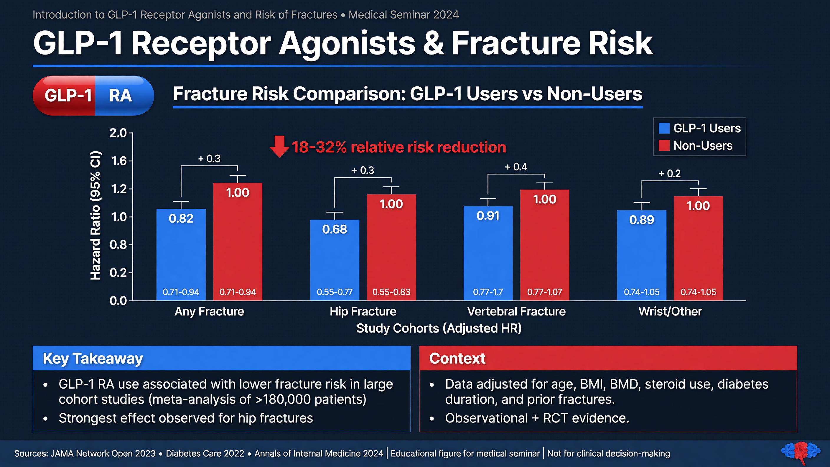
Glucagon-like peptide-1 receptor agonists have transformed the treatment of type 2 diabetes and obesity. Liraglutide and semaglutide are true GLP-1 receptor agonists, whereas tirzepatide is a dual glucose-dependent insulinotropic polypeptide/GLP-1 receptor agonist and should not be treated as pharmacologically identical. At obesity-treatment doses, these agents reduce appetite, delay gastric emptying, improve glycemic control, and can produce weight loss of a magnitude previously associated mainly with metabolic surgery. STEP 1 established the efficacy of semaglutide 2.4 mg weekly in adults with overweight or obesity, while the SCALE program evaluated liraglutide 3 mg daily. These trials were designed principally around weight and cardiometabolic outcomes—not adjudicated, long-latency skeletal endpoints.
Typical semaglutide titration begins at 0.25 mg subcutaneously weekly and increases approximately every four weeks toward 2.4 mg weekly as tolerated. Liraglutide is commonly initiated at 0.6 mg daily and increased weekly toward 3 mg daily. Dose escalation matters clinically: persistent nausea, vomiting, reduced intake, dehydration, or excessive dietary restriction can accelerate loss of lean tissue and impair calcium, vitamin D, and protein intake. The prescription alone is therefore less informative than the medication trajectory, rate of weight loss, nutrition, strength, balance, and fall history.
MUST ACT: Do not interpret a fracture in a GLP-1 recipient as proof that the medication caused it. Establish the mechanism of injury and assess conventional skeletal risks, including age, prior fragility fracture, glucocorticoid exposure, menopause, hypogonadism, chronic kidney or liver disease, malabsorption, smoking, alcohol use, immobility, sarcopenia, and medications that increase falls.
Constantine et al. reported an approximately 14% higher relative fracture risk among GLP-1 receptor agonist users treated for obesity without diabetes, with stronger signals in adults older than 67 years and those with class 3 obesity [PMID: 41429015]. This is clinically important but must be interpreted as an association. Relative risk does not disclose the absolute excess risk, and observational comparisons remain vulnerable to confounding by frailty, intentional weight loss, baseline mobility, prior falls, healthcare contact, and differences in obesity severity.
Conversely, Alalwani et al. describe potentially favorable or neutral bone-mineral-density findings in diabetic populations receiving GLP-1 receptor agonists [PMID: 40751153]. Type 2 diabetes complicates interpretation because patients may have normal or elevated areal bone density yet remain fracture-prone because of altered bone material properties, advanced glycation, neuropathy, retinopathy, hypoglycemia, and falls. Findings in diabetic populations therefore cannot automatically be transferred to older adults without diabetes who are losing weight rapidly.
Nuance: Several competing mechanisms may operate simultaneously. Reduced body mass decreases mechanical loading, and weight loss commonly lowers hip bone mineral density. Loss of muscle can increase falls and reduce protective loading. In contrast, improved glycemia, reduced systemic inflammation, greater mobility, and lower fall-inducing medication burden may benefit skeletal health. Direct GLP-1 signaling in bone has biologic plausibility, but human outcomes are likely dominated by the patient’s net change in weight, muscle, nutrition, activity, and falls.
Framework: At initiation and during substantial weight loss, document fracture history, falls, dietary intake, resistance exercise, and weight-loss velocity. Obtain dual-energy X-ray absorptiometry when indicated by age or clinical risk rather than automatically for every user. In a fragility fracture or suspected metabolic bone disease, consider calcium, albumin, creatinine, alkaline phosphatase, 25-hydroxyvitamin D, complete blood count, thyroid-stimulating hormone, and parathyroid hormone when clinically appropriate; add tests for hypogonadism, myeloma, or malabsorption when suggested by the presentation.
Adults should generally meet age-appropriate elemental calcium requirements—preferably through food—and correct vitamin D deficiency without indiscriminate megadosing. A practical target during weight loss is adequate protein, often approximately 1.0–1.2 g/kg/day in older adults when renal and hepatic status permit, combined with progressive resistance and balance training two or three times weekly. Dietary targets must be individualized in chronic kidney disease, severe obesity, and frailty.
A low-trauma hip or vertebral fracture may establish osteoporosis regardless of the DXA T-score. Treatment options include alendronate 70 mg weekly, zoledronic acid 5 mg intravenously yearly, or denosumab 60 mg subcutaneously every six months in selected patients; very-high-risk patients may warrant an anabolic strategy. Renal function, dental risk, hypocalcemia, adherence, and the rebound vertebral-fracture risk after denosumab discontinuation must be addressed.
Decision Point: A fracture is usually a reason to reassess the entire treatment plan—not to discontinue GLP-1 therapy reflexively. Continued cardiometabolic benefit may outweigh an uncertain skeletal signal if nutrition, muscle loss, falls, and osteoporosis are actively managed.
Audience Poll: Which would most change your recommendation: a 20% weight loss over six months, recurrent vomiting, a prior vertebral fracture, new gait instability, or an isolated high-energy fracture with otherwise normal bone health?
Advanced Imaging Techniques in Orthopedic Surgery
Duration: 15 min
Advanced imaging is valuable only when it answers a question that will change management. The foundational study remains properly positioned radiography: at least two orthogonal views, inclusion of the joint above and below for long-bone injuries, and comparison views only when they solve a specific uncertainty. Before ordering CT or MRI, the surgeon should be able to complete the sentence, “I need this study to determine whether….” Imaging acquired without a decision target increases radiation, expense, delays, and incidental findings without necessarily improving care.
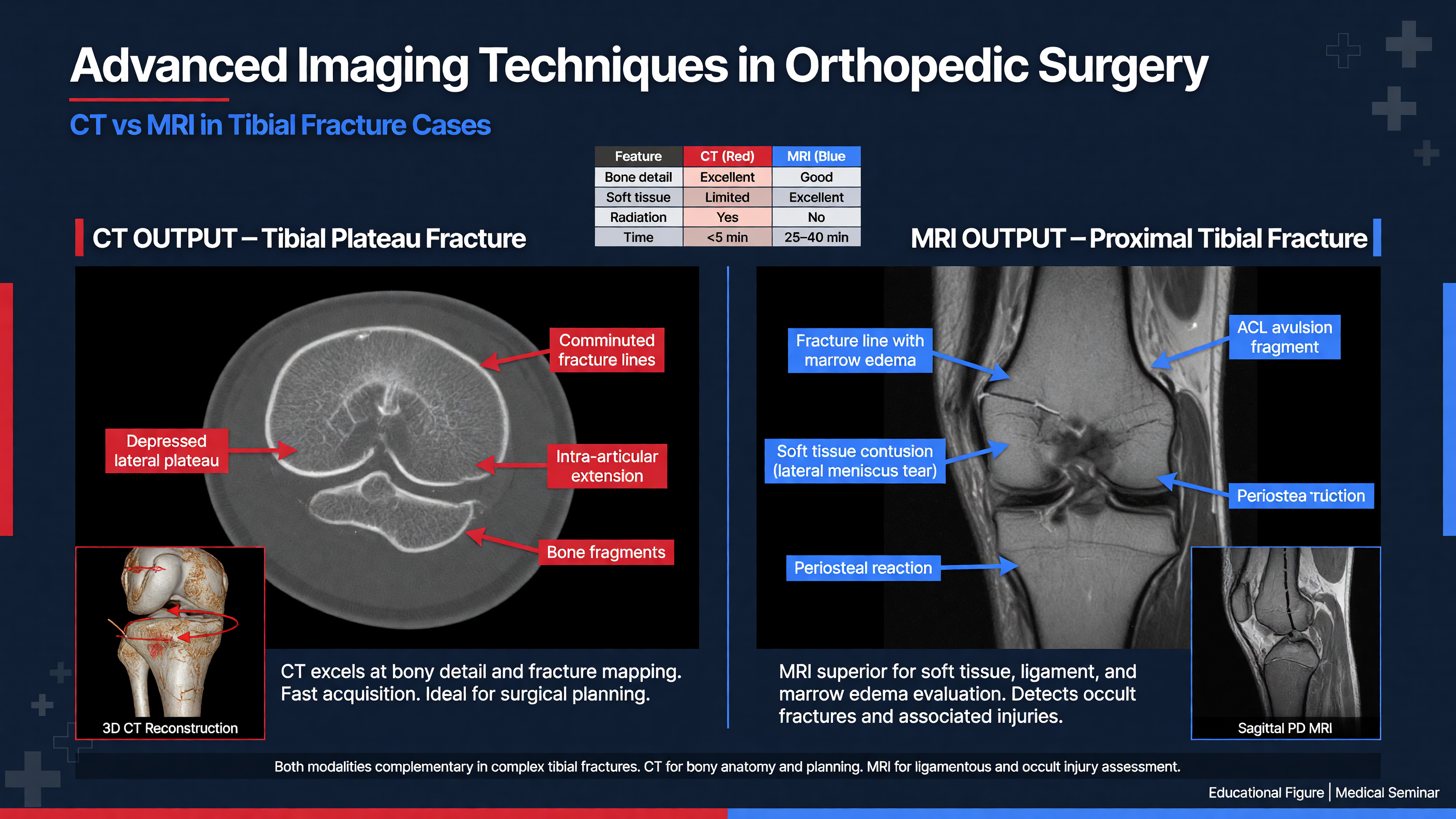
Framework: Organize imaging questions into five domains: osseous morphology, articular involvement, soft-tissue injury, vascular status, and operative execution. Plain radiographs establish alignment and gross morphology. CT defines complex bone anatomy. MRI evaluates marrow, cartilage, ligaments, tendons, and occult fractures. CT angiography evaluates arterial integrity. Fluoroscopy or intraoperative three-dimensional imaging tests whether reduction and implant placement match the plan.
CT is especially useful for intra-articular fractures, comminution, occult extension into an adjacent joint, pelvic and acetabular injury, and preoperative assessment when overlapping structures obscure radiographs. A thin-section acquisition—commonly reconstructed at approximately 0.5–1.25 mm—permits sagittal, coronal, and oblique multiplanar reformats. Three-dimensional surface renderings can improve spatial communication, but they must be reviewed alongside source images because rendering may hide nondisplaced fracture lines, thin fragments, or articular impaction. Bone and soft-tissue windows should both be inspected.
Teaching Point: CT is strongest when the question is “Where is the bone, and what is its three-dimensional geometry?” MRI is strongest when the question is “What has happened to marrow or soft tissue that radiography and CT cannot adequately show?”
MRI protocols should be tailored rather than indiscriminate. T1-weighted images define marrow replacement and anatomy; fluid-sensitive fat-suppressed or STIR sequences show edema, occult fracture, muscle injury, and ligament disruption. Small field-of-view imaging improves joint detail. MRI is particularly useful for radiographically occult hip fracture, stress injury, suspected pathological fracture, osteomyelitis, and multiligamentous knee injury after reduction and vascular assessment. Marrow edema alone is not a surgical diagnosis; its pattern must be integrated with symptoms, fracture lines, cortical integrity, and loading history.
Schmaranzer et al. illustrate the broader principle that CT and MRI contribute different, complementary information to orthopedic planning [PMID: 33474984]. Such modality-focused literature should be applied according to anatomy and clinical question rather than cited as proof that every complex fracture requires both studies.
CT angiography is not simply “another CT.” In trauma, its purpose is to detect occlusion, intimal injury, pseudoaneurysm, active extravasation, or impaired distal runoff. It is indicated when mechanism and examination suggest vascular injury, including an abnormal ankle-brachial index, asymmetric pulses, or concerning findings after knee dislocation. Conversely, hard signs such as active hemorrhage, an expanding pulsatile hematoma, distal ischemia, or a bruit with hemodynamic concern require immediate vascular and operative action; imaging must not create a harmful delay.
MUST ACT: Neither CT nor MRI excludes acute compartment syndrome. Compartment syndrome is diagnosed from evolving pain, pain with passive stretch, tense compartments, neurological change, and—when examination is unreliable—pressure measurement. A delta pressure of 30 mm Hg or less is concerning in the proper clinical context, but no single measurement replaces serial assessment.
Intraoperative imaging introduces its own failure modes. A technically perfect fluoroscopic image can still represent a malrotated limb if the wrong projection was accepted. Calibrate the C-arm, remove obstructing objects, obtain true orthogonal views, and compare femoral or tibial cortical profiles when rotational uncertainty remains. Intraoperative CT or cone-beam imaging can identify articular screw penetration or residual displacement in selected cases, but benefit is greatest when a predefined threshold will prompt immediate correction.
Nuance: Weight-bearing CT, low-dose biplanar imaging, metal-artifact-reduction techniques, navigation, patient-specific guides, and three-dimensional printed models may add value in selected deformity, foot-and-ankle, pelvis, and revision cases. Their availability should not lower the threshold for imaging that cannot alter treatment.
Decision Point: Before escalating imaging, ask whether the result could change reduction strategy, implant selection, fixation sequence, urgency, weight-bearing, or the need for vascular or subspecialty involvement. If none will change, advanced imaging may be interesting but not clinically necessary.
Audience Poll: For a complex fracture, which is the most common preventable imaging error in your practice: an incomplete radiographic series, failure to image the adjacent joint, CT without the correct field of view, delayed vascular imaging, or failure to review the source images personally?
Targets in Imaging for Managing Tibial Shaft Fractures
Duration: 10 min
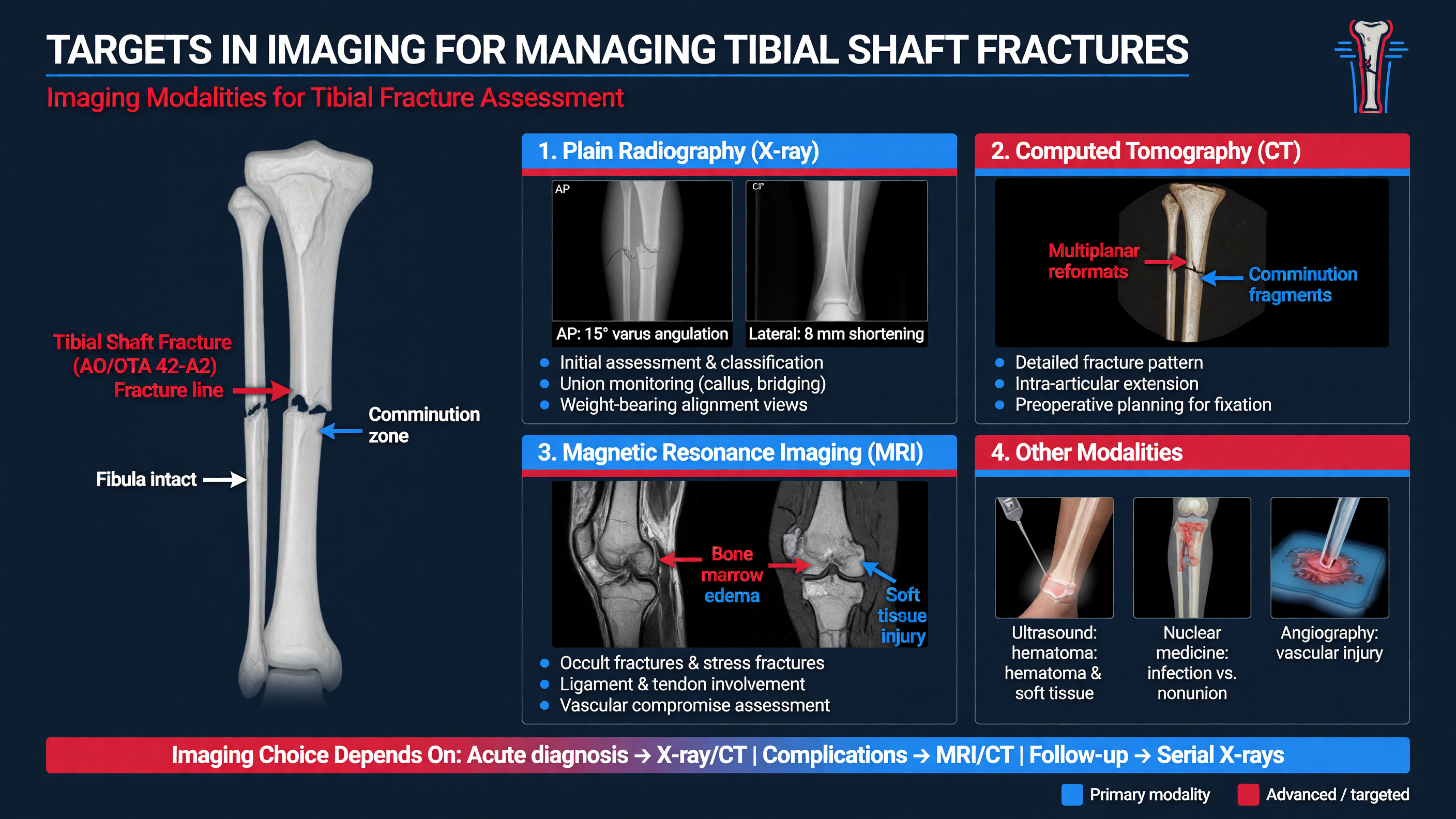
The imaging objective in a tibial shaft fracture is not merely to confirm that the tibia is broken. It is to characterize the injury sufficiently to choose treatment, anticipate instability, protect the joints and soft tissues, and establish a baseline against which reduction and healing can be judged. Initial imaging should include anteroposterior and lateral radiographs of the entire tibia, with the knee and ankle visible. Focused knee or ankle views are added when the fracture approaches a joint or symptoms suggest associated injury.
Framework: Read the study in a fixed sequence: location; simple, wedge, or complex morphology; open versus closed clinical status; displacement and angulation; shortening and rotation; comminution or segmental bone loss; fibular level; extension into the plateau or plafond; and signs of pathological bone. Then ask whether imaging explains the energy of injury and the patient’s examination.
Rotational deformity is easily underestimated on two-dimensional images. Compare the cortical widths and proximal and distal fragment profiles, but treat these as clues rather than exact measurements. Clinical assessment of patellar orientation, tibial tubercle, ankle position, and foot progression remains important. CT rotational profiling can be used when clinically significant malrotation is suspected after fixation, although protocols and reference axes must be consistent.
Distal-third spiral tibial fractures deserve special attention because an associated posterior malleolar fracture may be small or occult on routine films. Thin-section CT through the ankle can identify articular extension, fragment morphology, incisural involvement, and displacement that affect fixation sequence and postoperative weight-bearing. Proximal fractures similarly require scrutiny for tibial plateau extension. The CT field of view must include the joint in question; a scan centered too narrowly on the shaft can miss the very injury being sought.
MUST ACT: Imaging must not delay treatment of an open fracture, vascular compromise, or compartment syndrome. Cover an open wound with a sterile dressing, document neurovascular status, administer tetanus prophylaxis as indicated, and provide antibiotics promptly. A common initial regimen is cefazolin 2 g intravenously—3 g in adults weighing at least 120 kg—modified for allergy, contamination, local policy, and injury severity. Advanced imaging can follow stabilization and should not postpone urgent debridement or revascularization.
For nonoperative management, imaging determines whether alignment can be obtained and maintained. Commonly used acceptable parameters for selected closed, low-energy fractures include less than approximately 5 degrees of coronal angulation, less than 10 degrees of sagittal angulation, less than 1 cm shortening, substantial cortical apposition, and minimal rotational deformity. These are guides, not automatic indications. Patient reliability, soft tissues, body habitus, fracture level, occupational requirements, and capacity for close follow-up may be equally decisive.
A typical nonoperative pathway uses a well-molded long-leg splint or cast during swelling, followed by serial radiographs and transition to a patellar-tendon-bearing or functional brace when stability and soft tissues permit. Films are commonly repeated within one to two weeks after definitive casting and again during early healing, with timing individualized to instability. Varus drift, shortening, loss of cortical contact, or inability to maintain rotation should trigger reconsideration before deformity becomes established.
Intramedullary nailing remains the usual operative treatment for many displaced adult tibial shaft fractures. Imaging informs entry point, canal diameter, nail length, fracture reduction, need for adjunctive clamps or blocking screws, and whether a proximal or distal articular fragment requires separate fixation. Segmental fractures, open injuries, polytrauma, unacceptable alignment, and fractures that cannot be safely maintained nonoperatively commonly favor surgery.
MRI has a limited role in a straightforward acute shaft fracture. It becomes valuable for a radiographically occult stress fracture, pathological fracture, marrow lesion, infection, or persistent pain unexplained by radiography and CT. In stress injury, the MRI pattern and presence of a fracture line help distinguish a stress reaction from a completed fracture and guide protected weight-bearing.
Nuance: Patel et al. discuss contemporary ankle-fracture imaging and management principles [PMID: 38789168]. Because that work addresses ankle rather than tibial shaft fractures, it should be used to inform assessment of the distal articular interface—not cited as direct evidence for tibial-shaft treatment outcomes.
Decision Point: Order CT when detecting articular extension will change fixation or rehabilitation. Order CT angiography when vascular assessment is abnormal or unreliable. Order MRI when the suspected pathology is occult marrow or soft-tissue disease. Do not order all three simply because the fracture appears complex.
Audience Poll: In a distal spiral tibial shaft fracture with a normal-appearing ankle mortise, what finding would most strongly prompt CT: proximal fibular tenderness, inability to dorsiflex, a subtle posterior malleolar line, severe swelling alone, or patient preference?
Case Studies: Avoiding Complications with Modern Imaging
Duration: 15 min
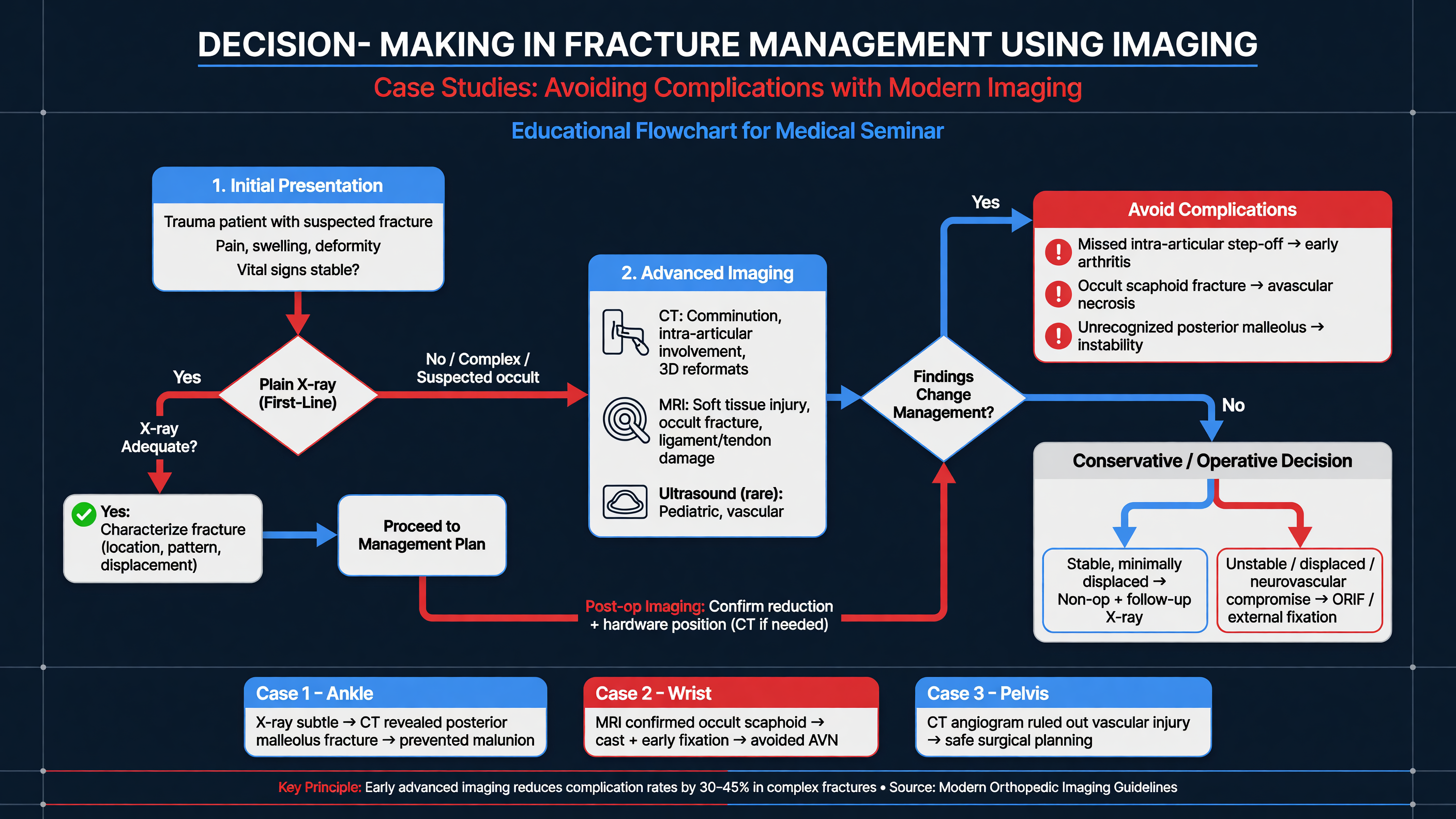
Modern imaging prevents complications when it is embedded in a disciplined clinical pathway. It cannot compensate for an incomplete examination, an incorrectly framed study, or failure to act on the result. Three recurring cases demonstrate how imaging changes management—and where reliance on imaging can itself become hazardous.
Case 1: The “Isolated” Distal Tibial Shaft Fracture
A 36-year-old skier sustains a closed distal-third spiral tibial fracture with a proximal fibular fracture. Ankle radiographs show no obvious displacement, and the first plan is isolated intramedullary nailing. Because the spiral line approaches the plafond and the injury pattern is associated with posterior malleolar extension, CT of the ankle is obtained. It reveals a nondisplaced posterior malleolar fragment extending into the incisura.
The CT changes the operative discussion: whether the fragment requires fixation, whether it should be stabilized before tibial manipulation, how distal locking screws will be positioned, and whether postoperative weight-bearing should be modified. Not every small posterior fragment requires fixation; fragment displacement, incisural involvement, syndesmotic stability, and anticipated displacement during nailing matter more than size alone.
Teaching Point: The complication prevented is not merely a “missed fracture.” It is secondary articular displacement, unrecognized syndesmotic instability, or a rehabilitation plan based on incomplete anatomy.
Case 2: The Reduced Knee Dislocation with Palpable Pulses
A 27-year-old motorcyclist arrives after an obvious knee deformity that reduced during transport. Radiographs confirm reduction but show only a small avulsion fragment. Both feet are warm and pedal pulses are palpable. This does not exclude popliteal artery intimal injury. A complete neurological examination, including common peroneal nerve function, is documented. The ankle-brachial index is 0.78 and differs from the contralateral side, prompting urgent CT angiography and vascular consultation. Imaging shows a focal popliteal arterial injury.
MUST ACT: Palpable pulses can persist despite a limb-threatening arterial lesion. After knee dislocation, perform and document serial vascular examinations. An ankle-brachial index below 0.9, asymmetric findings, or an unreliable examination generally warrants vascular imaging. Hard signs or an ischemic limb demand immediate operative coordination; CT angiography must not delay revascularization.
MRI is obtained only after reduction and vascular priorities are settled. It maps cruciate, collateral, posterolateral-corner, meniscal, and chondral injury for staged reconstruction. MRI does not determine timing in isolation: swelling, skin condition, vascular repair, nerve injury, range of motion, and patient factors influence whether treatment is acute, staged, or selective.
Case 3: Normal Hip Radiographs, Persistent Inability to Bear Weight
A 78-year-old taking a GLP-1 receptor agonist falls from standing. Pelvis and hip radiographs appear normal, but she cannot bear weight and has pain with axial loading and passive rotation. MRI demonstrates an occult nondisplaced femoral-neck fracture. Early diagnosis allows fixation before displacement. If MRI is unavailable or contraindicated, CT is a reasonable rapid next study; persistent clinical suspicion after a negative CT should still prompt MRI or continued protected management rather than diagnostic closure.
Nuance: Medication history raises the question of skeletal vulnerability but should not distract from the immediate diagnostic problem. The decisive clues are low-energy mechanism, age, examination, and inability to bear weight—not the GLP-1 prescription by itself.
Case 4: Post-nailing Malrotation Hidden by “Good” Fluoroscopy
A patient undergoes tibial nailing, and intraoperative AP and lateral views show acceptable length and coronal alignment. Postoperatively, the foot rests in excessive external rotation. Review reveals that rotational landmarks were never compared with the contralateral limb. CT rotational profiling confirms clinically important malrotation.
Framework: Prevent this complication before wound closure: establish a pre-reduction rotational reference, assess the patella and foot simultaneously, compare cortical profiles, evaluate the lesser trochanter or proximal tibiofibular relationship where applicable, and perform a final clinical examination with the limb uncovered. Fluoroscopy confirms projection and implant position; it does not independently guarantee rotation.
Audience Poll: Which case reflects the most dangerous cognitive error: accepting pulses as proof of vascular integrity, accepting a normal radiograph despite inability to bear weight, treating a three-dimensional fracture from a rendering alone, or assuming satisfactory AP and lateral views exclude malrotation?
Educational case resources, including the cited Orthobullets podcast discussion from March 5, 2026, can support pattern recognition and debate. They should be treated as teaching material rather than a substitute for primary studies, formal guidelines, or local trauma pathways.
Decision Point: When imaging changes anatomy but not the current plan, explicitly document why. When it changes urgency, fixation sequence, or weight-bearing, communicate that change to the entire team rather than leaving the finding buried in the report.
Surgical Planning: Combining Imaging with Real World Practice
Duration: 15 min
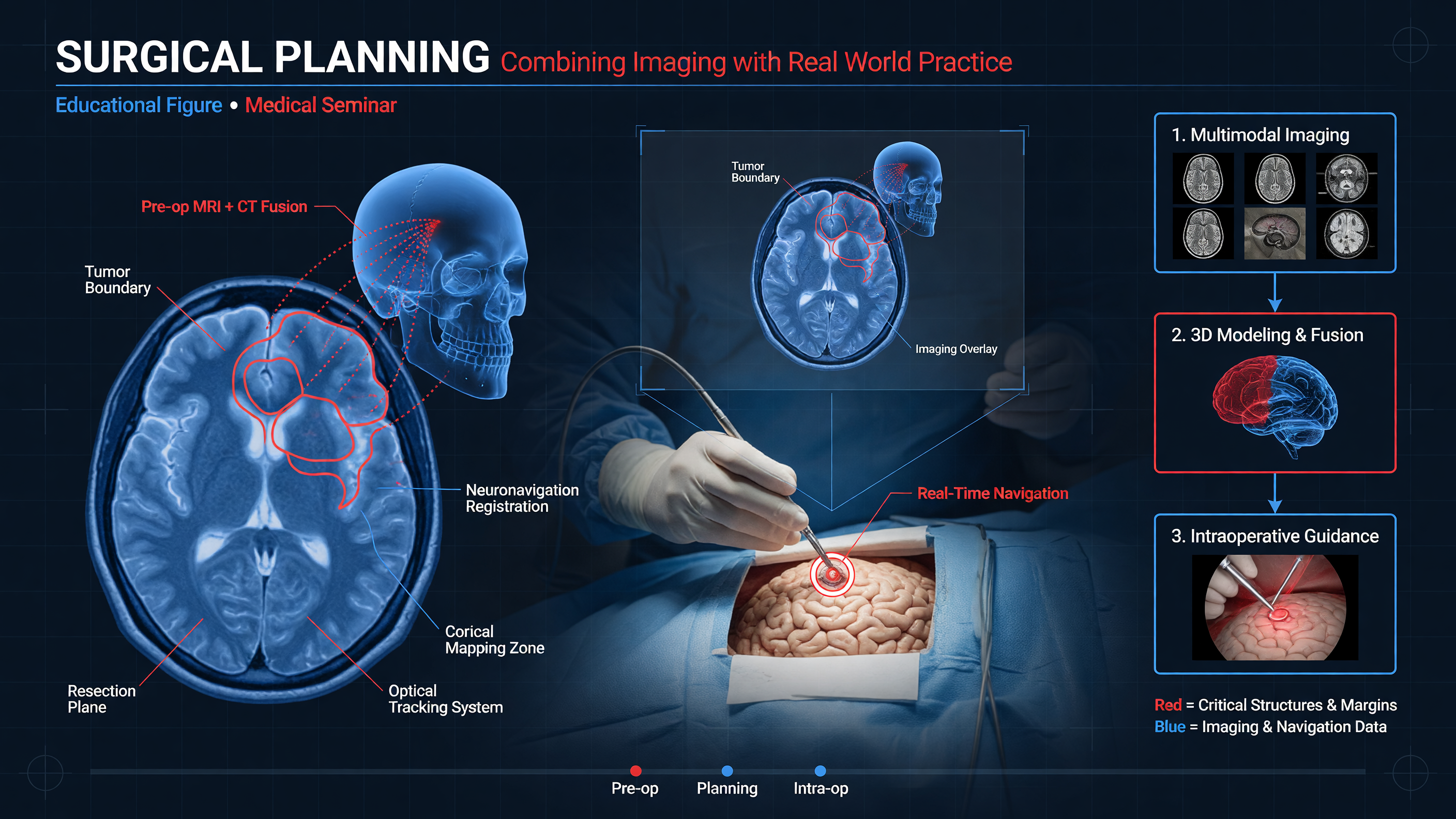
Surgical planning is the conversion of images into a sequence of actions. A radiology report may identify the injuries, but the operating surgeon must personally review the images, reconcile them with examination and soft-tissue status, and decide how reduction will be obtained and maintained. The output should be an executable primary plan, a backup plan, and explicit thresholds for changing course.
Framework: Build the plan in six layers: patient, soft tissue, fracture, reduction, fixation, and rehabilitation. Patient factors include physiology, anticoagulation, diabetes, nutrition, smoking, frailty, bone quality, and perioperative medication considerations. Soft-tissue assessment determines timing and approach. Fracture analysis defines every fragment that matters. Reduction planning determines traction, positioning, clamps, distractors, or temporary fixation. Fixation planning selects implant and sequence. Rehabilitation planning specifies range of motion, loading, and follow-up imaging.
For tibial intramedullary nailing, review the entire tibia and both adjacent joints. Estimate nail length and diameter while recognizing that digital templating is approximate. Identify canal narrowing, deformity, prior implants, proximal or distal comminution, and the available segment for interlocking screws. Plan the entry point, patient position, fluoroscopic access, and reduction aids. A proximal metaphyseal fracture may require a semiextended technique or blocking screws; a distal fracture may require multiple multiplanar distal interlocks and careful control of the short segment.
Teaching Point: Imaging should predict where the fracture will try to deform during fixation. The plan should state how that deforming tendency will be opposed before the nail or plate is inserted.
When CT shows posterior malleolar or plateau extension, decide whether articular stabilization precedes shaft fixation. The answer depends on fragment stability, displacement, screw trajectory, and the risk that reduction maneuvers will propagate or displace the articular component. A “small” fragment may be biomechanically important if it involves the incisura or compromises syndesmotic stability.
For periarticular plating, CT helps identify the dominant fragments and safe corridors, but approach selection must respect skin condition and blood supply. Three-dimensional overlays and printed models can improve understanding of selected acetabular, pelvic, deformity, and complex periarticular cases. They are adjuncts; segmentation errors, non-weight-bearing acquisition, interval displacement, and failure to display cartilage or soft tissue can produce a visually persuasive but incomplete model.
Knee dislocation illustrates multimodal planning. Radiographs confirm alignment and detect fracture. CT characterizes periarticular fragments. CT angiography addresses arterial integrity. MRI maps ligament, meniscal, and chondral injury after urgent threats are controlled. Stress examination under anesthesia may add functional information. A technically detailed MRI should not force immediate multiligament reconstruction through swollen or compromised soft tissues.
The ACL literature discussed by van Melick et al. reinforces the principle that imaging must be integrated with examination, functional goals, surgical technique, and rehabilitation rather than used as an isolated endpoint [PMID: 39925169]. That evidence concerns ACL reconstruction and should not be represented as a tibial-fracture fixation trial. Its transferable lesson is disciplined integration: morphology informs the plan, but function and biology determine whether the plan is appropriate.
MUST ACT: Conduct an imaging timeout before incision. Confirm patient and side, display the key radiographs and CT/MRI sequences, name associated injuries, verify implant availability, ensure the C-arm can obtain the required projections, and identify which finding would trigger the backup plan.
Intraoperatively, acquire views intentionally. For tibial nailing, confirm entry point, fracture alignment, nail depth, proximal and distal locking, and absence of joint penetration. Obtain final images that include the knee, fracture, and ankle when feasible. Check length, coronal and sagittal alignment, and rotation clinically. If a screw appears close to an articular surface, obtain an additional tangential or oblique projection rather than accepting an ambiguous standard view.
Nuance: Imaging precision does not overcome poor biology. Open injury, periosteal stripping, devascularized fragments, infection, smoking, malnutrition, and unstable fixation remain major determinants of nonunion. Conversely, perfect anatomical restoration is not always required for a diaphyseal fracture if length, alignment, rotation, stability, and biology are appropriately preserved.
Postoperative imaging establishes a baseline, but the schedule should reflect risk. Early films assess loss of reduction and hardware position; later films evaluate bridging callus on multiple cortices and progression over time. Radiographic union must be interpreted with pain, weight-bearing tolerance, examination, and time. CT for suspected nonunion should be reserved for cases in which radiographs and clinical findings are inconclusive and the result will alter intervention.
Decision Point: When new imaging arrives after a plan has been formulated, deliberately reopen the plan. Ask whether the study changes urgency, approach, fixation order, implant, need for another specialist, or rehabilitation. “Reviewed” is not the same as “integrated.”
Audience Poll: Which preoperative finding most often changes your operation: previously unrecognized articular extension, an unsafe soft-tissue corridor, vascular injury, inadequate distal fixation options, or bone quality incompatible with the planned construct?
Clinical Case: The Fracture Risk Reality
Presentation
A 68-year-old woman with class 3 obesity, hypertension, obstructive sleep apnea, and no diabetes presents after a low-energy twisting fall at home. Ten months earlier, she began semaglutide for obesity and titrated to 2.4 mg weekly. She has lost 18% of her body weight. During the preceding three months she has eaten little protein because of early satiety, stopped resistance exercise because of fatigue, and experienced two episodes of lightheadedness on standing. She has no prior DXA study and recalls losing approximately 4 cm in height.
She has distal tibial and ankle pain but no open wound. Compartments are compressible, pain is not disproportionate, and ankle-brachial indices are normal and symmetric. Radiographs of the complete tibia show a displaced distal-third spiral tibial shaft fracture and a proximal fibular fracture. A subtle line at the posterior plafond raises concern for articular extension.
MUST ACT: Repeat and document the neurovascular and compartment examination after splinting. Normal initial findings do not eliminate evolving compartment syndrome.
Thin-section CT of the ankle confirms a nondisplaced posterior malleolar fracture extending into the incisura. The finding changes the fixation sequence and postoperative discussion. Because reduction and nailing could displace the fragment, the team plans provisional or definitive stabilization of the posterior malleolar component, followed by tibial intramedullary nailing with careful distal interlocking. The exact sequence is selected according to the fragment trajectory and planned screw corridors.
Differential Reasoning
This is not automatically a “GLP-1 fracture.” The twisting mechanism explains the spiral pattern, but the low-energy fall and associated risk profile require investigation. Potential contributors include osteoporosis, vitamin D deficiency, sarcopenia during rapid weight loss, undernutrition, orthostatic hypotension after antihypertensive requirements changed, occult vertebral fractures, and reduced balance. A pathological fracture is less likely because imaging shows no focal lytic lesion, cortical destruction, or disproportionate periosteal reaction, but the images are reviewed for those features.
History clarifies that she has no prolonged glucocorticoid use, smoking, heavy alcohol intake, renal failure, bariatric surgery, inflammatory disease, or known malignancy. Examination identifies proximal muscle weakness and thoracic kyphosis without focal spinal tenderness. Laboratory assessment includes complete blood count, comprehensive metabolic panel, calcium, albumin, alkaline phosphatase, 25-hydroxyvitamin D, thyroid-stimulating hormone, and parathyroid hormone if calcium or vitamin D findings warrant it. DXA and vertebral-fracture assessment are arranged after acute stabilization.
Nuance: Constantine et al. support concern about fracture risk in older adults with severe obesity receiving GLP-1 therapy [PMID: 41429015], but the study does not establish individual causality. The clinically defensible conclusion is that her medication, rapid weight loss, nutrition, muscle loss, and fall risk deserve coordinated reassessment.
Perioperative Plan
The orthopedic, anesthesia, medical, and pharmacy teams reconcile the timing of her last semaglutide dose, current gastrointestinal symptoms, oral intake, and aspiration risk. GLP-1 therapy is not managed through an automatic one-size-fits-all cancellation rule. Active vomiting, severe bloating, recent dose escalation, or suspected gastroparesis would alter fasting and anesthetic planning. Her obstructive sleep apnea, changing antihypertensive needs, and postoperative opioid sensitivity are also addressed.
Surgery proceeds after soft-tissue assessment. The operating team performs an imaging timeout, confirms the posterior malleolar extension, positions the C-arm to obtain true ankle and tibial views, and plans a backup reduction strategy. Final imaging documents shaft alignment, distal fixation, ankle congruity, and absence of articular screw penetration. Rotation is checked clinically rather than inferred only from AP and lateral images.
Recovery and Secondary Prevention
Postoperatively, rehabilitation includes safe transfers, progressive loading according to construct and articular injury, ankle and knee motion when permitted, and resistance exercise adapted to healing. A dietitian addresses protein and micronutrient intake. Orthostatic vital signs prompt adjustment of blood-pressure therapy and hydration counseling. Home hazards, vision, footwear, and sedating medications are reviewed.
If evaluation confirms osteoporosis or an osteoporotic vertebral compression fracture, pharmacological therapy is discussed without waiting indefinitely for complete tibial union. Antiresorptive treatment initiated after acute fracture has not been shown to prevent routine fracture healing when appropriately used. Agent selection considers renal function, gastrointestinal tolerance, adherence, hypocalcemia risk, dental status, and overall fracture risk.
Teaching Point: The best outcome is not achieved by choosing between obesity treatment and bone health. It comes from preserving the cardiometabolic benefits of weight management while actively protecting muscle, nutrition, balance, and bone.
Decision Point: Semaglutide is temporarily interrupted or continued according to perioperative symptoms and institutional guidance, then reconsidered longitudinally. If resumed, the plan includes slower weight-loss targets if necessary, adequate protein, resistance training, fall monitoring, and coordinated osteoporosis care rather than unmonitored medication continuation.
Tonight on Shift
- Image the complete injury. Obtain orthogonal radiographs and include the joint above and below a long-bone fracture; add focused joint views when symptoms or fracture trajectory demand them.
- Search for the injury that changes urgency. Reassess skin, compartments, pulses, ankle-brachial index, motor function, and sensation after reduction or splinting. Do not let CT delay antibiotics, debridement, fasciotomy, or revascularization.
- Use CT for a defined bone question. In distal spiral tibial fractures, look specifically for posterior malleolar and incisural extension; in proximal fractures, exclude plateau involvement.
- Use MRI selectively. Choose it for occult fracture, stress injury, pathological marrow, infection, or ligamentous mapping—not as routine imaging for every tibial shaft fracture.
- Perform an imaging timeout before surgery. Name the associated injuries, reduction method, fixation sequence, implant options, required fluoroscopic views, and criteria for switching to the backup plan.
- Treat fracture risk as a systems problem. In an older GLP-1 recipient, assess weight-loss velocity, protein intake, muscle loss, falls, orthostasis, vitamin D status, DXA indications, and secondary causes before attributing the fracture to one medication.
Conclusion
Technological precision improves fracture care when it sharpens—not replaces—clinical reasoning. GLP-1 receptor agonists illustrate why fracture risk cannot be inferred from a prescription alone: medication effects intersect with age, obesity, diabetes, rapid weight loss, nutrition, muscle mass, falls, and baseline skeletal health. The reported fracture association in older adults without diabetes deserves attention [PMID: 41429015], while potentially neutral or favorable findings in diabetic populations emphasize that the evidence is heterogeneous [PMID: 40751153].
The same discipline applies to imaging. Radiographs establish the map, CT resolves osseous geometry, MRI reveals occult marrow and soft-tissue injury, CT angiography protects threatened limbs, and intraoperative imaging verifies execution. The decisive question is always whether the information changes urgency, reduction, fixation, rehabilitation, or prevention. When medication assessment, imaging targets, operative planning, and secondary fracture prevention are integrated, technological precision becomes clinically meaningful rather than merely technologically impressive.
Read this seminar as Markdown · All seminars · Lecture library · Question bank