Ophthalmic Innovations in Glaucoma Surgery: From MIGS to Neuroprotection
From MIGS to Neuroprotection
Ophthalmology · Seminar week 36 · released September 28, 2026 · includes a discussion video
Current trends in glaucoma treatment are shifting towards minimally invasive surgical options with a focus on patient-centered care and innovative strategies like…
Learning Objectives
- Differentiate the mechanisms, efficacy, and complications of trabeculectomy, tube shunts, cyclodestruction, and MIGS.
- Match a glaucoma procedure to disease severity, angle anatomy, target IOP, adherence, and life expectancy.
- Interpret the HORIZON trial’s pressure, medication, visual-field, and reoperation outcomes.
- Construct practical perioperative and postoperative plans for angle-based glaucoma surgery.
- Evaluate patient-level and practice-level economic consequences of medication reduction and surgical intervention.
- Appraise nicotinamide and other neuroprotective strategies without substituting experimental therapy for proven IOP control.
- Integrate randomized and real-world evidence into shared surgical decision-making.
Overview of Traditional Glaucoma Surgical Techniques
%%FIG0%% Traditional incisional surgery remains the reference standard when the required target pressure is lower than medication, laser, or angle surgery can predictably achieve. The objective is not merely to lower IOP, but to reach a pressure at which further retinal ganglion-cell loss is unlikely to cause meaningful disability during the patient’s lifetime. Target setting incorporates untreated pressure, central corneal thickness, pressure fluctuation, disc hemorrhage, measured OCT and visual-field progression, age, comorbidity, and the functional status of the fellow eye. A slowly progressive eye with early disease may tolerate the high teens; fixation-threatening progression at 14 mmHg may require a low-teens or occasionally single-digit target.
Trabeculectomy creates a guarded fistula from the anterior chamber beneath a partial-thickness scleral flap into a subconjunctival filtering bleb. Mitomycin C suppresses fibroblast proliferation and improves filtration. Concentration and exposure are individualized; approximately 0.2–0.4 mg/mL for one to several minutes is representative, not a universal protocol. Greater exposure may help an eye with young age, inflammation, prior conjunctival surgery, or aggressive scarring, but can produce a thin, avascular, leak-prone bleb. Trabeculectomy remains particularly valuable for advanced glaucoma because it can bypass both trabecular and distal outflow resistance.
MUST ACT: Pain, worsening vision, a shallow anterior chamber, Seidel-positive leakage, marked inflammation, choroidal elevation, or unexpectedly low IOP after filtering surgery requires urgent assessment. Do not label these findings “routine postoperative recovery.”
Postoperative management is an extension of the operation. A representative regimen is prednisolone acetate 1% every one to two hours while awake initially, followed by a response-guided taper over roughly 6–12 weeks, with a topical antibiotic four times daily for approximately one week. Flow may require titration with digital massage, releasable-suture removal, or laser suture lysis. Subconjunctival 5-fluorouracil—often 5 mg per injection—or bleb needling may be used when fibrosis develops. Early hazards include hypotony, choroidal effusion, wound leak, hyphema, malignant glaucoma, and rarely suprachoroidal hemorrhage. Late hazards include bleb dysesthesia, cataract, blebitis, endophthalmitis, and failure from fibrosis.
Tube shunts route aqueous through a silicone tube to a posterior plate, where the surrounding capsule supplies resistance. Valved devices such as the Ahmed restrict immediate flow; nonvalved devices such as the Baerveldt are usually ligated until a capsule forms, commonly opening after four to six weeks. Tubes are favored in many eyes with failed filtration, scarred conjunctiva, aphakia, neovascular glaucoma, or high anticipated trabeculectomy-failure risk. Complications include a hypertensive phase, tube obstruction or retraction, exposure, diplopia, endothelial loss, corneal decompensation, hypotony, and plate encapsulation.
The Primary Tube Versus Trabeculectomy Study illustrates why prior surgery matters. Among surgery-naïve eyes, five-year failure probabilities were not significantly different between Baerveldt implantation and trabeculectomy with mitomycin C; mean IOP was approximately 13 mmHg in both groups, but trabeculectomy required fewer medications (PMID: 35835337). In previously operated eyes, the original Tube Versus Trabeculectomy Study favored tube durability. These trials do not establish one universal winner; they define tradeoffs within specific ocular substrates.
Teaching Point: Trabeculectomy usually offers the clearest route to very low pressure. Tube implantation may offer greater predictability when conjunctiva is scarred or filtration failure is likely.
Cyclodestructive procedures reduce aqueous production. Continuous-wave transscleral cyclophotocoagulation has historically been reserved for refractory disease because inflammation, hypotony, and phthisis can occur. Micropulse transscleral treatment and endoscopic cyclophotocoagulation deliver energy differently and are increasingly used in selected seeing eyes, but variable durability and retreatment remain important.
Framework: Before selecting an operation, define the required pressure, urgency, conjunctival status, acceptable complication profile, and the patient’s ability to attend intensive follow-up.
Audience Poll: A monocular patient has severe POAG and confirmed progression at 14 mmHg. Is preserving conjunctiva more important than achieving a pressure near 10 mmHg?
Less invasive procedures have not made trabeculectomy or tubes obsolete. They have clarified when the efficacy of traditional surgery justifies its greater perioperative burden (PMID: 38108372).
Introduction to Minimally Invasive Glaucoma Surgery (MIGS)
%%FIG1%% MIGS describes heterogeneous procedures intended to lower IOP with limited tissue disruption, rapid recovery, and fewer vision-threatening complications than conventional filtration surgery. Most angle-based procedures are performed ab interno through a clear-corneal incision and preserve conjunctiva. The acronym does not imply equal efficacy: MIGS generally delivers modest pressure reduction or medication reduction, whereas trabeculectomy and tube surgery are more likely to reach low-teens targets. Device-specific comparators, washout protocols, endpoints, and definitions of success vary substantially (PMID: 34297650).
Trabecular bypass devices address the principal resistance site in open-angle glaucoma. Focal stents create a conduit through trabecular meshwork; the Hydrus Microstent combines trabecular bypass with Schlemm-canal scaffolding. Goniotomy, Trabectome, and excisional blades open or remove trabecular tissue. Canaloplasty systems catheterize and viscodilate Schlemm canal and may be combined with trabeculotomy. These approaches still depend on patent collector channels and are constrained by distal resistance and episcleral venous pressure. They cannot be expected to reproduce a functioning subconjunctival bleb’s pressure range.
Other procedures act through different pathways. Endoscopic cyclophotocoagulation reduces aqueous production. Subconjunctival microshunts create bleb-dependent drainage and require antimetabolite use, bleb surveillance, and sometimes needling; their risk profile is therefore distinct from angle surgery. The withdrawal of the suprachoroidal CyPass implant after excess endothelial-cell loss demonstrates that a small incision does not eliminate device-specific late harm.
The ELIOS platform performs implant-free excimer laser trabeculostomy, using a 308-nm excimer laser to create channels through trabecular meshwork with minimal thermal injury. Evidence includes small comparative and observational studies, but it should not be treated as interchangeable with Hydrus evidence. The completed US pivotal study evaluated ELIOS with cataract surgery in mild-to-moderate POAG, but results had not been posted as of June 2026; regulatory status and availability remain jurisdiction-specific (NCT04899063).
Nuance: “MIGS” is not a sufficient treatment plan. Name the target tissue—trabecular meshwork, Schlemm canal, subconjunctival space, suprachoroidal space, or ciliary body—and ask whether that pathway can physiologically achieve the target IOP.
The classic candidate has visually significant cataract, mild-to-moderate open-angle glaucoma, a target in the mid-to-high teens, and a meaningful reason to reduce medication. Gonioscopy is mandatory. The surgeon must identify angle closure, peripheral anterior synechiae, neovascularization, angle recession, heavy pigmentation, or congenital abnormalities that alter candidacy. Pseudoexfoliative and pigmentary glaucoma may respond to trabecular surgery, but greater pressure volatility may demand closer surveillance.
Decision Point: Cataract surgery does not automatically make MIGS appropriate. An eye progressing near fixation at 13–15 mmHg may need filtration surgery even when a safer angle procedure is technically convenient.
Combined-procedure counseling must separate phacoemulsification’s effect from the incremental MIGS effect. Cataract extraction alone can lower IOP, especially with higher baseline pressure or a crowded angle. The best trials therefore compare phaco-MIGS with phaco alone and assess medication-free control, rescue surgery, adverse events, and visual-field progression.
A typical postoperative regimen might include a topical antibiotic four times daily for one week and prednisolone acetate 1% four times daily tapered over two to four weeks. Exact protocols vary with inflammation and steroid response. Small reflux hyphemas are common and usually self-limited. Retained viscoelastic, steroid response, hyphema, device obstruction, malposition, or synechiae should be considered when IOP is unexpectedly elevated. Anticoagulant interruption should be individualized with the prescribing clinician.
Teaching Point: “Drop-free” is beneficial only if the eye remains at target without structural or functional progression.
MUST ACT: Document the preoperative target IOP, gonioscopy, baseline OCT and visual field, adherence, and the contingency plan if MIGS underperforms.
Audience Poll: In a patient controlled at 16 mmHg on three poorly tolerated agents, is the dominant goal lower pressure, fewer medications, less fluctuation, or preservation of future surgical options?
Clinical Outcomes and Applications of Hydrus Microstent Technology
%%FIG2%% The Hydrus Microstent is an approximately 8-mm nitinol scaffold occupying about 90 degrees of Schlemm canal. Its inlet bypasses trabecular resistance; its intracanalicular segment dilates the canal and provides access to multiple collector-channel ostia. It still depends on functioning conventional outflow and is not designed to create single-digit pressures. The US indication is implantation with cataract surgery to reduce IOP in adults with mild-to-moderate POAG (FDA PMA P170034). Standalone use, severe disease, and secondary glaucomas require separate appraisal of evidence, labeling, and payer policy.
HORIZON randomized 556 eyes with POAG and cataract in a 2:1 ratio to phacoemulsification plus Hydrus or phacoemulsification alone. Participants had no prior incisional glaucoma surgery and underwent medication washout for efficacy testing. At 24 months, 77.3% of Hydrus eyes versus 57.8% of controls achieved at least a 20% reduction in unmedicated diurnal IOP; approximately 78% versus 48% were medication-free (PMID: 29945799).
The benefit persisted at five years. Medication-free IOP of 18 mmHg or less occurred in 49.5% of Hydrus eyes versus 33.8% of controls. At least a 20% unmedicated IOP reduction occurred in 54.2% versus 32.8%. Mean medication use was 0.5 versus 0.9 agents, 66% versus 46% were medication-free, and cumulative incisional glaucoma surgery occurred in 2.4% versus 6.2% (PMID: 35218867). These are incremental benefits over cataract surgery, not comparisons with trabeculectomy, tubes, or maximal medical therapy.
Teaching Point: Similar medicated IOPs between study groups can conceal treatment benefit because clinicians add drops to reach target. Medication-free thresholds, rescue procedures, and progression rates often reveal more than mean medicated IOP.
A post hoc analysis of 2,966 reliable fields found mean progression of −0.26 dB/year after phaco-Hydrus and −0.49 dB/year after phaco alone. The difference was statistically significant, and fast progressors were more frequent in the control arm (PMID: 36813144). This is clinically compelling because preserving visual function is the ultimate goal. It remains post hoc, however, and does not demonstrate pressure-independent neuroprotection; medication use, pressure exposure, or other pathway effects may explain the difference.
Decision Point: Hydrus is best matched to an open-angle eye whose target is plausibly in the mid-teens and in whom medication reduction or delayed filtration surgery has meaningful value. It is poorly matched to fixation-threatening progression requiring 10–12 mmHg.
Real-world evidence broadens the population. In a consecutive series of 106 eyes with mild through severe open-angle glaucoma, IOP decreased from 18.9 ± 4.8 to 13.9 ± 2.3 mmHg and medications from 3.0 ± 1.2 to 2.0 ± 1.2 at 36 months. Structural and visual-field indices remained statistically stable, and 67% met the most stringent composite success definition (PMID: 36038108). The broader severity and medication burden improve clinical relevance, but the uncontrolled, single-surgeon design cannot establish causality or expand the label.
Practical success begins with preoperative gonioscopy and adequate visualization. After uncomplicated lens removal, the head and microscope are rotated to obtain a tangential nasal-angle view. The inlet should remain visible in the anterior chamber without iris or corneal contact, while the scaffold follows Schlemm canal. Blood reflux may support canal access but cannot replace visualization.
Postoperative care generally follows cataract protocols: for example, prednisolone acetate 1% four times daily tapered over three to four weeks, a short antibiotic course, and an NSAID when indicated. Check the wound, IOP, inflammation, hyphema, and implant position on day 1 and again during the early postoperative period. Glaucoma medications should be withdrawn sequentially when appropriate, not automatically stopped in a vulnerable eye.
Nuance: Focal peripheral anterior synechiae occurred more frequently in Hydrus-treated eyes, but five-year endothelial-cell loss after month 3 did not differ significantly from cataract surgery alone. Hyphema, obstruction, malposition, steroid response, and pressure spikes remain relevant.
MUST ACT: For unexpected postoperative IOP elevation, exclude retained viscoelastic, steroid response, hyphema, inflammatory debris, iris obstruction, and synechial closure before declaring device failure.
Audience Poll: Would you implant Hydrus in an eye with mean deviation −16 dB, IOP 17 mmHg on three agents, and a target of 11 mmHg? The issue is not whether Hydrus can lower IOP, but whether its expected floor matches the nerve’s requirement.
Economic Considerations: Practice Economics and Patient Medication Burden
%%FIG3%% Economic value is not synonymous with device price or practice reimbursement. Analysis must specify perspective—patient, payer, practice, or society—and time horizon. Up-front costs include the implant, facility time, training, gonioscopic equipment, and postoperative care. Potential downstream benefits include fewer prescriptions, less ocular-surface treatment, fewer medication-related systemic effects, delayed filtration surgery, and preserved independence. A procedure can be cost-effective without producing immediate budget savings.
Medication burden is financial, biological, and behavioral. A “three-drop” regimen may require a prostaglandin analogue nightly, a beta-blocker or carbonic-anhydrase inhibitor twice daily, and brimonidine two or three times daily. Copayments, formulary changes, shortages, poor dexterity, tremor, cognitive impairment, bottle-tip contamination, and caregiver time all generate friction. Pharmacy fills measure access, not successful administration. Adherence deteriorates with regimen complexity and when an asymptomatic patient perceives no immediate benefit (PMID: 37698824).
Chronic preservative exposure can aggravate tear-film instability, epithelial staining, conjunctival inflammation, and meibomian-gland dysfunction. Timolol may worsen bradycardia, heart block, hypotension, or bronchospasm; brimonidine commonly causes follicular allergy and may cause fatigue; topical carbonic-anhydrase inhibitors can sting or blur vision. Fixed combinations and preservative-free preparations can reduce burden but may increase copayments or encounter coverage restrictions.
Framework: Inventory five domains before recommending surgery: annual out-of-pocket cost, daily dosing events, preservative load, ability to instill drops, and the consequence of a missed dose.
Medication-free status is therefore substantive, not cosmetic. HORIZON’s 20-percentage-point absolute advantage in remaining drop-free at five years represents fewer administration tasks and less vulnerability to nonadherence, even though the mean between-group difference was only 0.4 medication (PMID: 35218867).
Formal economic models may favor appropriately selected trabecular MIGS, but assumptions dominate long-horizon results. One US Markov model projected 35-year costs of approximately $48,026 and 12.26 quality-adjusted life-years for Hydrus plus cataract surgery, compared with $54,409 and 12.04 QALYs for cataract surgery alone. Hydrus was cost-effective in 94.69% of probabilistic simulations at a $50,000/QALY threshold (PMID: 34563713). This is modeled evidence, not an invoice or guarantee. Results depend on device price, Medicare rates, durability, medication costs, progression assumptions, reoperation rates, discounting, and whether trial efficacy persists in practice.
Nuance: A payer saving may coexist with an unaffordable deductible, while a cost-effective procedure may increase the payer’s current-year budget. Always state perspective and time horizon when quoting “savings.”
Practice implementation requires competency-based training, standardized preoperative gonioscopy, inventory control, accurate staging, and payer-specific coverage verification. Coding, prior authorization, facility reimbursement, and bundling can change. Programs should track baseline and postoperative medications, target attainment, adverse events, reoperations, patient-reported burden, and visual-field slope. Device margin must not determine candidacy.
Selective laser trabeculoplasty and medication simplification belong in the comparison. SLT avoids an intraocular implant and may defer drops; fixed combinations or preservative-free formulations may solve the patient’s dominant problem without surgery. Conversely, advanced disease may justify the greater cost and risk of trabeculectomy because avoiding profound visual disability outweighs a smaller procedural saving.
Decision Point: A less expensive operation is poor value when it cannot reach the target pressure.
MUST ACT: During consent, state that MIGS reduces the probability of needing medication; it does not promise permanent medication independence. Drops may be restarted whenever IOP, OCT, disc findings, or visual fields require them.
Audience Poll: For a working patient with severe preservative-related disease, which outcome carries greatest value: 2 mmHg lower IOP, two fewer daily dosing events, reduced probability of filtration surgery, or the lowest current copayment?
Neuroprotective Approaches in Glaucoma: Nicotinamide and Other Agents
%%FIG4%% Glaucoma is a neurodegenerative optic neuropathy, not simply ocular hypertension. Laminar deformation, interrupted axonal transport, mitochondrial dysfunction, oxidative stress, excitotoxicity, glial activation, neuroinflammation, and impaired vascular autoregulation converge on retinal ganglion-cell somal, dendritic, and axonal injury. IOP reduction remains the only intervention conclusively shown to alter clinical risk, but progression despite apparently adequate pressure motivates pressure-independent strategies (PMID: 37331129).
Nicotinamide, the amide form of vitamin B3, is a precursor in the NAD salvage pathway. NAD is essential for redox reactions, ATP generation, DNA repair, and stress responses. Aging and experimental glaucoma can deplete retinal NAD, increasing vulnerability to metabolic stress. Axonal injury also activates pathways involving loss of the NAD-synthesizing enzyme NMNAT2 and SARM1-mediated axonal degeneration. In animal models, nicotinamide preserved retinal ganglion-cell bodies, dendrites, axons, mitochondrial function, and metabolomic homeostasis without requiring IOP reduction (PMID: 33932867; PMID: 37880000).
Early human data are promising but not practice-changing. In a small phase 2 trial, participants received escalating nicotinamide doses of 1–3 g/day with pyruvate 1.5–3 g/day. After a median follow-up of approximately 2.2 months, treated eyes had more visual-field locations improve beyond expected variability than placebo eyes, but global mean deviation and Visual Field Index did not significantly improve (PMID: 34792559). The short duration, small sample, learning effects, and neuroenhancement endpoint do not establish slower long-term progression.
A phase 3 trial has evaluated 3 g/day nicotinamide plus 1 g/day calcium pyruvate over 21 months in treated open-angle glaucoma. Its design incorporates fields, OCT, electrophysiology, and molecular biomarkers, but clinical outcome results remain necessary before routine adoption (PMID: 41461224).
MUST ACT: Nicotinamide is not an approved glaucoma therapy and must not replace pressure lowering. High-dose supplements should not be casually recommended as though they were dietary multivitamins.
Dose is a safety issue. Experimental regimens of 1.5–3 g/day greatly exceed ordinary nutritional intake. Nausea, diarrhea, fatigue, glycemic effects, thrombocytopenia, and hepatotoxicity are potential concerns, and drug-induced liver injury has occurred. The 2025 American Glaucoma Society–American Academy of Ophthalmology statement advises against doses of 3 g/day or more outside a monitored clinical trial. If a lower dose is nevertheless considered, it recommends primary-care collaboration and periodic liver-function testing (PMID: 39800263). Baseline liver disease, alcohol use, diabetes, renal impairment, interacting supplements, and hepatotoxic medications increase concern.
Nuance: Nicotinamide is not the same compound as nicotinic acid. They have different adverse-effect profiles, and supplement labels may use “niacin,” “niacinamide,” and “vitamin B3” imprecisely.
Other candidates remain investigational. The Low-Pressure Glaucoma Treatment Study reported less visual-field progression with brimonidine than timolol despite similar measured IOP, suggesting possible neuroprotection, but differential attrition and frequent brimonidine allergy limit causal inference. Large phase 3 memantine trials did not establish benefit. Citicoline, coenzyme Q10, nicotinamide riboside, pyruvate, calcium-channel modulation, complement inhibition, neurotrophic factors, SARM1 inhibition, gene therapy, and cell-based delivery remain under study. None should be represented as standard disease-modifying treatment.
Framework: When glaucoma progresses at “controlled” IOP, first verify the premise. Repeat unreliable fields; compare event and trend analyses; account for the OCT floor; examine adherence, diurnal variation, steroid exposure, corneal thickness, and disc hemorrhage. Review nocturnal hypotension, overtreatment of systemic blood pressure, sleep apnea, anemia, migraine, and vasospasm. If pallor exceeds cupping, color vision or acuity declines disproportionately, defects respect the vertical meridian, or progression is unusually rapid or asymmetric, reconsider compressive, ischemic, inflammatory, hereditary, or toxic optic neuropathy.
Decision Point: Confirmed progression usually justifies a lower target and proven IOP-lowering escalation before experimental supplementation.
Teaching Point: Neuroprotection is an adjunctive research strategy. It is not permission to tolerate an unsafe pressure.
Audience Poll: Would you offer high-dose nicotinamide to a patient with progressing normal-tension glaucoma and fatty liver disease? The biologic rationale is strong; the current efficacy and safety evidence is not.
Comparison of Randomized Controlled Trials Versus Real-World Data
%%FIG5%% Randomized trials estimate causal efficacy under a defined protocol. Randomization balances measured and unmeasured prognostic factors; standardized washout, follow-up, endpoint definitions, and adverse-event adjudication improve internal validity. In device trials, however, masking the surgeon is impossible, highly selected surgeons may outperform routine practice, and restrictive eligibility can exclude advanced disease, secondary glaucoma, poor adherence, or complex ocular histories.
HORIZON is a strong example. Its cataract-surgery control isolates the incremental effect of Hydrus, while medication washout distinguishes physiologic IOP reduction from clinician-driven medication changes. Five-year retention and rescue-surgery reporting strengthen interpretation. Yet its conclusions apply most directly to cataractous eyes resembling the enrolled mild-to-moderate POAG population—not automatically to standalone implantation, severe glaucoma, angle closure, or a target of 10 mmHg.
Real-world evidence estimates effectiveness across broader patients, surgeons, and workflows. Electronic records, claims, registries, and consecutive series capture medication intolerance, comorbidity, secondary glaucoma, varying disease severity, and the learning curve. They may reveal rare complications or durability not evident in a pivotal trial. Their liabilities are confounding by indication, regression to the mean, incomplete washout, inconsistent medication documentation, missing fields and OCTs, loss to follow-up, and informative censoring when failures seek care elsewhere.
The Salimi Hydrus series illustrates both value and limitation. Its 106 eyes included mild through severe open-angle glaucoma and carried a mean baseline burden of three medications. At three years, mean IOP and medication use were substantially reduced (PMID: 36038108). This supports transportability to a busier clinical population, but without a phaco-alone control it cannot distinguish Hydrus effect from cataract extraction, regression to the mean, medication changes, or surgeon selection.
Framework: Compare studies using six questions: Who was enrolled? What treatment was actually delivered? Was baseline IOP medicated or washed out? How was failure defined? Were additional procedures counted as failures? Who was missing at the endpoint?
A mean IOP change is particularly vulnerable to misinterpretation. Higher baseline IOP mechanically permits a larger absolute reduction, while a heavily medicated baseline can make postoperative medication reduction appear more impressive. “Success” may mean IOP below 21, below 18, a 20% reduction, no additional surgery, fewer medications, or a composite. Two studies using different definitions should not be ranked by their success percentages.
Teaching Point: Visual-field slope, medication-free target attainment, rescue surgery, and safety are often more clinically meaningful than a single mean IOP.
Patient-reported outcomes require similar discipline. Cataract extraction itself improves vision-related quality of life. A combined-surgery study must distinguish improved acuity from reduced medication burden and glaucoma control. Drop freedom may be valuable even when generic quality-of-life instruments are insensitive to it.
Nuance: Randomized and observational evidence answer different questions. An RCT asks whether assigning an intervention changes outcomes under protocol conditions. Real-world evidence asks what happens after clinicians select that intervention in practice.
Decision Point: Use randomized evidence to establish whether the device adds benefit, then real-world evidence to judge whether the benefit travels to the patient in front of you. Do not use an uncontrolled series to erase the eligibility boundaries of a pivotal trial.
MUST ACT: Before quoting a study to a patient, translate its endpoint into the patient’s goal: probability of reaching target, remaining off medication, avoiding further surgery, and preserving visual function.
Audience Poll: If an observational series shows a larger IOP reduction than HORIZON, is the device performing better—or did the study enroll higher-pressure eyes, use different washout rules, and lack a control group?
Case Studies: Integrating Innovative Methods in Patient Management
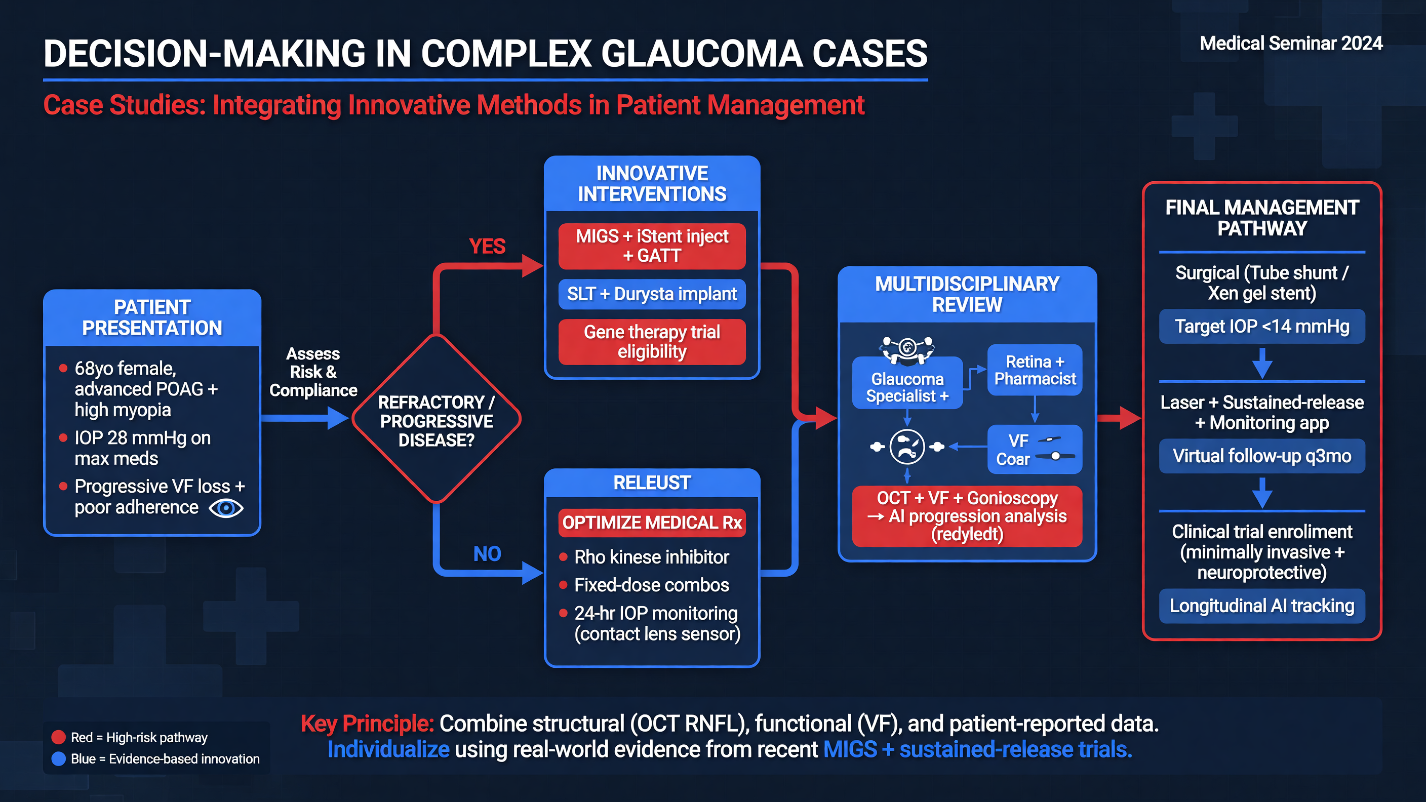
Case 1: Advanced disease with a low target
A 72-year-old monocular patient has POAG with a central island, mean deviation of −22 dB, and confirmed progression at IOPs of 13–15 mmHg despite four medication classes. Gonioscopy is open, and a cataract is present. Phaco-Hydrus would be technically feasible, but the clinical question is whether conventional outflow can reliably achieve a target near 10 mmHg.
Decision Point: Choose the operation that matches the target, not the operation that fits conveniently through the cataract incision. Trabeculectomy with mitomycin C may offer the greatest probability of very low pressure; a tube may be preferred if conjunctival scarring or failure risk is high. MIGS could reduce medication burden yet leave the optic nerve inadequately protected.
Perioperative planning includes counseling about wipeout anxiety without overstating its rarity, avoiding large pressure swings, arranging early follow-up, and ensuring that the patient can recognize bleb infection or hypotony symptoms. Cataract and glaucoma surgery may be combined or staged according to visual need, conjunctival risk, and surgeon judgment.
Case 2: Progressing normal-tension glaucoma
A 58-year-old has visual-field progression at office IOPs of 11–13 mmHg. Before labeling this “pressure-independent” disease, repeat fields, review OCT progression maps, examine disc photographs for hemorrhage, verify adherence, and obtain untreated baseline information if available. Ask about nocturnal antihypertensive dosing, sleep apnea, anemia, migraine, and Raynaud symptoms. Pallor, dyschromatopsia, rapid central loss, or vertical-meridian defects should trigger evaluation for a glaucoma mimic.
The Collaborative Normal-Tension Glaucoma Study supports an approximate 30% reduction from untreated baseline in selected progressing eyes. The difficulty is that a very low surgical target increases hypotony risk. Trabeculectomy with careful flow titration may be required; trabecular MIGS usually cannot create a large reduction from an already low baseline.
Nuance: Normal-tension glaucoma is not “normal-risk glaucoma.” Its treatment still begins with pressure, while vascular and metabolic factors are investigated without unproven attempts to raise systemic blood pressure.
The patient asks about nicotinamide. Explain the biologic rationale, ongoing trials, hepatotoxicity concern, and absence of proven long-term neuroprotection. Trial enrollment is preferable to unsupervised high-dose supplementation.
Case 3: Medication intolerance with a realistic MIGS target
A 67-year-old has mild-to-moderate POAG, IOP 16 mmHg on latanoprost and dorzolamide-timolol, severe preservative-related ocular-surface disease, and a target of 15–17 mmHg. The angle is open without synechiae, fields are stable, and cataract limits driving.
Phaco-Hydrus is a good mechanistic fit: the target lies within conventional outflow’s plausible range, cataract surgery is independently indicated, and reducing drops has tangible value. Alternatives include phaco alone, SLT before surgery, another angle procedure, preservative-free medication, or continued therapy. Consent should state that Hydrus improves the probability of medication reduction but does not guarantee it.
Framework: Integrate five domains: disease threat, target pressure, outflow anatomy, treatment burden, and rescue options.
Teaching Point: The same pressure can mean different things. Sixteen millimeters of mercury may be satisfactory in stable moderate disease and unacceptable in rapidly progressing advanced disease.
A practical escalation pathway
First confirm diagnosis and progression. Second set a numerical target tied to rate of change. Third identify the failing component—adherence, aqueous production, trabecular resistance, distal outflow, or subconjunctival scarring. Fourth select an intervention capable of reaching the target. Fifth define surveillance and rescue thresholds before surgery.
MUST ACT: Do not delay filtration surgery through serial low-efficacy interventions when the remaining visual field cannot tolerate continued progression.
Audience Poll: Which patient benefits most from MIGS: the eye needing single-digit pressure, the stable eye seeking medication reduction, or the eye with uncontrolled neovascular glaucoma? Only the second has a target and anatomy aligned with trabecular MIGS.
Case: A 65-Year-Old with Uncontrolled Open-Angle Glaucoma
A 65-year-old presents with visually significant cataract and POAG. Best-corrected acuity is 20/60 in the right eye. IOP is 21 mmHg on latanoprost nightly and dorzolamide-timolol twice daily, although refill history and discussion reveal missed morning doses. Central corneal thickness is 518 μm. Gonioscopy shows a Shaffer grade 3–4 angle without neovascularization or peripheral anterior synechiae. OCT demonstrates superior and inferior RNFL loss; the 24-2 field has a reproducible arcuate defect with mean deviation of −7.2 dB and a trend of approximately −0.6 dB/year. The target is revised to 14–16 mmHg.
The differential for apparent treatment failure includes nonadherence, poor instillation, pressure fluctuation, steroid exposure, and true progression despite therapy. Repeat testing confirms the field trend, and disc photographs show a new inferotemporal hemorrhage. Phacoemulsification alone may lower IOP but provides no assurance of medication reduction. Trabeculectomy would probably exceed the pressure-lowering requirement while introducing disproportionate bleb risk. Combined phaco-Hydrus is consistent with the labeled population and target.
Audience Poll: Is the primary indication uncontrolled IOP, progression, cataract, medication burden, or all four? The answer is all four, but progression and target compatibility determine whether MIGS is sufficient.
Consent covers cataract-surgery risks, hyphema, pressure spike, steroid response, malposition, synechiae, failure to reduce drops, and later need for SLT or filtration surgery. The patient understands that HORIZON estimates group probabilities, not an individual guarantee.
After uncomplicated phaco-Hydrus, a representative regimen is prednisolone acetate 1% four times daily tapered over four weeks, a topical antibiotic four times daily for one week, and an NSAID according to cataract protocol. Because the nerve is progressing, dorzolamide-timolol is initially continued while latanoprost is held; medication withdrawal is based on measured IOP rather than a preset desire for drop freedom. Follow-up occurs on day 1, at one week and one month, and then according to glaucoma risk. Gonioscopy is performed if pressure control is worse than expected.
At 12 months in this teaching scenario, acuity is 20/20, IOP is 14–15 mmHg on no medication, and OCT and fields are stable. This is a successful outcome only if stability persists. If IOP rises to 18 mmHg with renewed progression, drops or SLT should be restarted promptly, with filtration surgery considered if the revised target cannot be maintained.
The patient asks about 3 g/day nicotinamide. The appropriate response is not to add an unmonitored supplement to an otherwise evidence-based plan. Explain that high-dose nicotinamide is investigational, carries liver-injury risk, and should not substitute for surveillance or pressure control.
MUST ACT: Surgical success is preservation of useful vision at an acceptable burden—not implantation, a low pressure at one visit, or temporary medication freedom.
Tonight on Shift Checklist
- Define the diagnosis, stage, progression rate, and numerical target IOP before discussing a procedure.
- Perform gonioscopy and reject angle-based MIGS when anatomy, mechanism, or target pressure is incompatible.
- Treat pain, vision loss, bleb leak, shallow chamber, marked inflammation, hyphema with pressure elevation, or a postoperative IOP spike urgently.
- Interpret Hydrus evidence using medication-free target attainment, visual-field slope, adverse events, and rescue surgery—not mean IOP alone.
- Ask explicitly about adherence, instillation technique, cost, preservatives, systemic adverse effects, and caregiver burden.
- Keep neuroprotection investigational: optimize proven IOP lowering, exclude glaucoma mimics, and discourage unsupervised high-dose nicotinamide.
Read this seminar as Markdown · All seminars · Lecture library · Question bank