Understanding Vaccine-Induced Immune Thrombocytopenia and Thrombosis: Mechanisms and Clinical Implications
Mechanisms and Clinical Implications
Microbiology · Seminar week 22 · released August 10, 2026 · includes a discussion video
This seminar addresses the critical need to understand VITT, a serious adverse event linked to adenovirus vector-based COVID-19 vaccines. A NEJM study (P92) elucidates the…
Learning Objectives
By the end of this seminar, learners should be able to:
- Recognize the temporal, clinical, and laboratory phenotype of vaccine-induced immune thrombocytopenia and thrombosis (VITT).
- Explain how adenoviral core protein VII, platelet factor 4, and FcγRIIa-mediated cellular activation produce immunothrombosis.
- Interpret the roles of IGLV3-2102/03 and the acquired K31E somatic mutation without overstating the value of genetic screening.
- Differentiate VITT from heparin-induced thrombocytopenia, thrombotic thrombocytopenic purpura, disseminated intravascular coagulation, immune thrombocytopenia, and ordinary venous thromboembolism.
- Apply a time-critical diagnostic and treatment pathway using PF4 ELISA testing, intravenous immunoglobulin, non-heparin anticoagulation, and targeted supportive care.
- Evaluate how VITT changed vaccine pharmacovigilance, regulatory policy, informed consent, and adenoviral-vector design.
- Identify unanswered questions concerning penetrance, long-term recurrence, natural adenovirus-associated disease, and safer vector engineering.
Introduction to VITT and its Clinical Significance
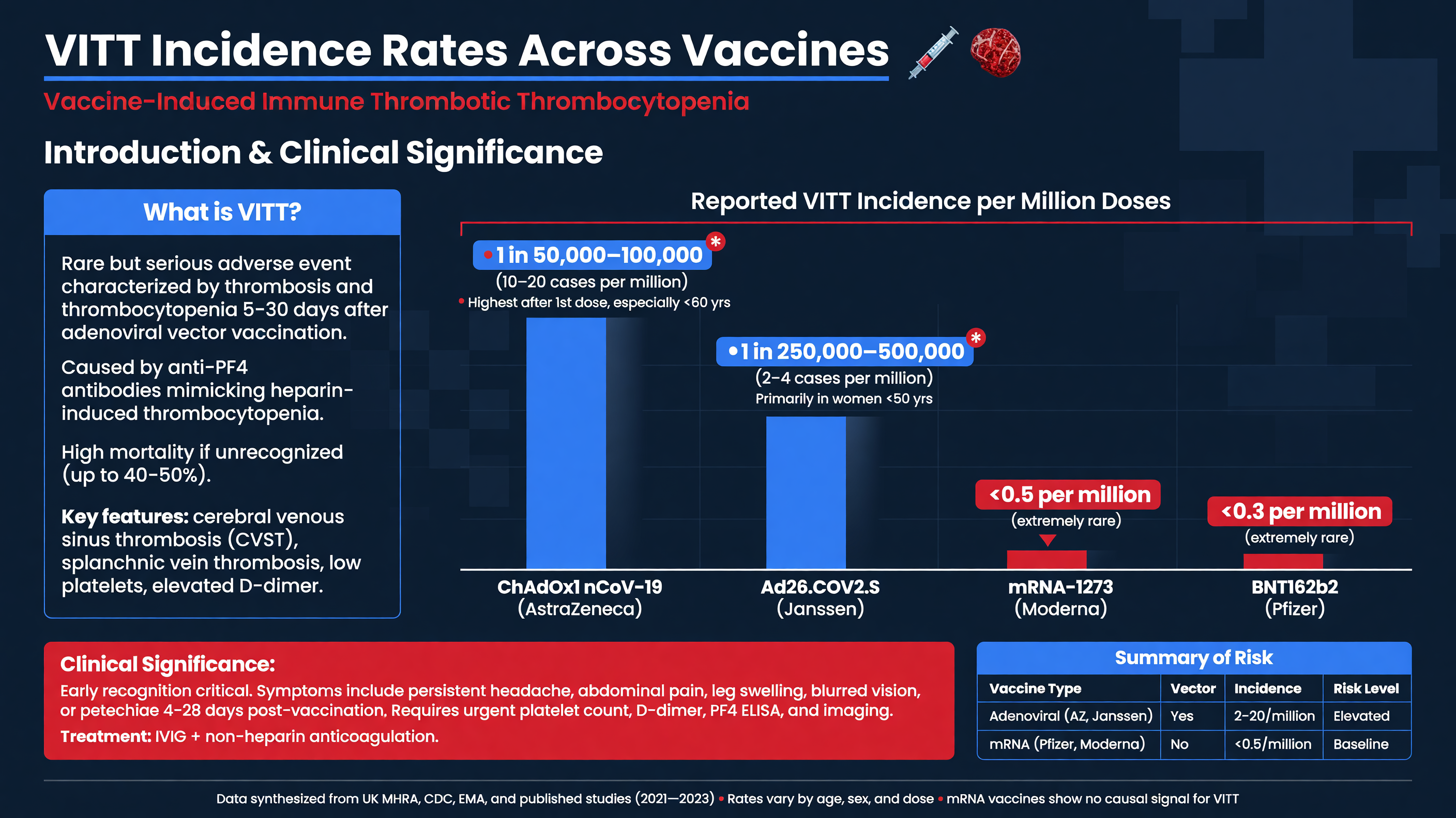
Vaccine-induced immune thrombocytopenia and thrombosis—more commonly termed vaccine-induced immune thrombotic thrombocytopenia—is an antibody-mediated thromboinflammatory syndrome initially recognized in 2021 after the adenoviral-vector COVID-19 vaccines ChAdOx1 nCoV-19, developed by Oxford–AstraZeneca, and Ad26.COV2.S, developed by Janssen. The defining combination is not merely “a clot after vaccination.” It is thrombosis, often in an unusual vascular bed, accompanied by platelet consumption, markedly increased D-dimer, and platelet-activating IgG antibodies against platelet factor 4 (PF4). The first reports established its resemblance to autoimmune heparin-induced thrombocytopenia despite the absence of preceding heparin exposure (Greinacher et al., NEJM 2021, PMID: 33835769; Schultz et al., NEJM 2021, PMID: 33835768; Scully et al., NEJM 2021, PMID: 33861525).
Teaching Point: VITT is narrower than thrombosis with thrombocytopenia syndrome, or TTS. TTS is a surveillance phenotype that can include cancer-associated thrombosis, sepsis-associated disseminated intravascular coagulation, thrombotic microangiopathy, antiphospholipid syndrome, and other disorders. VITT denotes the characteristic post-adenoviral exposure, anti-PF4 immune syndrome.
Symptoms usually begin 5–30 days after vaccination; an interval extending to 42 days is often accepted for isolated deep-vein thrombosis or pulmonary embolism. Median presentation in the large UK cohort was approximately 14 days. Severe or progressive headache, focal neurologic symptoms, visual disturbance, seizures, persistent abdominal or back pain, dyspnea, chest pain, leg swelling, limb ischemia, petechiae, or spontaneous bruising should trigger evaluation. Expected fever, fatigue, myalgia, and headache during the first 24–72 hours after vaccination usually represent ordinary reactogenicity. A headache that begins or intensifies several days later, persists despite analgesia, or accompanies neurologic or abdominal symptoms is qualitatively different.
MUST ACT: A patient in the risk window with severe headache, abdominal pain, dyspnea, neurologic dysfunction, or signs of thrombosis needs an urgent complete blood count, D-dimer, fibrinogen, coagulation studies, PF4-polyanion IgG ELISA, and symptom-directed vascular imaging. A normal noncontrast head CT does not exclude cerebral venous sinus thrombosis.
Cerebral venous sinus thrombosis and splanchnic-vein thrombosis are especially characteristic, but pulmonary embolism, conventional deep-vein thrombosis, adrenal-vein thrombosis, ischemic stroke, myocardial infarction, and peripheral arterial thrombosis also occur. Multiple vascular territories may be involved simultaneously. Thrombocytopenia can produce bleeding, yet the immediate biological threat is usually uncontrolled thrombosis. The large UK series of 220 definite or probable cases reported approximately 22% mortality; cerebral venous thrombosis, lower platelet count, lower fibrinogen, and higher D-dimer were associated with death (Pavord et al., NEJM 2021, PMID: 34379914).
Incidence estimates depend heavily on case definition, dose number, age distribution, ascertainment, and denominator quality. Mature UK pharmacovigilance estimated roughly 16 reports per million first or unknown ChAdOx1 doses overall, with a higher reported rate in younger adults. Published estimates for Ad26.COV2.S generally clustered around two to four cases per million doses. VITT was substantially less frequent after a second ChAdOx1 dose than after the first. Apparent early predominance among younger women was partly influenced by occupational rollout and vaccine allocation; no demographic or comorbidity profile is sufficiently predictive for bedside exclusion.
Nuance: Temporal association alone is weak evidence because ordinary venous thromboembolism, stroke, immune thrombocytopenia, and headache occur commonly in vaccinated populations. Conversely, waiting for the full pentad of thrombosis, thrombocytopenia, high D-dimer, low fibrinogen, and positive PF4 ELISA can miss “pre-VITT,” in which the immune process is active before imaging demonstrates a clot.
Framework: Think in three linked domains: exposure within a plausible interval, a consumptive coagulation phenotype, and PF4-dependent platelet activation. The more strongly those domains align, the lower the threshold for immediate treatment.
Audience Poll: A patient presents 11 days after an adenoviral-vector vaccine with a new refractory headache, platelets of 162 × 10⁹/L, and a D-dimer eight times the upper limit of normal. Would you discharge after a normal noncontrast CT, repeat testing within hours, or initiate a VITT pathway now?
Mechanism of Action: Adenoviral P7 Protein and PF4 Molecular Mimicry
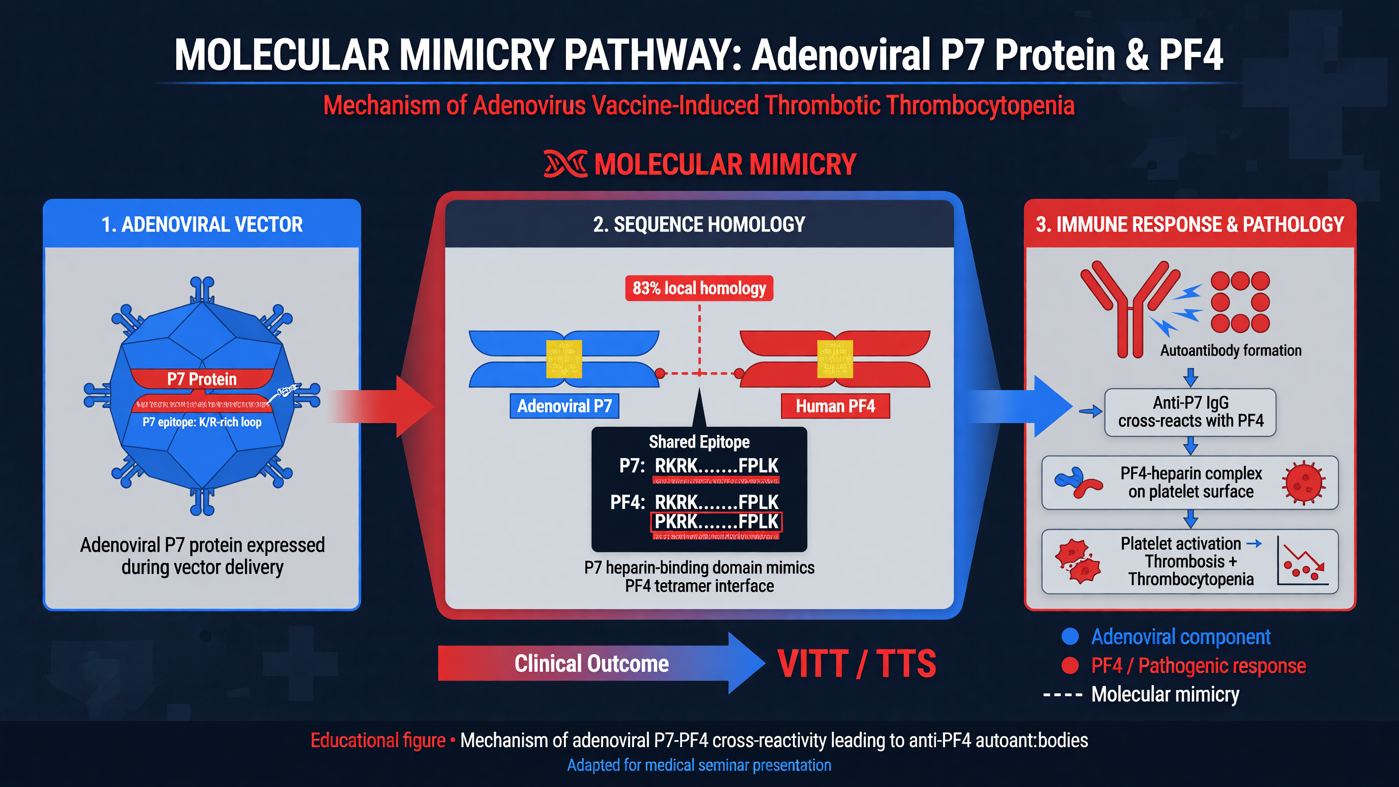
The relevant adenoviral protein is core protein VII, conventionally written pVII and sometimes informally rendered “P7.” It is a highly basic, histone-like protein that packages and condenses adenoviral DNA inside the virion. It is not the SARS-CoV-2 spike protein. Earlier mechanistic models proposed that negatively charged vaccine components—including adenoviral DNA, capsid-derived material, and other polyanions—could bind the positively charged chemokine PF4, create immunogenic complexes, and break B-cell tolerance. Those models explained how PF4 might become antigenic but did not identify the initiating adenoviral antigen with precision.
PF4, also called CXCL4, is stored in platelet alpha granules and released during platelet activation. It forms cationic tetramers that avidly bind polyanions. In classical heparin-induced thrombocytopenia, antibodies recognize conformational epitopes on PF4–heparin complexes. In VITT, high-titer IgG generally binds PF4 in the absence of pharmacologic heparin, targets a region that overlaps the heparin-binding surface, and can activate platelets when additional PF4 is present. This distinction explains why a rapid HIT immunoassay can be negative even when a PF4-polyanion ELISA is strongly positive.
A 2026 New England Journal of Medicine study moved the model from plausible association toward a defined antigenic pathway. Investigators used antibody proteomics in patients with VITT, immunoglobulin sequencing, affinity purification against adenoviral proteins, epitope mapping, recombinant antibodies, and human PF4/FcγRIIa mouse models. Only antibodies purified against pVII contained species matching the characteristic VITT anti-PF4 antibody fingerprint. Cross-reactivity mapped to a basic linear epitope on pVII. When the critical antibody mutation was experimentally reverted to its germline residue, PF4 binding and prothrombotic activity were lost while preference for pVII was restored (Wang et al., NEJM 2026, PMID: 41671482).
Framework: The emerging sequence is adenoviral pVII exposure → activation of a pVII-reactive B-cell clone → somatic mutation and antigenic shift toward PF4 → anti-PF4 IgG production → FcγRIIa-dependent immunothrombosis. “Molecular mimicry” here is therefore more sophisticated than two proteins sharing an identical sequence. It involves a cross-reactive antigenic surface, electrostatic complementarity, and mutation of the antibody paratope.
Once pathogenic IgG binds PF4, immune complexes cross-link FcγRIIa receptors on platelets. Activated platelets release additional PF4 and procoagulant microparticles, creating a feed-forward loop. Monocytes activated through Fcγ receptors express tissue factor; neutrophils release neutrophil extracellular traps that provide scaffolds for coagulation; endothelial activation promotes adhesion and local thrombin generation; and complement may amplify cellular injury. Platelets and fibrinogen are consumed as thrombin generation accelerates, accounting for the paradox of thrombosis, thrombocytopenia, hypofibrinogenemia, and occasional bleeding.
Teaching Point: The platelet count is not simply falling because antibodies clear platelets in the spleen, as in conventional immune thrombocytopenia. Platelets are being activated and incorporated into a multisystem thromboinflammatory process. This is why raising the platelet count without suppressing Fc-mediated activation and treating thrombosis is inadequate.
VITT and HIT converge at PF4-dependent FcγRIIa activation but differ in their inciting antigens, antibody epitopes, heparin exposure, and laboratory behavior. High heparin concentrations may inhibit some VITT antibodies in vitro, and retrospective data have not conclusively shown that heparin always worsens VITT. Nevertheless, non-heparin anticoagulants remain preferred because alternatives are available and inadvertent enhancement cannot be confidently excluded. This is a risk-management recommendation, not proof that every heparin exposure causes deterioration.
Nuance: The pVII mechanism does not establish that every adenoviral vector, formulation, route, or dose has an identical VITT risk. Vector serotype, pVII sequence, particle integrity, manufacturing impurities, tissue distribution, and innate inflammatory signals may modify antigen availability. Nor does the mechanism implicate mRNA vaccination or the encoded spike antigen as equivalent triggers.
MUST ACT: Molecular insight should sharpen recognition, not delay care. Neither anti-pVII testing nor antibody sequencing is a clinical emergency test. The actionable biomarker remains a compatible syndrome with a strongly positive PF4 IgG ELISA, supported when necessary by a PF4-enhanced functional platelet-activation assay.
Audience Poll: Which intervention most directly interrupts the immediate pathogenic loop: platelet transfusion, aspirin alone, high-dose intravenous immunoglobulin, or vitamin K administration?
Genetic Predisposition: The Role of IGLV3-21 and Somatic Hypermutation
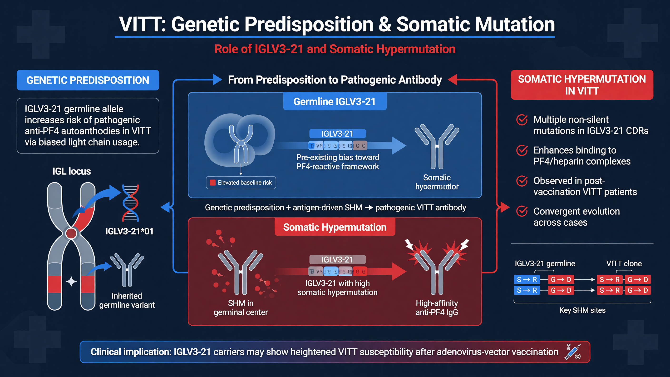
The striking rarity of VITT despite administration of millions of adenoviral-vector vaccine doses suggested that exposure was necessary but insufficient. Antibody sequencing has now identified a highly stereotyped immunoglobulin signature. The 2026 study found that VITT antibodies used the lambda light-chain variable allele IGLV3-2102 or the closely related 03 allele and carried a critical lysine-to-glutamic-acid substitution at position 31, designated K31E. Proteomic sequencing examined anti-PF4 antibodies from 21 patients, while genes encoding the light-chain hypervariable region were evaluated in 100 patients. The convergence on a shared light-chain architecture was exceptional for an acquired autoimmune syndrome (PMID: 41671482).
The pathogenic antibodies also display an unusually acidic antigen-binding surface. Conserved acidic residues in the light-chain complementarity-determining regions, together with an acidic motif in the heavy-chain CDR3, create a negatively charged paratope suited to binding highly cationic PF4. Heavy-chain variable-gene usage can differ, so the antibodies are not all descendants of one universal clone, but their structural solutions are remarkably similar. Serum repertoires are often monoclonal or oligoclonal rather than broadly polyclonal.
Teaching Point: IGLV3-21 is an inherited immunoglobulin gene segment; K31E is not. K31E arises through somatic hypermutation in an activated B cell. The predisposition is therefore a two-stage interaction between germline repertoire and a stochastic acquired mutation.
Somatic hypermutation normally occurs during antigen-driven B-cell maturation, introducing nucleotide substitutions into immunoglobulin variable regions. Most mutations are neutral or reduce affinity; a small number improve binding and are selected during clonal expansion. In the proposed VITT pathway, the germline K31 antibody recognizes pVII preferentially. Replacement of positively charged lysine with negatively charged glutamic acid changes the electrostatic surface of the antibody and redirects binding toward PF4. Experimentally back-mutating E31 to the germline lysine eliminated PF4 binding and platelet-activating activity in vitro and in vivo while restoring preferential pVII recognition. This is functional evidence that K31E is not merely a molecular passenger.
Framework: Susceptibility requires at least four events: possession of a compatible germline allele, exposure of the relevant pVII epitope, activation of an appropriate B-cell lineage, and acquisition plus selection of the K31E mutation. Additional inflammatory and host factors probably determine whether the clone expands enough to produce disease.
This multistep model explains low penetrance. Many people carry an IGLV3-21 allele yet never develop VITT; most will never generate the relevant mutation in the correct clone and immunologic context. Allele carriage therefore has poor positive predictive value when the outcome occurs only a few times per million vaccinations. A screening program would label a vastly larger group “at risk” than would ever become ill, with uncertain psychological and public-health consequences.
Decision Point: Do not order IGLV3-21 sequencing in the emergency evaluation of suspected VITT, and do not use allele status to withhold clinically indicated vaccination outside a validated research protocol. The platelet count, D-dimer, fibrinogen, imaging, PF4 IgG ELISA, and clinical trajectory determine acute care.
VITT-like anti-PF4 syndromes have also been described after natural adenovirus infection. Antibody fingerprints in these cases can closely resemble vaccine-associated VITT, supporting adenovirus rather than spike as the upstream trigger and prompting proposals for the broader term virus-induced immune thrombocytopenia and thrombosis. Other infections can produce anti-PF4 disorders through different antibody clonotypes, however, so not every infection-associated PF4 antibody represents the pVII–IGLV3-21 pathway.
Nuance: The observed genetic convergence should not be confused with a simple Mendelian thrombophilia. Family screening is not established, conventional inherited thrombophilia panels do not diagnose VITT, and a history of factor V Leiden, estrogen exposure, prior venous thromboembolism, or autoimmunity neither confirms nor reliably excludes the syndrome.
Research priorities include determining population frequencies and penetrance of 02 and 03 alleles, identifying the B-cell compartment in which K31E arises, explaining the rapid 5–30-day time course, and establishing whether prior adenovirus exposure creates memory clones that can be recalled by vaccination. Prospective biobanking before IVIG will be essential because treatment rapidly alters functional antibody assays.
Audience Poll: Even if an IGLV3-21 assay became inexpensive, would screening be justified without a validated absolute-risk estimate, an effective alternative for every patient, and evidence that knowing the result improves outcomes?
Diagnostic Criteria and Current Management Strategies
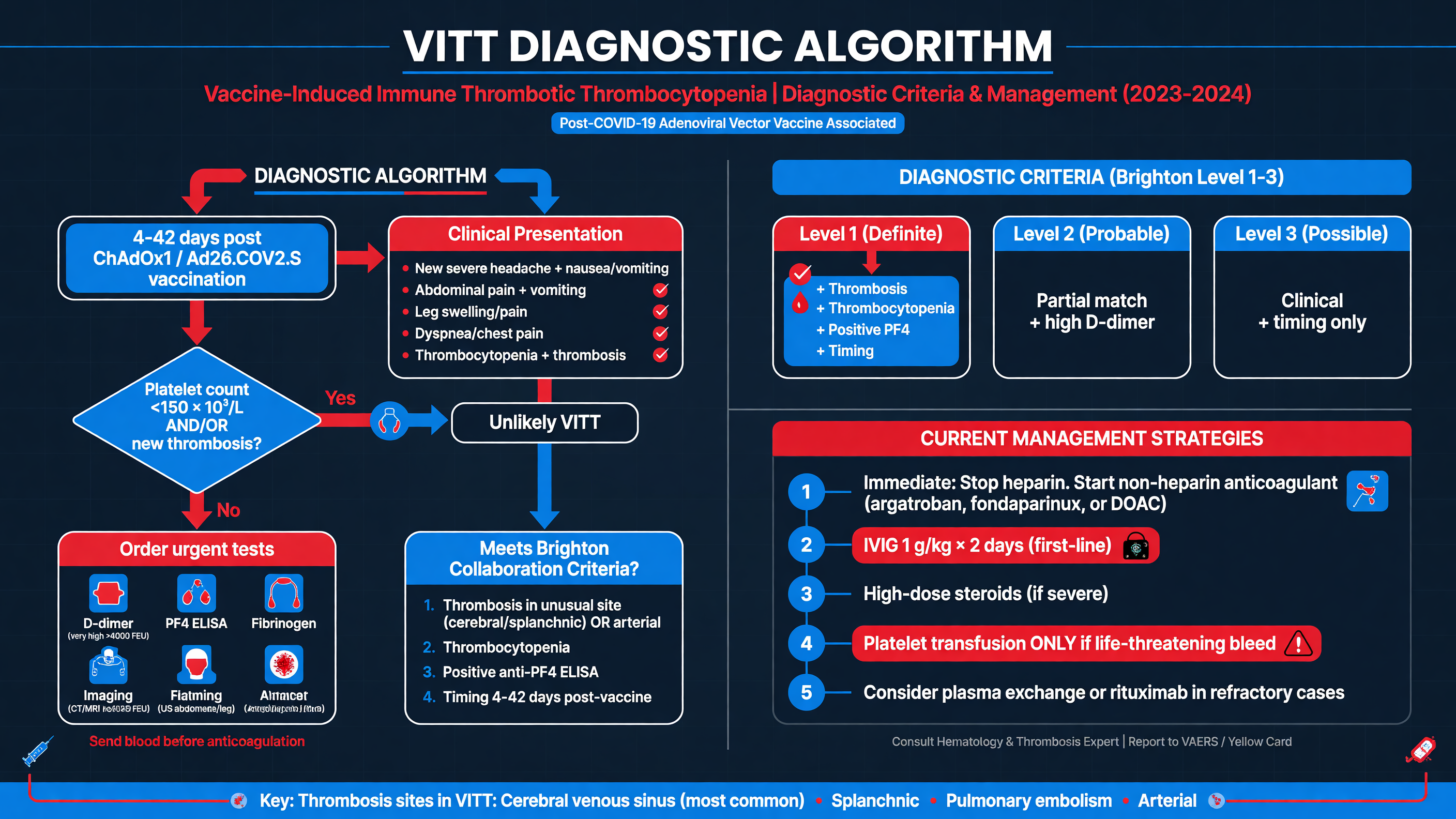
VITT is a time-critical clinicopathologic diagnosis. Commonly used criteria for definite disease include symptom onset 5–30 days after vaccination, or up to 42 days for isolated deep-vein thrombosis or pulmonary embolism; objectively confirmed thrombosis; platelet count below 150 × 10⁹/L; D-dimer above 4,000 fibrinogen-equivalent units, approximately eight times a typical upper limit; and a positive PF4-polyanion IgG ELISA. Probable disease may lack one element, particularly when thrombosis has not yet become radiographically visible or the platelet count is falling from a higher baseline.
MUST ACT: Draw blood for complete blood count with smear, D-dimer, fibrinogen, PT/INR, aPTT, renal and hepatic function, and PF4 ELISA before IVIG when feasible—but never delay treatment in a high-probability, deteriorating patient. Repeat the platelet count within 12–24 hours if initial results are normal but suspicion remains high.
Imaging must follow symptoms. Obtain CT or MR venography for persistent or severe headache, focal deficits, seizure, papilledema, or altered consciousness; contrast-enhanced abdominal imaging for persistent abdominal or back pain; CT pulmonary angiography for suspected pulmonary embolism; and compression ultrasonography for limb symptoms. A noncontrast head CT may detect hemorrhage or venous infarction but cannot reliably exclude cerebral venous thrombosis.
Framework: The central differential is organized by mechanism. HIT requires a plausible heparin exposure and typically PF4–heparin-dependent antibodies. Thrombotic thrombocytopenic purpura produces microangiopathic hemolysis, schistocytes, and severe ADAMTS13 deficiency, usually without extreme D-dimer or low fibrinogen. Sepsis-associated disseminated intravascular coagulation has an infectious or shock context and more generalized coagulation-factor consumption. Immune thrombocytopenia generally causes isolated thrombocytopenia and bleeding rather than profound D-dimer elevation and unusual-site thrombosis. Catastrophic antiphospholipid syndrome, active COVID-19, malignancy, paroxysmal nocturnal hemoglobinuria, drug-induced thrombocytopenia, and pseudothrombocytopenia remain context-dependent alternatives.
Use a PF4-polyanion IgG ELISA rather than relying on rapid latex, particle-gel, or chemiluminescent HIT assays, which may be falsely negative. A strong ELISA optical-density signal supports VITT but is not independently diagnostic. Specialized laboratories can perform PF4-enhanced serotonin-release or heparin-induced platelet-activation assays when the phenotype and ELISA disagree.
Start high-dose IVIG promptly in probable or confirmed VITT. A widely used regimen is 1 g/kg intravenously on day 1 and again approximately 24 hours later, although some guidelines begin with 1 g/kg and repeat only for inadequate laboratory or clinical response. IVIG saturates Fc receptors and rapidly reduces VITT-antibody-mediated platelet activation; improvement in platelet activation after IVIG was demonstrated in early clinical observations (Bourguignon et al., NEJM 2021, PMID: 34107198).
Decision Point: Begin therapeutic non-heparin anticoagulation as soon as thrombosis risk outweighs active bleeding risk—even when the platelet count is low. Options include argatroban or bivalirudin for unstable patients needing a rapidly titratable infusion, fondaparinux for stable patients with adequate renal function, and a direct oral anticoagulant when absorption is reliable and no urgent procedure is expected. Typical fondaparinux treatment is 5 mg daily below 50 kg, 7.5 mg daily at 50–100 kg, and 10 mg daily above 100 kg. Standard venous-thromboembolism loading regimens may be used for apixaban or rivaroxaban once clinically appropriate. Argatroban often begins below the usual 2 micrograms/kg/min in critical illness or hepatic dysfunction and is titrated using aPTT and institutional protocols.
Avoid acute warfarin initiation until platelets have recovered. Heparin is generally avoided while VITT remains likely, although lifesaving anticoagulation should not be withheld if no alternative is immediately available. Aspirin is not an adequate substitute for anticoagulation and IVIG.
Replace fibrinogen with cryoprecipitate or fibrinogen concentrate when markedly reduced, commonly targeting at least 1.5 g/L in bleeding, cerebral venous thrombosis, or procedural settings. Avoid routine platelet transfusion because additional platelets can fuel activation; transfuse for life-threatening bleeding or urgent surgery after hematology and procedural consultation. Severe or refractory disease—especially progressive thrombosis, cerebral venous thrombosis, platelets below 30 × 10⁹/L, or failure to respond to IVIG—may require daily one-plasma-volume exchange with plasma replacement. High-dose corticosteroids are adjunctive rather than primary therapy; rituximab is reserved for exceptional refractory or relapsing disease.
Nuance: Intracranial hemorrhage secondary to cerebral venous thrombosis does not automatically preclude anticoagulation. Management requires hematology, neurology, neurosurgery, critical care, and thrombosis expertise, with rapid transfer to a center capable of decompressive surgery or endovascular rescue when deterioration occurs.
Continue anticoagulation for at least three months after thrombosis and often three to six months, individualized to vascular site, recanalization, bleeding risk, platelet recovery, and persistence of platelet-activating antibodies. ELISA positivity may persist after functional activity wanes. Patients require serial platelet, D-dimer, and fibrinogen monitoring, recurrence education, adverse-event reporting, and specialist guidance regarding subsequent vaccination.
Audience Poll: If PF4 ELISA results will not return until tomorrow, which finding would most strongly justify immediate IVIG tonight: isolated platelets of 135 × 10⁹/L, ordinary headache 24 hours after vaccination, or progressive headache on day 9 with falling platelets and D-dimer above 20,000 FEU?
Impact of VITT on Vaccine Development and Regulatory Policies
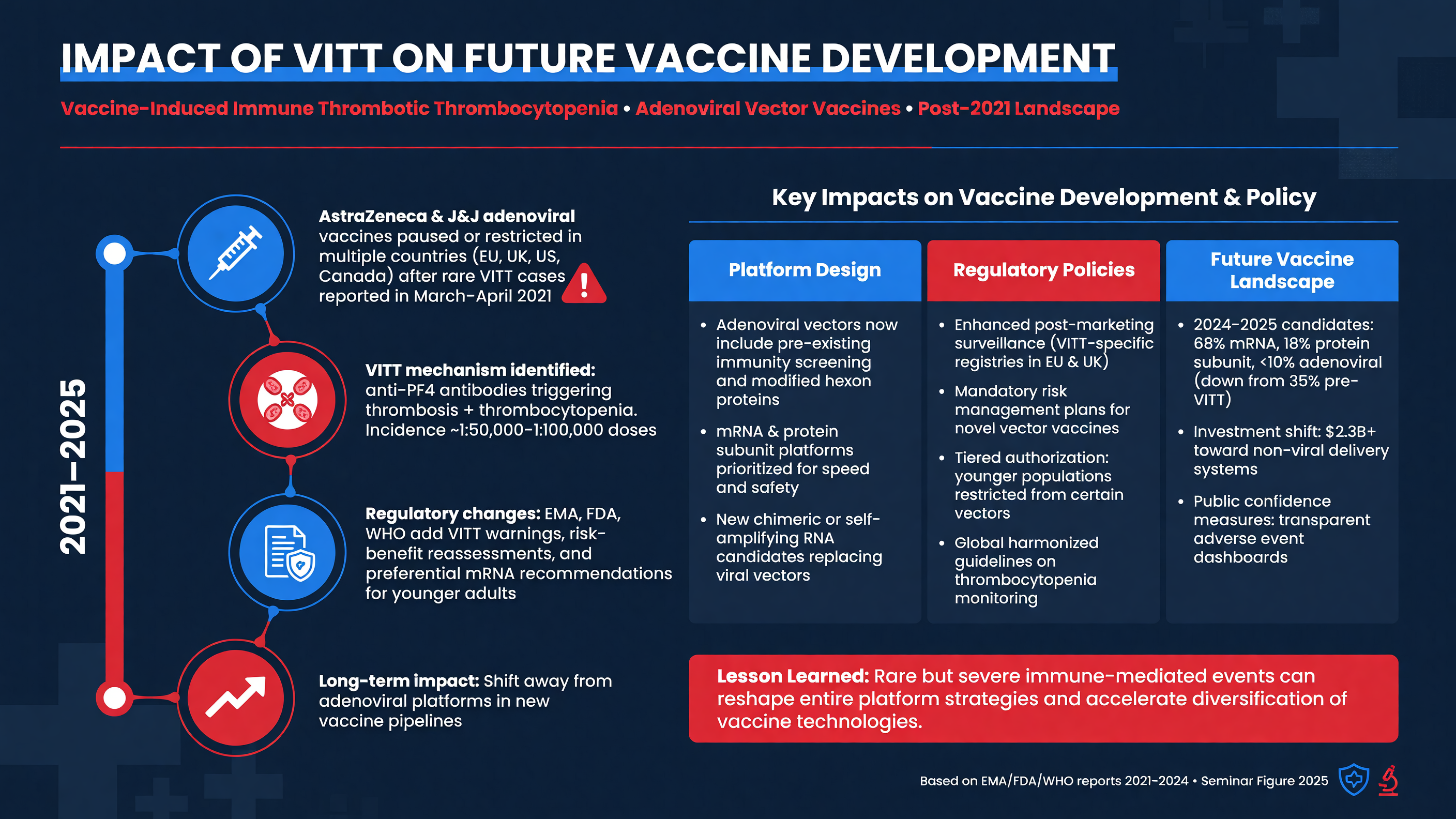
VITT demonstrated why vaccine safety cannot end with randomized efficacy trials. A complication occurring between one per tens of thousands and one per several hundred thousand exposures may not appear, or may appear only once without a recognizable pattern, in a preauthorization trial. Recognition required millions of administered doses, clinicians noticing an unusual combination of cerebral or splanchnic thrombosis and thrombocytopenia, rapid international case sharing, and mechanistic laboratory confirmation.
Teaching Point: Passive surveillance generates signals, not incidence by itself. Reports must be adjudicated against standardized definitions, linked to reliable dose denominators, compared with background rates, and stratified by time since exposure. A case of ordinary pulmonary embolism after vaccination is not automatically VITT; failure to distinguish TTS from PF4-positive VITT can distort platform comparisons.
Regulators responded in stages: temporary pauses, label warnings, age-based restrictions, enhanced informed consent, preferential use of alternative platforms, and active follow-up. In December 2021, the US Advisory Committee on Immunization Practices preferentially recommended mRNA COVID-19 vaccines over Janssen’s vaccine because of both effectiveness and safety considerations, including TTS. The Janssen emergency use authorization was revoked in June 2023 at the manufacturer’s request after US supplies expired, demand ceased, and no strain update was planned—not as a new finding that VITT alone made the product unacceptable. In the European Union, the Vaxzevria authorization was withdrawn in March 2024 at AstraZeneca’s request for commercial reasons; Jcovden’s authorization was withdrawn in August 2024. These distinctions matter when interpreting regulation as evidence of causation or comparative risk (FDA Janssen regulatory history; EMA withdrawn COVID-19 products).
Nuance: Risk–benefit conclusions are contextual. During intense pandemic transmission, before alternatives were widely available, a single-dose, refrigerator-stable adenoviral vaccine could prevent far more hospitalizations and deaths than the number of VITT cases it caused. As mRNA and protein-based alternatives became abundant and viral strains evolved, the incremental benefit of the older products declined while their rare platform-specific risk remained.
The pVII finding creates concrete development questions. Developers can examine whether modifying the cross-reactive pVII epitope, reducing free core protein or disrupted particles, improving purification, changing vector dose, altering formulation, or modifying tissue distribution reduces PF4-directed immunogenicity. Yet pVII is a structural DNA-condensing protein, so simply deleting it may impair virion assembly or function. Any redesign must preserve vector yield, stability, transduction, and vaccine immunogenicity while demonstrating reduced anti-pVII/PF4 cross-reactivity.
Framework: A rational preclinical safety program would combine biochemical PF4-binding studies, anti-pVII antibody mapping, human serum and B-cell repertoire analysis, FcγRIIa-dependent platelet-activation assays, and human PF4/FcγRIIa animal models. Clinical development should prespecify thrombosis-with-thrombocytopenia events, retain acute biospecimens, and continue active surveillance after authorization.
The implications extend beyond COVID-19 vaccination. Adenoviral vectors are used or investigated in vaccines, cancer immunotherapy, and gene delivery. Risk may vary profoundly with serotype, route, dose, replication competence, manufacturing process, and patient population. The correct response is not to classify all adenoviral technology as unsafe; it is to test whether the pVII epitope and pathogenic antibody pathway are conserved in each product.
Decision Point: Regulators must decide whether a rare, biologically plausible event warrants restriction based on absolute risk, disease burden, alternative products, population age, logistics, and the ability to recognize and treat the complication. No single risk threshold is appropriate in every epidemic or health system.
Risk communication should use absolute numbers and a defined interval: for example, “approximately several cases per million doses, concentrated in the first few weeks,” rather than “increased clot risk.” Patients should hear both the warning symptoms and the expected first-day vaccine effects. Overstatement can undermine trust; minimization can delay care.
Audience Poll: If a redesigned adenoviral vector eliminates measurable PF4 cross-reactivity but offers major cold-chain and access advantages, what evidence would you require before widespread use: preclinical assays alone, a large trial, postauthorization active surveillance, or all three?
Case Studies and Future Research Directions
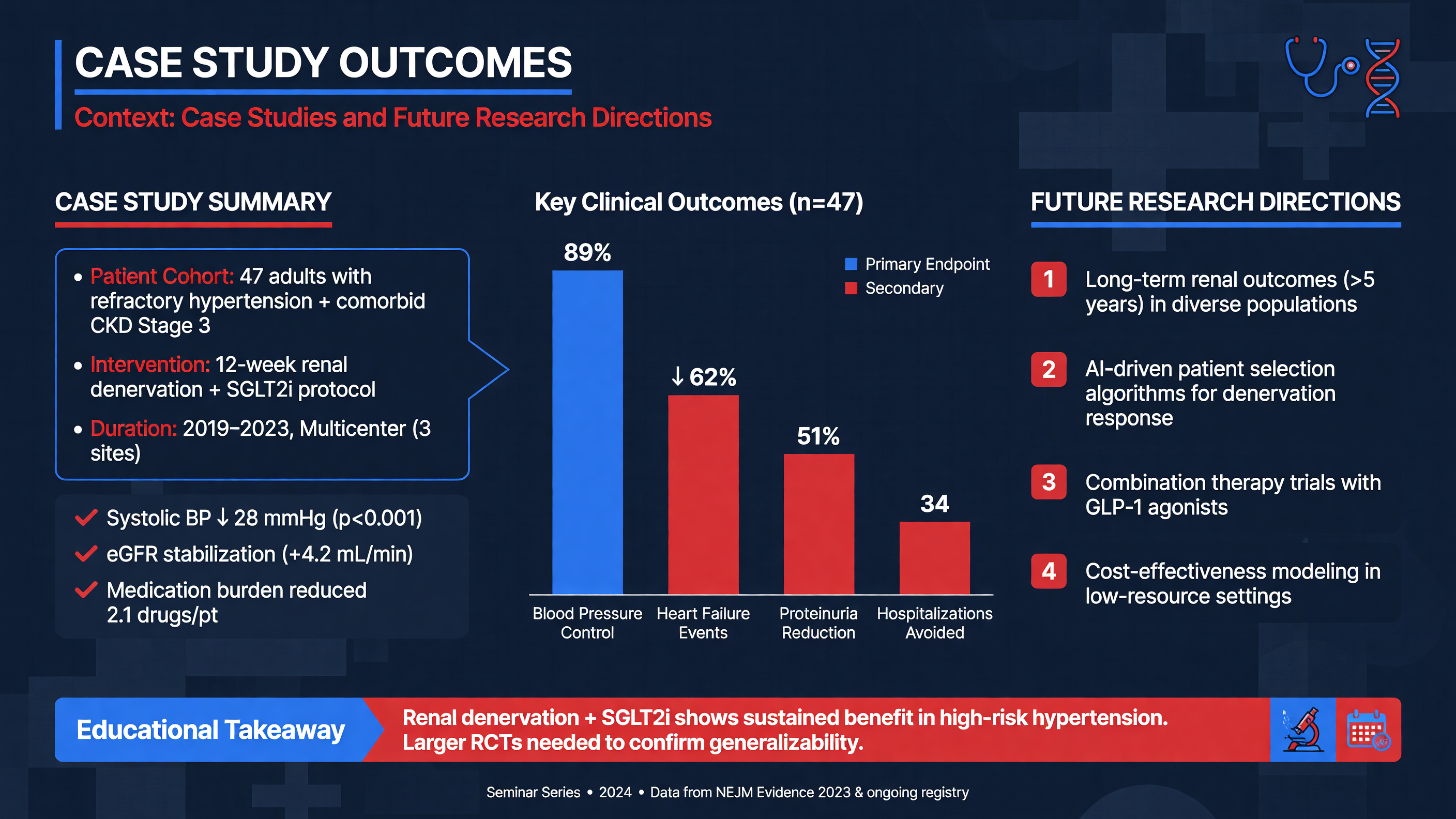
The early VITT literature illustrates how clinical recognition and mechanism developed together. Greinacher and colleagues described patients with unusual thrombosis, thrombocytopenia, high D-dimer, and PF4-reactive antibodies despite no heparin exposure. Schultz and colleagues reported five patients presenting 7–10 days after ChAdOx1 vaccination, several with cerebral venous thrombosis and intracranial injury. Scully and colleagues independently established a pathogenic PF4-dependent syndrome. The later UK cohort converted these observations into a reproducible clinical definition and identified prognostic markers (PMIDs: 33835769, 33835768, 33861525, and 34379914).
Teaching Point: The historical progression was bedside phenotype → PF4 immunology → treatment adaptation → antibody structure → inciting antigen. This is a model for investigating rare immune-mediated drug and vaccine reactions.
Three clinical phenotypes deserve particular attention. In classic VITT, thrombocytopenia, extreme D-dimer elevation, PF4 antibodies, and imaging-confirmed thrombosis appear together. In pre-VITT, severe headache or other warning symptoms accompany falling platelets and high D-dimer before thrombosis is visible; early IVIG may prevent progression. In persistent or relapsing VITT, platelet-activating antibodies or thrombocytopenia recur after initial improvement, sometimes requiring renewed IVIG, corticosteroids, plasma exchange, or B-cell-directed therapy. A fourth category—VITT-like illness after natural adenovirus infection—may illuminate the same pVII pathway without vaccination.
Framework: Future studies should separate six outcomes: generation of anti-PF4 antibodies, functional platelet activation, clinical thrombocytopenia, radiographic thrombosis, organ injury, and death. These are related but not interchangeable. An intervention might suppress platelet activation without immediately clearing ELISA antibodies, or improve survival without rapidly recanalizing thrombosed vessels.
Treatment evidence remains largely observational. There are no adequately powered randomized trials comparing IVIG regimens, anticoagulants, plasma exchange, or immunosuppressive strategies. Apparent improvements over time may reflect earlier diagnosis, changes in case mix, more aggressive neurocritical care, and avoidance of harmful delays as well as treatment efficacy. International registries should capture exact treatment timing, serial laboratory values, antibody function, imaging burden, and neurologic outcomes.
Long-term cohorts indicate that anti-PF4 ELISA reactivity can persist after platelet-activating capacity declines. Recurrent thrombosis appears uncommon after recovery and appropriate anticoagulation, and subsequent non-adenoviral vaccination has generally been tolerated, but numbers remain limited (Schönborn et al., long-term outcomes, PMID: 37394120). Survivorship research should also address chronic headache, post-thrombotic symptoms, neurocognitive disability, anxiety, and vaccine-related psychological trauma.
Nuance: The K31E discovery does not yet answer why the mutation emerges rapidly in only certain people, whether prior adenovirus infection primes the responsible clone, or why some antibody-positive patients develop cerebral thrombosis while others develop splanchnic or arterial disease. Vascular-bed biology, local PF4 concentration, endothelial activation, complement, and neutrophil extracellular traps may determine organ tropism.
Priority research directions include rapid PF4-enhanced functional assays available outside reference centers; prospective germline and B-cell repertoire studies with appropriate population controls; structural comparison of pVII across vector serotypes; measurement of intact versus disrupted particles in vaccine preparations; and validation of redesigned vectors. Experimental blockade of FcγRIIa, complement, or downstream cellular activation is scientifically attractive but is not standard clinical therapy.
Decision Point: Genetic or antibody screening should be introduced only if it predicts clinically meaningful disease at a useful positive predictive value and leads to an intervention that improves outcomes. Detecting a biologic susceptibility without an actionable threshold can cause more harm than benefit.
Audience Poll: Which research gap is most likely to change practice first: population genetic screening, a rapid functional anti-PF4 assay, pVII-modified vector design, or a randomized comparison of immunomodulatory treatments?
Case Study: 35-Year-Old Female with Post-Vaccine Thrombosis
This composite case reflects the phenotype described in published VITT cohorts. A previously healthy 35-year-old woman presents 10 days after a first ChAdOx1 vaccination. She had fever and myalgia for 36 hours after vaccination, recovered, and then developed a new diffuse headache on day 7. During the next three days, the headache became severe and positional, with transient blurred vision, vomiting, and poorly localized upper abdominal pain. She has scattered lower-extremity petechiae but no meningismus or focal weakness. She has not received heparin, takes no estrogen, and has no history of thrombosis.
Laboratory studies show platelets of 54 × 10⁹/L, down from 238 × 10⁹/L six months earlier; D-dimer above 35,000 FEU; fibrinogen 0.8 g/L; INR 1.3; and mildly increased transaminases. Hemoglobin is initially normal, the smear shows no schistocyte burden, and creatinine is normal. A noncontrast head CT is interpreted as showing no acute hemorrhage.
MUST ACT: A negative noncontrast CT does not end this evaluation. The combination of delayed severe headache, abdominal pain, profound D-dimer elevation, hypofibrinogenemia, and a greater than 75% platelet fall creates a high pretest probability of VITT.
Blood is collected immediately for PF4-polyanion IgG ELISA and, if available, a PF4-enhanced functional assay. CT venography shows superior sagittal and right transverse sinus thrombosis with a small venous infarct but no major hemorrhage. Contrast-enhanced abdominal CT demonstrates portal and superior mesenteric vein thrombosis. The PF4 ELISA later returns strongly positive.
The differential initially includes thrombotic thrombocytopenic purpura, disseminated intravascular coagulation, antiphospholipid syndrome, acute COVID-19, and catastrophic intra-abdominal infection. TTP becomes less likely because there is no microangiopathic hemolytic anemia, renal injury, or severe ADAMTS13 deficiency. Sepsis-associated DIC is less likely without infection or shock. Isolated immune thrombocytopenia cannot explain the extreme D-dimer and multisite thrombosis. The exposure interval and PF4 result complete the VITT phenotype.
Decision Point: Treatment begins before PF4 confirmation. She receives IVIG 1 g/kg immediately, repeated the following day. Because she has extensive cerebral and splanchnic thrombosis with possible need for an urgent procedure, an argatroban infusion is started at a reduced critical-illness dose and titrated using aPTT and close clinical assessment. Cryoprecipitate is given to maintain fibrinogen above 1.5 g/L. Platelets are not transfused because she has no life-threatening bleeding or planned operation.
She is transferred to a neurocritical-care center. Neurologic examinations are performed frequently, and neurosurgery and interventional neuroradiology are alerted because patients with VITT-associated cerebral venous thrombosis can deteriorate abruptly. Antiseizure therapy is used only if clinically indicated. If she developed malignant venous infarction, impending herniation, or a large hemorrhage, decompressive surgery would be considered; anticoagulation decisions would be individualized rather than reflexively abandoned.
Within 36 hours, her platelet count rises to 92 × 10⁹/L, fibrinogen reaches 1.7 g/L, abdominal pain improves, and no new thrombosis appears. By day 5, platelets are 168 × 10⁹/L. She transitions to an oral factor Xa inhibitor after procedural risk and enteral absorption are reassessed. Had the platelet count continued to fall or thrombosis progressed despite IVIG, daily one-plasma-volume exchange would have been initiated, with corticosteroids as an adjunct. Rituximab would be considered only for refractory or relapsing disease.
Teaching Point: The management target is not a particular platelet number. Success means suppression of Fc-mediated platelet activation, prevention of new thrombosis, restoration of fibrinogen and platelets, and protection of threatened organs.
At discharge, she receives a planned three- to six-month course of anticoagulation, hematology and neurology follow-up, serial platelet and coagulation testing, and explicit instructions to return for recurrent headache, abdominal pain, dyspnea, bleeding, or neurologic symptoms. The event is reported to the appropriate pharmacovigilance system. Future vaccination decisions are made with a vaccine specialist and hematologist; a non-adenoviral platform is generally favored.
Several common errors would have changed her outcome: attributing the headache to ordinary vaccine reactogenicity, accepting a normal noncontrast CT, ordering only a rapid HIT assay, waiting for PF4 ELISA before giving IVIG, withholding all anticoagulation because platelets were low, using aspirin alone, or transfusing platelets routinely. The case demonstrates that VITT is survivable when the syndrome is recognized before catastrophic venous infarction, bowel ischemia, or multisystem thrombosis develops.
Tonight on Shift
- [ ] Recognize the window: Ask specifically about adenoviral-vector vaccination or recent adenovirus illness when thrombosis, severe headache, abdominal pain, or thrombocytopenia occurs approximately 5–30 days later.
- [ ] Order the complete VITT panel: Obtain CBC with smear, D-dimer, fibrinogen, PT/INR, aPTT, renal and hepatic studies, and a PF4-polyanion IgG ELISA; repeat platelets promptly when suspicion remains high.
- [ ] Image the symptomatic vascular bed: Use CT or MR venography for concerning headache or neurologic symptoms and contrast-enhanced imaging for abdominal, pulmonary, or limb presentations; do not rely on a normal noncontrast head CT.
- [ ] Treat probable disease immediately: Give IVIG, usually 1 g/kg with protocol-directed repeat dosing, and start therapeutic non-heparin anticoagulation when the benefit outweighs bleeding risk; do not wait for confirmatory ELISA results.
- [ ] Avoid predictable pitfalls: Do not use a negative rapid HIT assay to exclude VITT, aspirin as sole therapy, warfarin during acute thrombocytopenia, or routine platelet transfusion without major bleeding or an urgent procedure.
- [ ] Escalate and follow through: Involve hematology early, transfer cerebral venous thrombosis to a neuroscience-capable center, replace severely low fibrinogen, consider plasma exchange for refractory disease, report the event, and arrange long-term anticoagulation and follow-up.
Read this seminar as Markdown · All seminars · Lecture library · Question bank