# Revolutionizing Caries Management with Silver Diamine Fluoride: Practical Integration and Current Evidence
## Learning Objectives
1. Explain the antimicrobial, remineralizing, and matrix-preserving mechanisms of 38% silver diamine fluoride.
2. Select appropriate lesions and patients for SDF after excluding pulpal and periapical disease.
3. Apply SDF using a reproducible, dose-conscious protocol that minimizes soft-tissue exposure and unintended staining.
4. Interpret contemporary clinical trials and guidelines without overstating the certainty of the evidence.
5. Integrate SDF with glass ionomer, SMART, ART, and definitive restorative strategies.
6. Document, code, and seek reimbursement for SDF within contemporary caries-management pathways.
7. Conduct shared decision-making that addresses staining, retreatment, prognosis, and reasonable alternatives.
---
## Introduction to Silver Diamine Fluoride and its Mechanism of Action
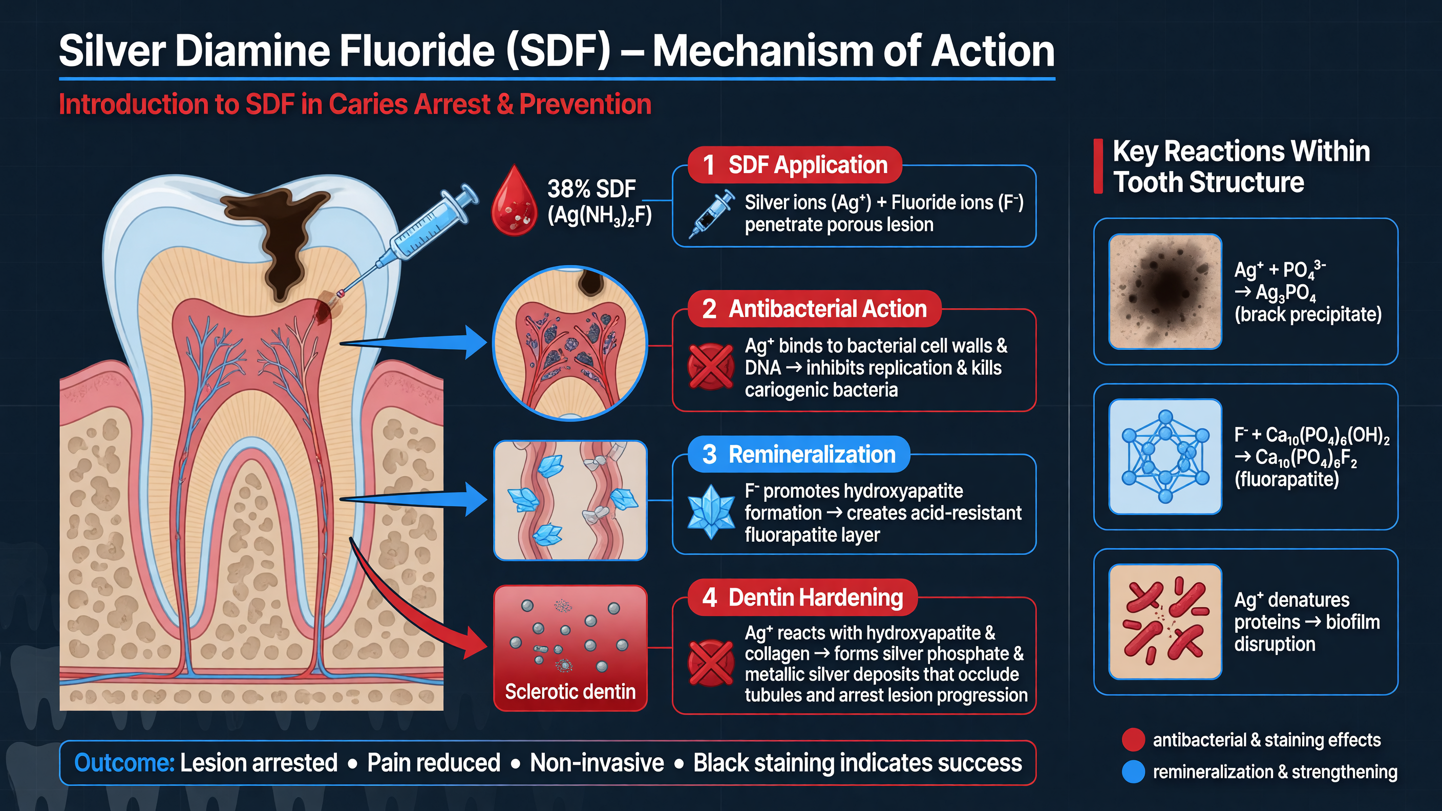 Silver diamine fluoride is best understood as a topical disease-control agent rather than a miniature filling. In the United States, commercially available SDF is generally a 38% weight/volume alkaline solution of diamine-silver fluoride, Ag(NH₃)₂F. It contains approximately 24%–29% silver, 5%–6% fluoride—about 44,800 parts per million fluoride—and 8% ammonia, with a pH commonly between 10 and 13. Ammonia stabilizes the silver-fluoride complex. The US Food and Drug Administration has cleared SDF as a Class II device for dentinal hypersensitivity; using it to arrest caries is an accepted but off-label application requiring diagnosis, informed consent, and longitudinal monitoring.
**Teaching Point:** SDF does not simply “kill the cavity.” It shifts an active lesion toward inactivity through three interacting effects: antimicrobial activity, preservation of the dentin matrix, and promotion of mineral deposition.
Silver ions act at multiple microbial targets. They disrupt bacterial cell membranes, bind sulfhydryl groups in metabolic enzymes, interfere with electron transport, denature proteins, and interact with nucleic acids. This broad activity reduces the viability and metabolic output of acidogenic organisms within the treated lesion. The ecological effect is not sterilization: viable organisms and a cariogenic oral environment may remain. The clinical objective is to suppress lesion-level activity while simultaneously changing the behavioral and environmental drivers of disease.
Fluoride contributes differently. In the presence of calcium and phosphate, it supports mineral redeposition and creates fluoride-rich reservoirs that can release fluoride during subsequent acid challenges. Laboratory studies demonstrate increased mineral density and reduced demineralization in SDF-treated dentin. Precipitates involving calcium fluoride-like material, silver phosphate, silver oxide, and other silver compounds can reduce dentin permeability and occlude tubules, explaining both desensitization and the hardening observed in arrested lesions. Fluoride should therefore be viewed as promoting a more acid-resistant lesion surface, not rebuilding missing tooth anatomy.
SDF also protects dentin’s organic scaffold. Caries progression involves not only dissolution of hydroxyapatite but degradation of exposed collagen by host and microbial proteases, including matrix metalloproteinases and cysteine cathepsins. Silver and fluoride inhibit several collagen-degrading enzymes. Preserved collagen provides a framework into which mineral can redeposit. The result is a surface layer that is harder, less permeable, and more resistant to further enzymatic breakdown.
**Nuance:** The characteristic black color is chemically related to the treatment but is not, by itself, proof of arrest. Silver compounds darken as they precipitate or are reduced within porous demineralized tissue. A successfully arrested dentin lesion is generally dark and hard on gentle assessment, with no clinical or radiographic progression. A black but soft lesion remains active or inadequately controlled.
Sound, mature enamel usually does not turn black because it lacks the porosity needed for substantial silver penetration. Demineralized enamel, developmental defects, open margins, exposed dentin, and active white-spot lesions may stain. SDF therefore must be confined to the intended lesion rather than painted indiscriminately across an esthetic surface.
**Framework:** Think of SDF as “lesion control plus time.” It can stabilize disease while a child develops coping skills, medical status improves, definitive treatment is scheduled, additional teeth erupt, or access barriers are addressed. In selected primary teeth or root surfaces, repeated SDF may remain the definitive nonrestorative strategy. In structurally compromised teeth, however, arrest does not restore contour, contact, cleansability, or fracture resistance.
Emerging laboratory work continues to explore ways to enhance fluoride uptake. Elsegaey and colleagues found greater fluoride retention and dentinal-tubule occlusion when SDF-treated dentin was subsequently irradiated with selected diode or frequency-doubled Nd:YAG laser parameters. This was an in vitro experiment, not evidence that lasers improve patient-centered caries outcomes; thermal safety and clinical effectiveness remain unestablished ([PMID: 41894090](https://pubmed.ncbi.nlm.nih.gov/41894090/)).
**MUST ACT:** Never let a biologically plausible mechanism substitute for diagnosis. SDF cannot treat irreversible pulpitis, pulp necrosis, abscess, cellulitis, or a tooth that is unrestorable. Spontaneous or lingering pain, swelling, sinus tract, pathologic mobility, percussion tenderness, pulp exposure, or furcation/periapical pathology requires a pulpal, endodontic, surgical, or extraction pathway.
**Audience Poll:** When you see a black SDF-treated lesion at recall, what additional finding do you require before documenting arrest—hardness, symptom resolution, radiographic stability, or all three?
---
## Current Clinical Evidence and Guidelines for SDF Use
Silver diamine fluoride is best understood as a topical disease-control agent rather than a miniature filling. In the United States, commercially available SDF is generally a 38% weight/volume alkaline solution of diamine-silver fluoride, Ag(NH₃)₂F. It contains approximately 24%–29% silver, 5%–6% fluoride—about 44,800 parts per million fluoride—and 8% ammonia, with a pH commonly between 10 and 13. Ammonia stabilizes the silver-fluoride complex. The US Food and Drug Administration has cleared SDF as a Class II device for dentinal hypersensitivity; using it to arrest caries is an accepted but off-label application requiring diagnosis, informed consent, and longitudinal monitoring.
**Teaching Point:** SDF does not simply “kill the cavity.” It shifts an active lesion toward inactivity through three interacting effects: antimicrobial activity, preservation of the dentin matrix, and promotion of mineral deposition.
Silver ions act at multiple microbial targets. They disrupt bacterial cell membranes, bind sulfhydryl groups in metabolic enzymes, interfere with electron transport, denature proteins, and interact with nucleic acids. This broad activity reduces the viability and metabolic output of acidogenic organisms within the treated lesion. The ecological effect is not sterilization: viable organisms and a cariogenic oral environment may remain. The clinical objective is to suppress lesion-level activity while simultaneously changing the behavioral and environmental drivers of disease.
Fluoride contributes differently. In the presence of calcium and phosphate, it supports mineral redeposition and creates fluoride-rich reservoirs that can release fluoride during subsequent acid challenges. Laboratory studies demonstrate increased mineral density and reduced demineralization in SDF-treated dentin. Precipitates involving calcium fluoride-like material, silver phosphate, silver oxide, and other silver compounds can reduce dentin permeability and occlude tubules, explaining both desensitization and the hardening observed in arrested lesions. Fluoride should therefore be viewed as promoting a more acid-resistant lesion surface, not rebuilding missing tooth anatomy.
SDF also protects dentin’s organic scaffold. Caries progression involves not only dissolution of hydroxyapatite but degradation of exposed collagen by host and microbial proteases, including matrix metalloproteinases and cysteine cathepsins. Silver and fluoride inhibit several collagen-degrading enzymes. Preserved collagen provides a framework into which mineral can redeposit. The result is a surface layer that is harder, less permeable, and more resistant to further enzymatic breakdown.
**Nuance:** The characteristic black color is chemically related to the treatment but is not, by itself, proof of arrest. Silver compounds darken as they precipitate or are reduced within porous demineralized tissue. A successfully arrested dentin lesion is generally dark and hard on gentle assessment, with no clinical or radiographic progression. A black but soft lesion remains active or inadequately controlled.
Sound, mature enamel usually does not turn black because it lacks the porosity needed for substantial silver penetration. Demineralized enamel, developmental defects, open margins, exposed dentin, and active white-spot lesions may stain. SDF therefore must be confined to the intended lesion rather than painted indiscriminately across an esthetic surface.
**Framework:** Think of SDF as “lesion control plus time.” It can stabilize disease while a child develops coping skills, medical status improves, definitive treatment is scheduled, additional teeth erupt, or access barriers are addressed. In selected primary teeth or root surfaces, repeated SDF may remain the definitive nonrestorative strategy. In structurally compromised teeth, however, arrest does not restore contour, contact, cleansability, or fracture resistance.
Emerging laboratory work continues to explore ways to enhance fluoride uptake. Elsegaey and colleagues found greater fluoride retention and dentinal-tubule occlusion when SDF-treated dentin was subsequently irradiated with selected diode or frequency-doubled Nd:YAG laser parameters. This was an in vitro experiment, not evidence that lasers improve patient-centered caries outcomes; thermal safety and clinical effectiveness remain unestablished ([PMID: 41894090](https://pubmed.ncbi.nlm.nih.gov/41894090/)).
**MUST ACT:** Never let a biologically plausible mechanism substitute for diagnosis. SDF cannot treat irreversible pulpitis, pulp necrosis, abscess, cellulitis, or a tooth that is unrestorable. Spontaneous or lingering pain, swelling, sinus tract, pathologic mobility, percussion tenderness, pulp exposure, or furcation/periapical pathology requires a pulpal, endodontic, surgical, or extraction pathway.
**Audience Poll:** When you see a black SDF-treated lesion at recall, what additional finding do you require before documenting arrest—hardness, symptom resolution, radiographic stability, or all three?
---
## Current Clinical Evidence and Guidelines for SDF Use
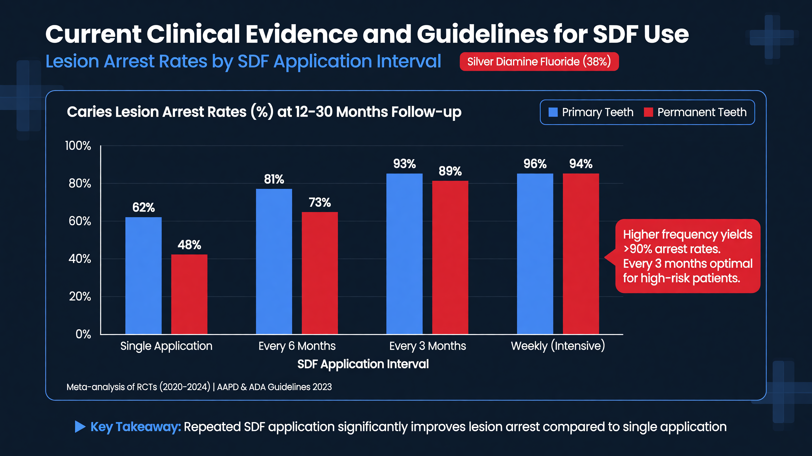 The evidence for SDF has an important apparent contradiction: many trials report clinically useful arrest, yet systematic reviewers continue to rate much of the evidence as low or very low certainty. Both statements can be true. Dark staining makes operator and examiner blinding difficult; studies use different concentrations, application schedules, surfaces, co-interventions, and definitions of arrest; lesions are clustered within participants; and several trials are small or conducted at a single center.
The 2017 American Academy of Pediatric Dentistry guideline conditionally recommended 38% SDF for arresting cavitated lesions in primary teeth as part of comprehensive caries management. Its pooled estimate suggested that approximately 68% of treated lesions would be arrested at two years, but the confidence interval was extremely wide and the supporting evidence was judged low quality. The 2018 American Dental Association guideline prioritized biannual 38% SDF for advanced cavitated coronal lesions in primary teeth and extrapolated that option to permanent teeth when nonrestorative management is appropriate. Neither guideline makes SDF a universal replacement for restorative treatment.
**Teaching Point:** The most defensible claim is that 38% SDF can arrest a meaningful proportion of accessible, asymptomatic cavitated dentin lesions. Claims about the single best interval, superiority to every restorative alternative, or durable success after one application are less certain.
The 2024 Cochrane review included 29 randomized trials and 13,036 participants, predominantly children but also older adults. Compared with placebo or no treatment, SDF may increase the number of arrested surfaces in the primary dentition, but certainty was low. Evidence for many outcomes in permanent coronal lesions was very uncertain. One comparatively stronger finding was prevention of new root caries: SDF reduced root-surface caries increment by a mean of approximately 0.79 surfaces versus placebo or no treatment, supported by moderate-certainty evidence. The review could not establish an optimal application schedule or confidently determine whether SDF, fluoride varnish, ART, or other comparators were superior across settings ([PMID: 39508296](https://pubmed.ncbi.nlm.nih.gov/39508296/)).
The Schroth open-label randomized trial provides useful but narrower interval data. Eighty-four young children with early childhood caries contributed 505 ICDAS 5 or 6 lesions. All groups received two applications of 38% SDF plus 5% sodium fluoride varnish, with treatment visits separated by one, four, or six months. At the final assessment, arrest was reported in 98% of lesions in the one-month group, 95.8% in the four-month group, and 72% in the six-month group. This supports early reassessment and a second application for severe disease; it does not prove that every high-risk child requires indefinite monthly SDF. The trial was small, open-label, and included fluoride varnish as a co-intervention ([PMID: 39227897](https://pubmed.ncbi.nlm.nih.gov/39227897/)).
**Nuance:** A one-month and a four-month interval performed similarly in the Schroth trial. “Monthly is always best” is therefore an overinterpretation. Interval selection should incorporate lesion activity, surface accessibility, plaque stagnation, disease burden, reliability of follow-up, and the consequence of failure.
A 2026 randomized study of 45 children compared an SDF-based minimally invasive approach with papain-based chemomechanical excavation and ceramic-bur excavation before glass-ionomer restoration. SDF reduced the need for local anesthesia and was faster than the papain approach, although not faster than ceramic-bur excavation. Parent-reported adverse events—including discoloration within a composite outcome—were more common in the SDF group, and follow-up was only three months. The study supports feasibility, not long-term superiority ([PMID: 41542896](https://pubmed.ncbi.nlm.nih.gov/41542896/)).
**Decision Point:** Select SDF when the lesion is active, cavitated, accessible to the microbrush, and free of clinical or radiographic pulpal pathology—especially when conventional care is delayed, poorly tolerated, or disproportionately burdensome. Common candidates include young children with early childhood caries, patients with intellectual or physical disabilities, medically complex patients, frail older adults with root caries, people with xerostomia, and patients facing geographic or financial barriers.
Before treatment, distinguish active from inactive disease. Active dentin is often soft or leathery, matte, plaque-retentive, and located in a stagnation area. Arrested dentin is hard or firm, frequently shiny, and clinically stable. Obtain bitewing or periapical imaging when feasible for deep or proximal lesions. A lesion approaching the pulp can still be asymptomatic, but the closer it is to the pulp, the greater the need for careful testing, radiographic interpretation, and a contingency plan.
Arrest is generally more predictable on anterior and smooth surfaces than on plaque-retentive posterior or inaccessible proximal surfaces. SDF cannot work well where the clinician cannot deliver it or the patient cannot keep the site reasonably clean. Orthodontic separators may improve proximal access, but radiographic-only lesions with no brush access are poor targets.
**MUST ACT:** Reassess rather than “apply and forget.” Current AAPD chairside guidance permits follow-up between approximately two weeks and four months after the initial treatment. Document hardness, plaque retention, symptoms, new cavitation, and radiographic behavior. Reapply when the lesion remains soft or active, and consider a restorative seal or different treatment when repeated applications do not achieve control.
Safety experience is reassuring when small topical doses are used correctly. The expected adverse effects are black staining of carious tissue, transient metallic or bitter taste, temporary skin staining, and superficial mucosal irritation after accidental contact. Serious systemic events have not emerged as a common clinical signal. Avoid SDF in patients with a confirmed silver allergy and defer application over ulcerative gingivitis, stomatitis, or open mucosal lesions that prevent safe isolation.
**Framework:** Evidence strength should calibrate the promise. Tell patients that SDF often arrests lesions and may defer drilling, anesthesia, sedation, or extraction—not that it guarantees permanent arrest. Baghdadi’s 2026 evidence-calibration framework makes the same distinction: evidence that SDF has an effect is stronger than evidence defining its optimal implementation conditions ([PMID: 42194156](https://pubmed.ncbi.nlm.nih.gov/42194156/)).
**Audience Poll:** Which would make you change from repeat SDF to restoration: persistent softness, food impaction, loss of marginal ridge, unreliable follow-up, or any one of these?
---
## Technique and Application Protocols: Addressing Missteps
The evidence for SDF has an important apparent contradiction: many trials report clinically useful arrest, yet systematic reviewers continue to rate much of the evidence as low or very low certainty. Both statements can be true. Dark staining makes operator and examiner blinding difficult; studies use different concentrations, application schedules, surfaces, co-interventions, and definitions of arrest; lesions are clustered within participants; and several trials are small or conducted at a single center.
The 2017 American Academy of Pediatric Dentistry guideline conditionally recommended 38% SDF for arresting cavitated lesions in primary teeth as part of comprehensive caries management. Its pooled estimate suggested that approximately 68% of treated lesions would be arrested at two years, but the confidence interval was extremely wide and the supporting evidence was judged low quality. The 2018 American Dental Association guideline prioritized biannual 38% SDF for advanced cavitated coronal lesions in primary teeth and extrapolated that option to permanent teeth when nonrestorative management is appropriate. Neither guideline makes SDF a universal replacement for restorative treatment.
**Teaching Point:** The most defensible claim is that 38% SDF can arrest a meaningful proportion of accessible, asymptomatic cavitated dentin lesions. Claims about the single best interval, superiority to every restorative alternative, or durable success after one application are less certain.
The 2024 Cochrane review included 29 randomized trials and 13,036 participants, predominantly children but also older adults. Compared with placebo or no treatment, SDF may increase the number of arrested surfaces in the primary dentition, but certainty was low. Evidence for many outcomes in permanent coronal lesions was very uncertain. One comparatively stronger finding was prevention of new root caries: SDF reduced root-surface caries increment by a mean of approximately 0.79 surfaces versus placebo or no treatment, supported by moderate-certainty evidence. The review could not establish an optimal application schedule or confidently determine whether SDF, fluoride varnish, ART, or other comparators were superior across settings ([PMID: 39508296](https://pubmed.ncbi.nlm.nih.gov/39508296/)).
The Schroth open-label randomized trial provides useful but narrower interval data. Eighty-four young children with early childhood caries contributed 505 ICDAS 5 or 6 lesions. All groups received two applications of 38% SDF plus 5% sodium fluoride varnish, with treatment visits separated by one, four, or six months. At the final assessment, arrest was reported in 98% of lesions in the one-month group, 95.8% in the four-month group, and 72% in the six-month group. This supports early reassessment and a second application for severe disease; it does not prove that every high-risk child requires indefinite monthly SDF. The trial was small, open-label, and included fluoride varnish as a co-intervention ([PMID: 39227897](https://pubmed.ncbi.nlm.nih.gov/39227897/)).
**Nuance:** A one-month and a four-month interval performed similarly in the Schroth trial. “Monthly is always best” is therefore an overinterpretation. Interval selection should incorporate lesion activity, surface accessibility, plaque stagnation, disease burden, reliability of follow-up, and the consequence of failure.
A 2026 randomized study of 45 children compared an SDF-based minimally invasive approach with papain-based chemomechanical excavation and ceramic-bur excavation before glass-ionomer restoration. SDF reduced the need for local anesthesia and was faster than the papain approach, although not faster than ceramic-bur excavation. Parent-reported adverse events—including discoloration within a composite outcome—were more common in the SDF group, and follow-up was only three months. The study supports feasibility, not long-term superiority ([PMID: 41542896](https://pubmed.ncbi.nlm.nih.gov/41542896/)).
**Decision Point:** Select SDF when the lesion is active, cavitated, accessible to the microbrush, and free of clinical or radiographic pulpal pathology—especially when conventional care is delayed, poorly tolerated, or disproportionately burdensome. Common candidates include young children with early childhood caries, patients with intellectual or physical disabilities, medically complex patients, frail older adults with root caries, people with xerostomia, and patients facing geographic or financial barriers.
Before treatment, distinguish active from inactive disease. Active dentin is often soft or leathery, matte, plaque-retentive, and located in a stagnation area. Arrested dentin is hard or firm, frequently shiny, and clinically stable. Obtain bitewing or periapical imaging when feasible for deep or proximal lesions. A lesion approaching the pulp can still be asymptomatic, but the closer it is to the pulp, the greater the need for careful testing, radiographic interpretation, and a contingency plan.
Arrest is generally more predictable on anterior and smooth surfaces than on plaque-retentive posterior or inaccessible proximal surfaces. SDF cannot work well where the clinician cannot deliver it or the patient cannot keep the site reasonably clean. Orthodontic separators may improve proximal access, but radiographic-only lesions with no brush access are poor targets.
**MUST ACT:** Reassess rather than “apply and forget.” Current AAPD chairside guidance permits follow-up between approximately two weeks and four months after the initial treatment. Document hardness, plaque retention, symptoms, new cavitation, and radiographic behavior. Reapply when the lesion remains soft or active, and consider a restorative seal or different treatment when repeated applications do not achieve control.
Safety experience is reassuring when small topical doses are used correctly. The expected adverse effects are black staining of carious tissue, transient metallic or bitter taste, temporary skin staining, and superficial mucosal irritation after accidental contact. Serious systemic events have not emerged as a common clinical signal. Avoid SDF in patients with a confirmed silver allergy and defer application over ulcerative gingivitis, stomatitis, or open mucosal lesions that prevent safe isolation.
**Framework:** Evidence strength should calibrate the promise. Tell patients that SDF often arrests lesions and may defer drilling, anesthesia, sedation, or extraction—not that it guarantees permanent arrest. Baghdadi’s 2026 evidence-calibration framework makes the same distinction: evidence that SDF has an effect is stronger than evidence defining its optimal implementation conditions ([PMID: 42194156](https://pubmed.ncbi.nlm.nih.gov/42194156/)).
**Audience Poll:** Which would make you change from repeat SDF to restoration: persistent softness, food impaction, loss of marginal ridge, unreliable follow-up, or any one of these?
---
## Technique and Application Protocols: Addressing Missteps
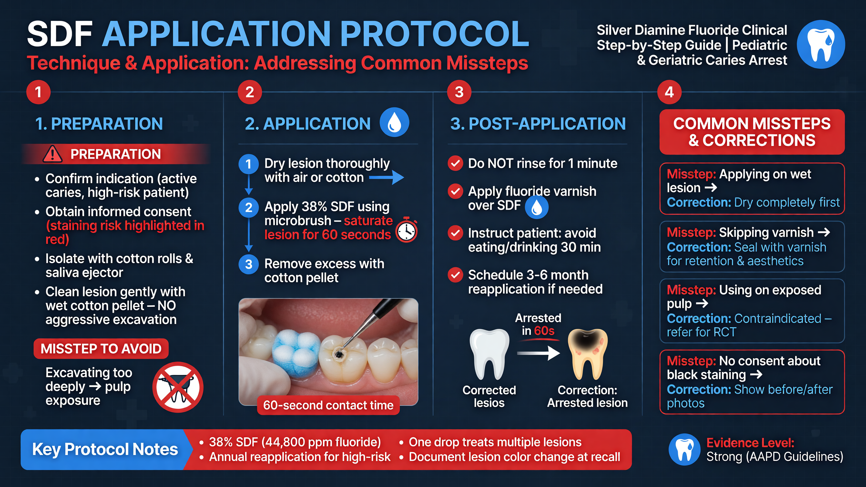 A successful SDF visit begins before the bottle is opened. Confirm the diagnosis, determine whether the tooth is restorable, assess symptoms and pulp status, obtain appropriate imaging, and record baseline lesion activity. Show the patient or caregiver representative photographs of expected staining. Consent should cover off-label caries arrest, permanent darkening of treated porous tissue, temporary soft-tissue staining, possible mucosal irritation, uncertain duration of arrest, likely reapplication, and alternatives such as fluoride varnish, sealant, restoration, crown, pulp therapy, extraction, or observation where appropriate.
**MUST ACT:** Do not place SDF simply because a patient cannot cooperate with drilling. A child with spontaneous nocturnal pain and a furcation radiolucency has a source-control problem, not an application-technique problem.
Use appropriate personal protective equipment and protect counters, clothing, and instruments. SDF can permanently stain fabrics and many clinical surfaces. Dispense only the amount needed into a disposable plastic well; do not work directly from the bottle. Keep the bottle away from light and follow its storage and expiration instructions. A bent microbrush allows controlled access while keeping the operator’s hand outside the direct line of the lips and tongue.
The current AAPD chairside guide estimates that one drop contains approximately 1.7 mg fluoride and 8.5 mg silver. Actual drop volume varies with the dispenser and product. Age, weight, total lesion burden, and the manufacturer’s instructions should determine how many drops are used at a visit. One carefully managed drop is often sufficient for several small lesions. Stage treatment rather than repeatedly dispensing solution without tracking the dose.
### A reproducible chairside sequence
1. **Prepare the field.** Apply petroleum jelly or another barrier to exposed lips and nearby skin. A thin gingival barrier may be useful, but do not coat the carious dentin; contamination prevents SDF contact. Isolate with cotton rolls, dry angles, suction, or a rubber dam when practical.
2. **Establish access.** Remove food, plaque, and gross debris with gauze, an excavator, or a toothbrush. Routine carious dentin excavation is unnecessary for arrest and may increase treatment burden. Limited excavation can be considered to improve cleansability, restorative form, or esthetics, provided pulpal risk is respected. Use a separator before a later appointment if a proximal lesion cannot be reached.
3. **Dry the lesion.** Gentle air drying improves control and helps the small volume remain on target. Complete desiccation is unnecessary. Maintain suction and prevent tongue contact.
4. **Control the liquid.** Dip the microbrush into the drop, then dab it against the side of the well so it is damp rather than dripping. Apply only to the intended dentin. Avoid sound enamel, gingiva, adjacent white-spot lesions, restorations, and exposed permanent anterior teeth.
5. **Maintain contact.** Keep SDF in contact with the entire lesion for at least one minute when patient safety and cooperation permit. Large posterior lesions require deliberate coverage of all accessible walls. A shorter application may be necessary in a very young or movement-prone patient; document the limitation and plan closer follow-up.
6. **Remove excess and dry.** Blot residual liquid with gauze, cotton, or a pellet to minimize ingestion and spread. Gently air dry while maintaining isolation. Follow the product instructions regarding rinsing and post-application care. Fluoride varnish may subsequently be applied to untreated teeth and noncavitated surfaces as part of the broader preventive plan.
**Teaching Point:** More liquid does not mean more arrest. Flooding the lesion increases metallic taste, mucosal exposure, ingestion, and unintended staining without establishing a dose-response benefit.
If SDF touches oral mucosa, remove it promptly, rinse or irrigate, and document the event. A superficial white or brown lesion may develop and usually resolves rapidly. Skin contact can produce a painless, henna-like stain that fades as epithelial cells exfoliate, often over several days. Eye exposure requires immediate copious irrigation and appropriate urgent assessment. Contaminated clothing may remain permanently stained.
**Nuance:** Potassium iodide applied after SDF can initially reduce darkening by forming a light silver-iodide precipitate, but the esthetic effect may diminish with light exposure and time. Studies have not established that it reliably prevents long-term staining, and uncertainty remains about whether it changes silver availability or arrest. Discuss it as a product- and protocol-specific option, not as a guaranteed cosmetic antidote.
At follow-up, gently evaluate lesion hardness with minimal force; avoid repeatedly gouging the surface. Compare photographs and radiographs when available. A hard, dark, asymptomatic, stable lesion can enter maintenance, commonly with six-month surveillance and reapplication according to activity and risk. If the lesion remains soft, first ask why: Was contact time inadequate? Was the surface inaccessible? Is plaque stagnating? Has the child’s sugar exposure remained frequent? Is the lesion deeper than initially recognized? Reapplication without addressing the cause may merely repeat the failure.
**Framework:** Separate technical failure from disease failure. A missed proximal wall, contaminated lesion, or insufficient contact is technical. New lesions elsewhere, persistent plaque, and continued frequent sugar exposure indicate uncontrolled disease. Both require correction, but only the first is solved at the microbrush.
Common missteps include treating an undiagnosed pulpal lesion, using SDF on intact noncavitated enamel when another fluoride therapy is better indicated, coding multiple surfaces on one tooth as separate D1354 units, promising that one application is definitive, and failing to document the black stain before a caregiver later interprets it as worsening decay.
**Decision Point:** If a lesion becomes hard but remains a food trap, the biologic objective has been achieved but the functional objective has not. Restore or seal the defect rather than continuing SDF solely because the dentin is arrested.
**Audience Poll:** Which technique error is most common in your setting—overloading the brush, inadequate isolation, poor proximal access, inadequate follow-up, or failure to assess the pulp?
---
## Integration with Restorative Approaches: The SMART Technique and Beyond
A successful SDF visit begins before the bottle is opened. Confirm the diagnosis, determine whether the tooth is restorable, assess symptoms and pulp status, obtain appropriate imaging, and record baseline lesion activity. Show the patient or caregiver representative photographs of expected staining. Consent should cover off-label caries arrest, permanent darkening of treated porous tissue, temporary soft-tissue staining, possible mucosal irritation, uncertain duration of arrest, likely reapplication, and alternatives such as fluoride varnish, sealant, restoration, crown, pulp therapy, extraction, or observation where appropriate.
**MUST ACT:** Do not place SDF simply because a patient cannot cooperate with drilling. A child with spontaneous nocturnal pain and a furcation radiolucency has a source-control problem, not an application-technique problem.
Use appropriate personal protective equipment and protect counters, clothing, and instruments. SDF can permanently stain fabrics and many clinical surfaces. Dispense only the amount needed into a disposable plastic well; do not work directly from the bottle. Keep the bottle away from light and follow its storage and expiration instructions. A bent microbrush allows controlled access while keeping the operator’s hand outside the direct line of the lips and tongue.
The current AAPD chairside guide estimates that one drop contains approximately 1.7 mg fluoride and 8.5 mg silver. Actual drop volume varies with the dispenser and product. Age, weight, total lesion burden, and the manufacturer’s instructions should determine how many drops are used at a visit. One carefully managed drop is often sufficient for several small lesions. Stage treatment rather than repeatedly dispensing solution without tracking the dose.
### A reproducible chairside sequence
1. **Prepare the field.** Apply petroleum jelly or another barrier to exposed lips and nearby skin. A thin gingival barrier may be useful, but do not coat the carious dentin; contamination prevents SDF contact. Isolate with cotton rolls, dry angles, suction, or a rubber dam when practical.
2. **Establish access.** Remove food, plaque, and gross debris with gauze, an excavator, or a toothbrush. Routine carious dentin excavation is unnecessary for arrest and may increase treatment burden. Limited excavation can be considered to improve cleansability, restorative form, or esthetics, provided pulpal risk is respected. Use a separator before a later appointment if a proximal lesion cannot be reached.
3. **Dry the lesion.** Gentle air drying improves control and helps the small volume remain on target. Complete desiccation is unnecessary. Maintain suction and prevent tongue contact.
4. **Control the liquid.** Dip the microbrush into the drop, then dab it against the side of the well so it is damp rather than dripping. Apply only to the intended dentin. Avoid sound enamel, gingiva, adjacent white-spot lesions, restorations, and exposed permanent anterior teeth.
5. **Maintain contact.** Keep SDF in contact with the entire lesion for at least one minute when patient safety and cooperation permit. Large posterior lesions require deliberate coverage of all accessible walls. A shorter application may be necessary in a very young or movement-prone patient; document the limitation and plan closer follow-up.
6. **Remove excess and dry.** Blot residual liquid with gauze, cotton, or a pellet to minimize ingestion and spread. Gently air dry while maintaining isolation. Follow the product instructions regarding rinsing and post-application care. Fluoride varnish may subsequently be applied to untreated teeth and noncavitated surfaces as part of the broader preventive plan.
**Teaching Point:** More liquid does not mean more arrest. Flooding the lesion increases metallic taste, mucosal exposure, ingestion, and unintended staining without establishing a dose-response benefit.
If SDF touches oral mucosa, remove it promptly, rinse or irrigate, and document the event. A superficial white or brown lesion may develop and usually resolves rapidly. Skin contact can produce a painless, henna-like stain that fades as epithelial cells exfoliate, often over several days. Eye exposure requires immediate copious irrigation and appropriate urgent assessment. Contaminated clothing may remain permanently stained.
**Nuance:** Potassium iodide applied after SDF can initially reduce darkening by forming a light silver-iodide precipitate, but the esthetic effect may diminish with light exposure and time. Studies have not established that it reliably prevents long-term staining, and uncertainty remains about whether it changes silver availability or arrest. Discuss it as a product- and protocol-specific option, not as a guaranteed cosmetic antidote.
At follow-up, gently evaluate lesion hardness with minimal force; avoid repeatedly gouging the surface. Compare photographs and radiographs when available. A hard, dark, asymptomatic, stable lesion can enter maintenance, commonly with six-month surveillance and reapplication according to activity and risk. If the lesion remains soft, first ask why: Was contact time inadequate? Was the surface inaccessible? Is plaque stagnating? Has the child’s sugar exposure remained frequent? Is the lesion deeper than initially recognized? Reapplication without addressing the cause may merely repeat the failure.
**Framework:** Separate technical failure from disease failure. A missed proximal wall, contaminated lesion, or insufficient contact is technical. New lesions elsewhere, persistent plaque, and continued frequent sugar exposure indicate uncontrolled disease. Both require correction, but only the first is solved at the microbrush.
Common missteps include treating an undiagnosed pulpal lesion, using SDF on intact noncavitated enamel when another fluoride therapy is better indicated, coding multiple surfaces on one tooth as separate D1354 units, promising that one application is definitive, and failing to document the black stain before a caregiver later interprets it as worsening decay.
**Decision Point:** If a lesion becomes hard but remains a food trap, the biologic objective has been achieved but the functional objective has not. Restore or seal the defect rather than continuing SDF solely because the dentin is arrested.
**Audience Poll:** Which technique error is most common in your setting—overloading the brush, inadequate isolation, poor proximal access, inadequate follow-up, or failure to assess the pulp?
---
## Integration with Restorative Approaches: The SMART Technique and Beyond
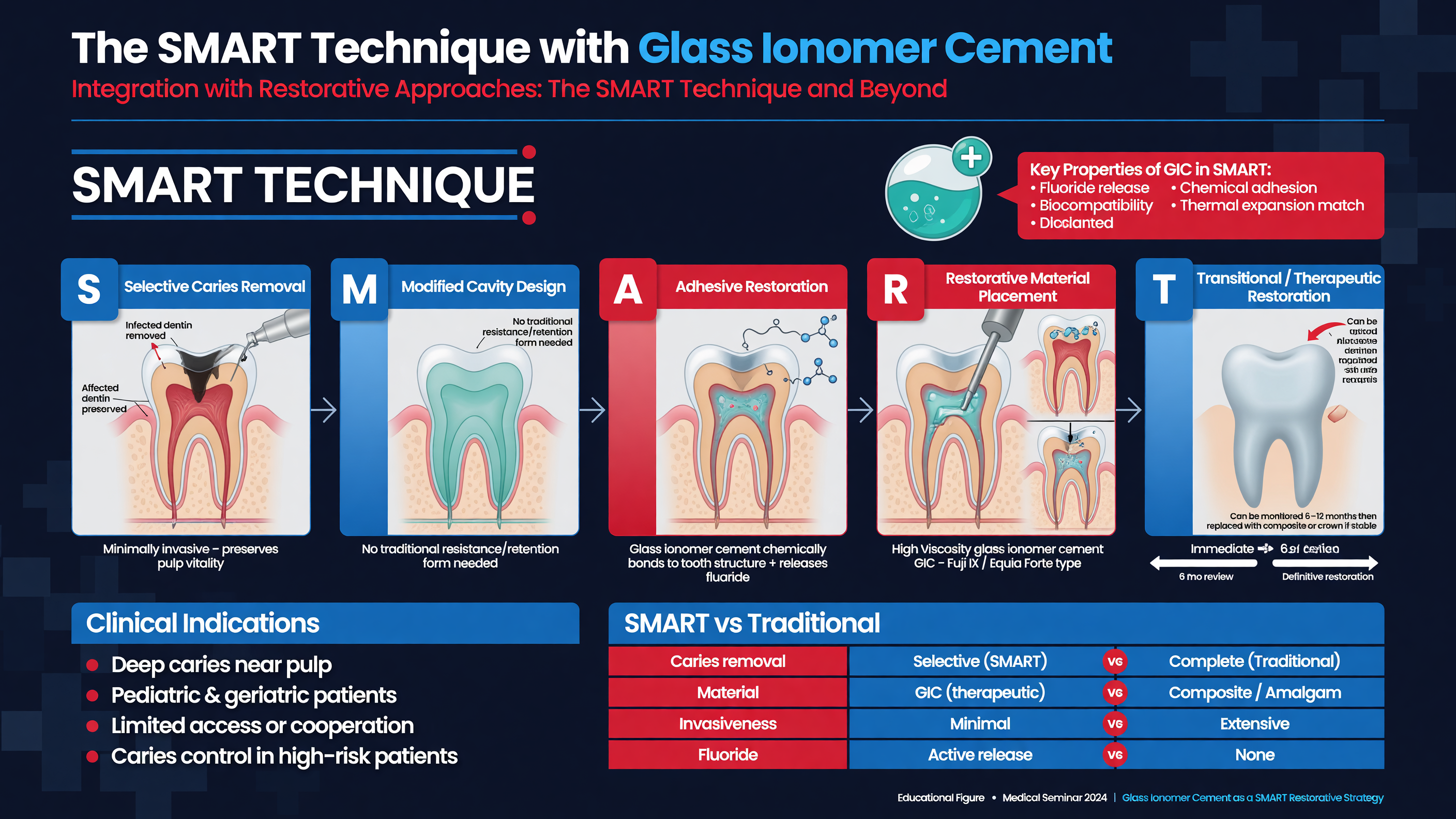 Silver-modified atraumatic restorative treatment, or SMART, combines SDF-mediated lesion control with a glass ionomer restoration. The concept is attractive because the components address different problems: SDF suppresses lesion activity and promotes hardening, while glass ionomer cement seals the lesion, restores contour, reduces food impaction, and partly masks discoloration. SMART should not be reduced to “SDF plus any filling,” nor should it be assumed superior to established restorative care in every cavity.
**Teaching Point:** Arrest and restoration are distinct endpoints. SDF can make diseased dentin inactive; glass ionomer can provide shape and a seal. Neither guarantees pulpal health or structural survival when case selection is poor.
SMART is particularly useful for accessible, asymptomatic single-surface lesions in primary molars; root-surface lesions requiring contour; patients who cannot tolerate rotary instrumentation; and lesions being stabilized during staged comprehensive care. It is less predictable when the marginal ridge is lost, occlusal load is high, the cavity cannot retain or support glass ionomer, moisture control is impossible, or radiographic and symptom findings suggest pulpal disease.
A practical sequence is to remove plaque and gross debris, selectively remove unsupported or heavily infected tissue when required for access and restoration form, apply a controlled amount of 38% SDF, blot excess, and manage the surface according to the selected glass-ionomer manufacturer’s instructions. High-viscosity glass ionomer is then placed, adapted, protected during maturation, and adjusted for occlusion. Some protocols place the restoration during the same visit; others arrest the lesion first and restore it after reassessment. Both models are used, and the evidence does not establish one universal sequence.
**Nuance:** Glass-ionomer conditioning, rinsing, and SDF timing vary among clinical studies. Aggressively generalizing a laboratory bonding protocol to every commercial material is unsafe. SDF does not appear to meaningfully impair conventional glass-ionomer bonding in aggregate evidence, but the operator should follow the restorative material’s instructions and avoid leaving a visibly pooled contaminant beneath the restoration.
SMART differs from conventional ART. ART generally involves removal of soft carious tissue with hand instruments followed by high-viscosity glass ionomer. SMART adds SDF, often allowing a more conservative approach to excavation. The distinction matters because the two approaches may differ in chair time, lesion biology, staining, and cost even when their restorations appear similar.
In the Aly randomized trial, 67 children aged five to nine years with asymptomatic single-surface primary-molar lesions received SMART or ART. At 12 months, mean restoration survival was comparable—11.8 months for SMART and 11.6 months for ART—with no significant survival difference. SMART took a mean of 7.8 minutes versus 15 minutes for ART and had lower calculated treatment costs. These findings support efficiency in a narrow case type; they do not establish superiority for multisurface lesions or long-term survival ([PMID: 36460236](https://pubmed.ncbi.nlm.nih.gov/36460236/)).
A 2026 split-mouth trial by Solh and colleagues included 32 children and 68 primary molars with ICDAS 4 or 5 lesions. Six-month clinical success was 70.5% for SMART and 67.6% for ART, a nonsignificant difference. Class I restorations performed better than Class II restorations, and wear and marginal-integrity loss were important failure modes. The correct interpretation is comparable short-term performance with a possible, unproven arrest advantage—not that SMART is categorically more effective than ART ([PMID: 41632901](https://pubmed.ncbi.nlm.nih.gov/41632901/)).
**Decision Point:** Choose the least invasive option that reliably achieves all necessary endpoints:
- Use **SDF alone** when arrest is the main goal and the cavity is cleansable, structurally acceptable, and esthetically acceptable.
- Use **SMART or an interim therapeutic restoration** when the lesion needs arrest plus sealing, contour, or improved cleansability.
- Use **ART** when hand excavation and glass ionomer can predictably restore the cavity without a specific need for SDF.
- Use a **preformed metal crown, conventional restoration, pulp therapy, or extraction** when structural loss, pulpal status, occlusion, or prognosis makes a topical strategy insufficient.
The Hall technique is an important alternative for suitable primary molars: a preformed metal crown seals the lesion without conventional caries removal. It may provide more durable full-coronal coverage than SMART in a heavily compromised molar, although it requires appropriate occlusal and radiographic selection. SDF can also serve as an interim stabilizer before a crown, but it should not delay definitive source control in a symptomatic tooth.
Resin-based restoration after SDF is possible, but the evidence is less straightforward than for glass ionomer. Residual silver precipitates and the subsequent adhesive protocol can influence bond testing. When resin is planned, remove pooled SDF, follow an evidence-informed rinsing and adhesive protocol, and ensure adequate enamel margins and isolation. Do not assume that black dentin at a margin represents recurrent caries; evaluate hardness, seal, symptoms, and radiographic progression.
**MUST ACT:** Restore function when function is compromised. A hard lesion beneath a fractured marginal ridge can still trap food, injure the periodontium, permit tooth migration, or fracture further. “Arrested” is not synonymous with “finished.”
Glass ionomer’s fluoride release and relative moisture tolerance are useful, but its limitations remain: early moisture sensitivity, desiccation, occlusal wear, marginal breakdown, and reduced survival in large multisurface preparations. Protect the restoration during initial setting and plan repair or replacement criteria. A failed restoration over arrested dentin may be repairable; a failed restoration over soft progressing dentin requires renewed disease assessment.
**Framework:** Use a staged treatment architecture: stabilize disease, verify arrest, restore form where needed, and maintain risk reduction. The stages may occur in one appointment or over months, but each should have a defined endpoint and escalation trigger.
**Audience Poll:** For a cooperative six-year-old with an asymptomatic two-surface primary molar lesion and a weakened marginal ridge, would you choose SMART, a Hall crown, conventional restoration, or extraction—and what finding would change your answer?
---
## Navigating Coding, Reimbursement, and Legacy Practice Models
Silver-modified atraumatic restorative treatment, or SMART, combines SDF-mediated lesion control with a glass ionomer restoration. The concept is attractive because the components address different problems: SDF suppresses lesion activity and promotes hardening, while glass ionomer cement seals the lesion, restores contour, reduces food impaction, and partly masks discoloration. SMART should not be reduced to “SDF plus any filling,” nor should it be assumed superior to established restorative care in every cavity.
**Teaching Point:** Arrest and restoration are distinct endpoints. SDF can make diseased dentin inactive; glass ionomer can provide shape and a seal. Neither guarantees pulpal health or structural survival when case selection is poor.
SMART is particularly useful for accessible, asymptomatic single-surface lesions in primary molars; root-surface lesions requiring contour; patients who cannot tolerate rotary instrumentation; and lesions being stabilized during staged comprehensive care. It is less predictable when the marginal ridge is lost, occlusal load is high, the cavity cannot retain or support glass ionomer, moisture control is impossible, or radiographic and symptom findings suggest pulpal disease.
A practical sequence is to remove plaque and gross debris, selectively remove unsupported or heavily infected tissue when required for access and restoration form, apply a controlled amount of 38% SDF, blot excess, and manage the surface according to the selected glass-ionomer manufacturer’s instructions. High-viscosity glass ionomer is then placed, adapted, protected during maturation, and adjusted for occlusion. Some protocols place the restoration during the same visit; others arrest the lesion first and restore it after reassessment. Both models are used, and the evidence does not establish one universal sequence.
**Nuance:** Glass-ionomer conditioning, rinsing, and SDF timing vary among clinical studies. Aggressively generalizing a laboratory bonding protocol to every commercial material is unsafe. SDF does not appear to meaningfully impair conventional glass-ionomer bonding in aggregate evidence, but the operator should follow the restorative material’s instructions and avoid leaving a visibly pooled contaminant beneath the restoration.
SMART differs from conventional ART. ART generally involves removal of soft carious tissue with hand instruments followed by high-viscosity glass ionomer. SMART adds SDF, often allowing a more conservative approach to excavation. The distinction matters because the two approaches may differ in chair time, lesion biology, staining, and cost even when their restorations appear similar.
In the Aly randomized trial, 67 children aged five to nine years with asymptomatic single-surface primary-molar lesions received SMART or ART. At 12 months, mean restoration survival was comparable—11.8 months for SMART and 11.6 months for ART—with no significant survival difference. SMART took a mean of 7.8 minutes versus 15 minutes for ART and had lower calculated treatment costs. These findings support efficiency in a narrow case type; they do not establish superiority for multisurface lesions or long-term survival ([PMID: 36460236](https://pubmed.ncbi.nlm.nih.gov/36460236/)).
A 2026 split-mouth trial by Solh and colleagues included 32 children and 68 primary molars with ICDAS 4 or 5 lesions. Six-month clinical success was 70.5% for SMART and 67.6% for ART, a nonsignificant difference. Class I restorations performed better than Class II restorations, and wear and marginal-integrity loss were important failure modes. The correct interpretation is comparable short-term performance with a possible, unproven arrest advantage—not that SMART is categorically more effective than ART ([PMID: 41632901](https://pubmed.ncbi.nlm.nih.gov/41632901/)).
**Decision Point:** Choose the least invasive option that reliably achieves all necessary endpoints:
- Use **SDF alone** when arrest is the main goal and the cavity is cleansable, structurally acceptable, and esthetically acceptable.
- Use **SMART or an interim therapeutic restoration** when the lesion needs arrest plus sealing, contour, or improved cleansability.
- Use **ART** when hand excavation and glass ionomer can predictably restore the cavity without a specific need for SDF.
- Use a **preformed metal crown, conventional restoration, pulp therapy, or extraction** when structural loss, pulpal status, occlusion, or prognosis makes a topical strategy insufficient.
The Hall technique is an important alternative for suitable primary molars: a preformed metal crown seals the lesion without conventional caries removal. It may provide more durable full-coronal coverage than SMART in a heavily compromised molar, although it requires appropriate occlusal and radiographic selection. SDF can also serve as an interim stabilizer before a crown, but it should not delay definitive source control in a symptomatic tooth.
Resin-based restoration after SDF is possible, but the evidence is less straightforward than for glass ionomer. Residual silver precipitates and the subsequent adhesive protocol can influence bond testing. When resin is planned, remove pooled SDF, follow an evidence-informed rinsing and adhesive protocol, and ensure adequate enamel margins and isolation. Do not assume that black dentin at a margin represents recurrent caries; evaluate hardness, seal, symptoms, and radiographic progression.
**MUST ACT:** Restore function when function is compromised. A hard lesion beneath a fractured marginal ridge can still trap food, injure the periodontium, permit tooth migration, or fracture further. “Arrested” is not synonymous with “finished.”
Glass ionomer’s fluoride release and relative moisture tolerance are useful, but its limitations remain: early moisture sensitivity, desiccation, occlusal wear, marginal breakdown, and reduced survival in large multisurface preparations. Protect the restoration during initial setting and plan repair or replacement criteria. A failed restoration over arrested dentin may be repairable; a failed restoration over soft progressing dentin requires renewed disease assessment.
**Framework:** Use a staged treatment architecture: stabilize disease, verify arrest, restore form where needed, and maintain risk reduction. The stages may occur in one appointment or over months, but each should have a defined endpoint and escalation trigger.
**Audience Poll:** For a cooperative six-year-old with an asymptomatic two-surface primary molar lesion and a weakened marginal ridge, would you choose SMART, a Hall crown, conventional restoration, or extraction—and what finding would change your answer?
---
## Navigating Coding, Reimbursement, and Legacy Practice Models
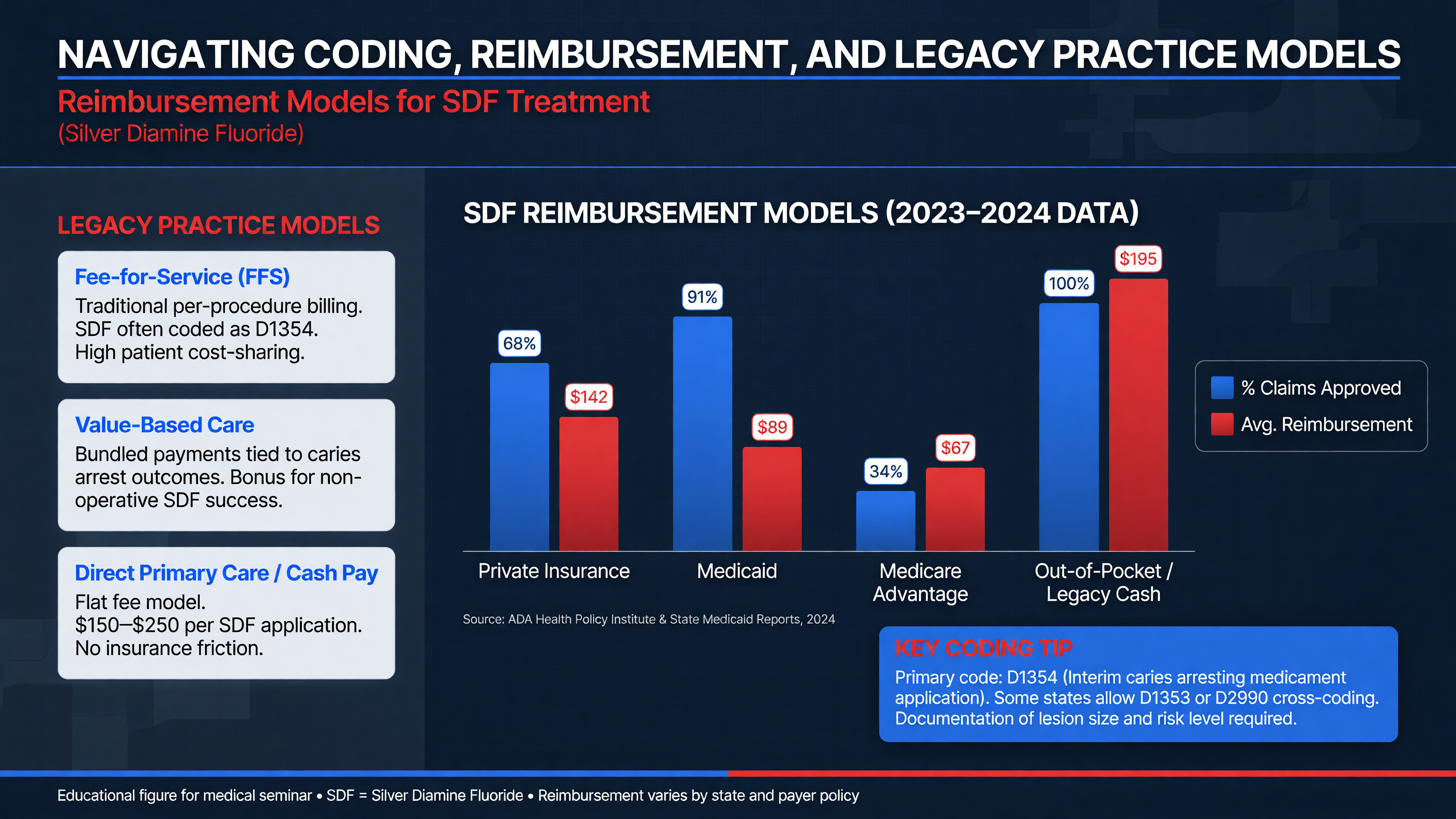 In the United States, the principal CDT code for caries arrest is D1354, “interim caries arresting medicament application—per tooth.” The descriptor refers to conservative topical treatment of an active, nonsymptomatic carious lesion without mechanical removal of sound tooth structure. The code is not limited to one brand or necessarily to SDF, although SDF is its most familiar use. It has been effective since 2016 and is reported by tooth, not by lesion or surface.
**MUST ACT:** Three lesions on one tooth remain one D1354 unit for that date of service. Three treated teeth generally support three units. Do not multiply units by the number of surfaces.
D1354 is not interchangeable with a preventive fluoride-varnish code. SDF applied to an active cavitated lesion is a disease-arrest procedure; fluoride varnish delivered to prevent new lesions or manage noncavitated disease represents a different service. Likewise, a glass-ionomer restoration, interim therapeutic restoration, crown, or extraction should be reported using the code that accurately describes the procedure performed. Code selection must follow the current CDT manual, payer contract, state rules, and the actual clinical service—not whichever code is most likely to pay.
A defensible record includes the tooth and surfaces, lesion activity and depth, absence of symptoms suggesting pulpal disease, relevant radiographic findings, caries-risk status, reason for choosing SDF, alternatives discussed, consent to permanent staining, product and concentration, lot and expiration information when required by policy, amount dispensed, isolation, application time, adverse events, and follow-up plan. At reassessment, document whether the lesion is hard, soft, stable, symptomatic, restored, or retreated.
**Nuance:** Coverage is not uniform. A payer may restrict D1354 by age, tooth, diagnosis, frequency, provider type, or number of lifetime applications. Some Medicaid programs cover it with explicit per-tooth frequency limits; some commercial plans exclude it, bundle it with another procedure, or require narrative documentation. Verify the current plan rather than relying on last year’s adjudication.
When SMART is completed, the claim may include D1354 and the appropriate restorative procedure if both services are distinct, documented, and permitted by the payer. Some contracts bundle SDF into the restoration or deny same-day combinations. A denial does not retroactively change what was performed, and it should not prompt inaccurate coding. Obtain a pretreatment estimate when feasible and explain that an estimate is not a guarantee of payment.
**Framework:** Build a standardized SDF pathway: diagnostic examination, consent, per-tooth application, short-interval reassessment, risk-control services, and restoration or maintenance. Templates and recall flags reduce missed reassessments and make outcomes auditable.
Traditional fee-for-service models reward completed restorations more visibly than disease stabilization. SDF challenges that legacy model because a brief, low-material-cost encounter may prevent or defer a more highly reimbursed procedure. The appropriate economic comparison is not “one drop versus one filling”; it includes repeat visits, caregiver time, sedation or general-anesthesia exposure, operating-room access, restoration repair, and the consequences of untreated disease.
The SMART-versus-ART trial found comparable 12-month performance but shorter treatment time and lower calculated cost for SMART in single-surface primary molars ([PMID: 36460236](https://pubmed.ncbi.nlm.nih.gov/36460236/)). That economic result should not be generalized to every payer or lesion. Practices should calculate local chair time, staffing, material costs, reapplication rates, failed-appointment risk, and reimbursement.
**Decision Point:** If D1354 is not covered, present the patient’s clinically appropriate options and transparent self-pay cost. Do not convert an active lesion into a “preventive” diagnosis or proceed directly to a more invasive covered service solely because the benefit design is misaligned.
Delegation rules also vary. The ADA supports delegation to appropriately trained allied personnel when permitted by state law and performed under required supervision after a dentist’s diagnosis and patient-specific plan. Confirm scope-of-practice requirements before building hygiene-led or community programs.
**Teaching Point:** The most sustainable model treats SDF as one step in accountable caries management. Track arrest, reapplication, restoration, emergency visits, and loss to follow-up. A high application count with no outcome data is production, not disease control.
**Audience Poll:** Does your current workflow capture lesion hardness and disposition after D1354, or only that SDF was applied?
---
## Patient Communication: Addressing Concerns and Enhancing Acceptance
In the United States, the principal CDT code for caries arrest is D1354, “interim caries arresting medicament application—per tooth.” The descriptor refers to conservative topical treatment of an active, nonsymptomatic carious lesion without mechanical removal of sound tooth structure. The code is not limited to one brand or necessarily to SDF, although SDF is its most familiar use. It has been effective since 2016 and is reported by tooth, not by lesion or surface.
**MUST ACT:** Three lesions on one tooth remain one D1354 unit for that date of service. Three treated teeth generally support three units. Do not multiply units by the number of surfaces.
D1354 is not interchangeable with a preventive fluoride-varnish code. SDF applied to an active cavitated lesion is a disease-arrest procedure; fluoride varnish delivered to prevent new lesions or manage noncavitated disease represents a different service. Likewise, a glass-ionomer restoration, interim therapeutic restoration, crown, or extraction should be reported using the code that accurately describes the procedure performed. Code selection must follow the current CDT manual, payer contract, state rules, and the actual clinical service—not whichever code is most likely to pay.
A defensible record includes the tooth and surfaces, lesion activity and depth, absence of symptoms suggesting pulpal disease, relevant radiographic findings, caries-risk status, reason for choosing SDF, alternatives discussed, consent to permanent staining, product and concentration, lot and expiration information when required by policy, amount dispensed, isolation, application time, adverse events, and follow-up plan. At reassessment, document whether the lesion is hard, soft, stable, symptomatic, restored, or retreated.
**Nuance:** Coverage is not uniform. A payer may restrict D1354 by age, tooth, diagnosis, frequency, provider type, or number of lifetime applications. Some Medicaid programs cover it with explicit per-tooth frequency limits; some commercial plans exclude it, bundle it with another procedure, or require narrative documentation. Verify the current plan rather than relying on last year’s adjudication.
When SMART is completed, the claim may include D1354 and the appropriate restorative procedure if both services are distinct, documented, and permitted by the payer. Some contracts bundle SDF into the restoration or deny same-day combinations. A denial does not retroactively change what was performed, and it should not prompt inaccurate coding. Obtain a pretreatment estimate when feasible and explain that an estimate is not a guarantee of payment.
**Framework:** Build a standardized SDF pathway: diagnostic examination, consent, per-tooth application, short-interval reassessment, risk-control services, and restoration or maintenance. Templates and recall flags reduce missed reassessments and make outcomes auditable.
Traditional fee-for-service models reward completed restorations more visibly than disease stabilization. SDF challenges that legacy model because a brief, low-material-cost encounter may prevent or defer a more highly reimbursed procedure. The appropriate economic comparison is not “one drop versus one filling”; it includes repeat visits, caregiver time, sedation or general-anesthesia exposure, operating-room access, restoration repair, and the consequences of untreated disease.
The SMART-versus-ART trial found comparable 12-month performance but shorter treatment time and lower calculated cost for SMART in single-surface primary molars ([PMID: 36460236](https://pubmed.ncbi.nlm.nih.gov/36460236/)). That economic result should not be generalized to every payer or lesion. Practices should calculate local chair time, staffing, material costs, reapplication rates, failed-appointment risk, and reimbursement.
**Decision Point:** If D1354 is not covered, present the patient’s clinically appropriate options and transparent self-pay cost. Do not convert an active lesion into a “preventive” diagnosis or proceed directly to a more invasive covered service solely because the benefit design is misaligned.
Delegation rules also vary. The ADA supports delegation to appropriately trained allied personnel when permitted by state law and performed under required supervision after a dentist’s diagnosis and patient-specific plan. Confirm scope-of-practice requirements before building hygiene-led or community programs.
**Teaching Point:** The most sustainable model treats SDF as one step in accountable caries management. Track arrest, reapplication, restoration, emergency visits, and loss to follow-up. A high application count with no outcome data is production, not disease control.
**Audience Poll:** Does your current workflow capture lesion hardness and disposition after D1354, or only that SDF was applied?
---
## Patient Communication: Addressing Concerns and Enhancing Acceptance
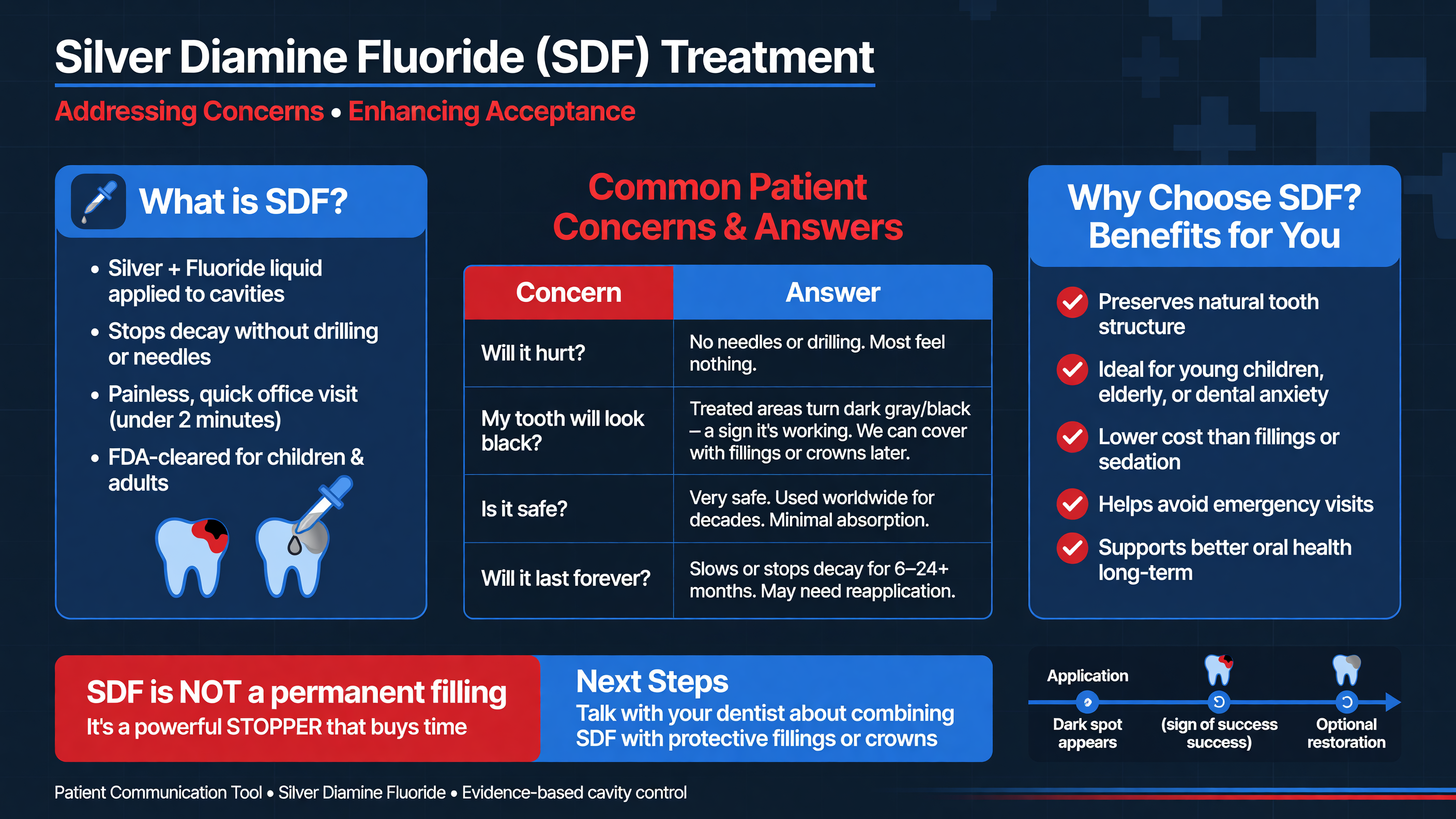 Acceptance improves when SDF is presented as a real clinical choice rather than a compromise imposed on families who cannot access “ideal” dentistry. Start with the diagnosis and treatment goal: “This area is active tooth decay. We can paint a medicine on the softened part to try to stop it from progressing. If it works, the decayed area becomes harder, but it also turns permanently dark.”
That explanation contains four essential facts: the lesion is active; SDF aims to arrest rather than remove it; clinical success requires follow-up; and blackening is expected. Avoid saying that SDF “heals,” “sterilizes,” or “fills” the cavity. It does not replace lost anatomy and may not eliminate the need for future restoration, crown, pulp treatment, or extraction.
**MUST ACT:** Show representative before-and-after photographs before consent, especially for anterior teeth. Verbal descriptions such as “some discoloration” routinely understate the appearance of arrested dentin.
Clarify the stain pattern. Porous diseased dentin usually becomes dark brown or black. Sound mature enamel generally remains unchanged, but demineralized enamel, cracks, developmental defects, and exposed dentin can stain. Gingiva or mucosa touched by SDF may develop a temporary white or brown area, and skin may show a temporary henna-like mark. Clothing and some surfaces may be permanently stained.
A balanced options discussion should cover anticipated burden as well as technical details. Alternatives may include fluoride varnish for appropriate noncavitated lesions, sealants, selective caries removal and restoration, SMART, ART, a Hall crown, conventional crown, pulp therapy, extraction, or monitored deferral. Discuss anesthesia, drilling, number of visits, behavior guidance, sedation, longevity, esthetics, cost, and consequences of doing nothing.
**Framework:** Use “choice talk, option talk, decision talk.” First signal that more than one reasonable pathway exists. Then compare benefits and harms in plain language. Finally, ask which outcome matters most to the patient: avoiding injections, minimizing visits, preserving appearance, restoring chewing, maximizing durability, or avoiding sedation.
Esthetic tolerance is contextual. Many caregivers accept posterior staining to avoid or defer sedation, while an anterior black lesion may be unacceptable even when SDF is biologically attractive. Some patients prefer the visible mark of arrested disease to ongoing destruction; others experience stigma or distress. Neither response is irrational. The clinician’s role is to identify the trade-off, not decide its value on the patient’s behalf.
**Audience Poll:** Would your recommendation change if the identical lesion were on a primary molar, a maxillary primary incisor, or an exposed root surface in an older adult?
Use teach-back rather than asking, “Do you understand?” A useful prompt is: “Please tell me what you expect the tooth to look like afterward and what follow-up it may still need.” If the caregiver says the tooth will return to its original color or never require treatment again, correct the misunderstanding before consent. Use qualified interpreters and translated materials rather than relying on a child or family member to translate a nuanced risk discussion.
**Nuance:** Potassium iodide may make the lesion look lighter immediately, but it cannot be promised to prevent long-term darkening. Restoring an arrested lesion with an opaque glass ionomer can improve appearance, although dark dentin may remain visible at thin or open margins. An esthetic plan should be described as camouflage or coverage, not reversal of the silver reaction.
The 2025 narrative review of fluoride therapies identified SDF’s high arrest potential and esthetic trade-off as a recurring acceptance issue while emphasizing the need to individualize fluoride strategies ([PMID: 40941599](https://pubmed.ncbi.nlm.nih.gov/40941599/)). Baghdadi’s 2026 viewpoint similarly argues for calibrated guidance: clinicians should be confident about SDF’s preservation role while remaining transparent about uncertainties in scheduling and implementation ([PMID: 42194156](https://pubmed.ncbi.nlm.nih.gov/42194156/)).
For an older adult with root caries, communication may focus less on black staining and more on dexterity, xerostomia, recurrent disease at crown margins, and whether the surface remains cleansable. For a medically complex child, families may prioritize avoiding general anesthesia. For an adolescent with an anterior lesion, esthetics may dominate. The same evidence therefore supports different reasonable decisions.
**Decision Point:** If a patient rejects staining after an informed discussion, do not minimize the concern or imply that refusal is irresponsible. Reassess whether a restorative seal, crown, alternative topical treatment, or referral can meet the biologic and personal goals more acceptably.
Document the selected option and the reasons that mattered to the patient. A robust consent note records permanent lesion darkening, possible temporary tissue staining, anticipated reapplication, uncertainty of arrest, alternatives, and the agreed escalation plan. Communication is successful when the patient can predict what happens next—not merely when a signature is obtained.
**Teaching Point:** A useful closing statement is: “We will know this is working when the area is hard and stable, not just because it is black. We will check it again, and if it remains soft, painful, or structurally weak, we will change the plan.”
---
## Evidence-Based Clinical Case
### Staged Management of Severe Caries in a Six-Year-Old
A six-year-old child presents with multiple cavitated primary-tooth lesions, frequent grazing on sweetened snacks, brushing once daily without consistent adult assistance, and no established dental home. Previous attempts at operative care ended when the child became distressed during local-anesthetic administration. The caregiver’s priority is to relieve risk without general anesthesia but is concerned about visible staining.
Examination identifies an accessible active occlusal dentin lesion on the mandibular left second primary molar, a buccal lesion on the contralateral molar, and a dark proximal lesion on a maxillary primary incisor. The molar lesions are soft, matte, and plaque-retentive. Neither molar has spontaneous pain, percussion tenderness, swelling, sinus tract, or pathologic mobility. Bitewings show dentin involvement without furcation pathology. A separate first primary molar has a history of spontaneous nocturnal pain and a furcation radiolucency.
**MUST ACT:** Remove the symptomatic first primary molar from the SDF pathway. Its differential includes irreversible pulpitis and necrosis with radicular disease; it requires urgent pulp therapy or extraction based on restorability and resorption. Painting SDF on that lesion would delay source control.
The asymptomatic molars are reasonable SDF candidates. The anterior lesion requires a separate discussion because esthetic consequences differ. After viewing photographs, the caregiver accepts black staining on the posterior teeth but prefers definitive esthetic management for the incisor. The team documents tooth-specific choices rather than treating every lesion with the same modality.
One measured drop of 38% SDF is dispensed, with the total dose kept within the product instructions and current pediatric guidance. The posterior lesions are cleaned of debris, isolated with cotton rolls and suction, dried, and treated using a blotted microbrush for approximately one minute each. Excess is removed before isolation is released. Five-percent sodium fluoride varnish is applied to untreated at-risk surfaces. The family receives a written plan addressing twice-daily brushing with fluoridated toothpaste, parent-assisted nighttime brushing, elimination of bedtime sweetened drinks, reduced frequency of free-sugar exposures, and a four-week reassessment.
At four weeks, the buccal lesion is black and hard. The occlusal lesion is darker but remains leathery at its deepest accessible point. Rather than declaring failure based on color, the clinician identifies persistent plaque retention, reinforces brushing, and reapplies SDF. The tooth remains asymptomatic. The family now accepts a SMART restoration because the occlusal cavity traps food.
At the subsequent visit, the lesion is hard. Unsupported tissue is conservatively removed to improve form without approaching the pulp. SDF is reapplied in a controlled manner, excess is managed, and high-viscosity glass ionomer is placed according to the material instructions. Occlusion and marginal adaptation are checked. The buccal lesion remains under nonrestorative surveillance because it is hard, cleansable, and does not compromise contour.
**Decision Point:** The glass-ionomer restoration is not placed because SDF “failed.” It is added because the biologic goal of arrest has been achieved while the functional problem of food retention remains.
At six months, both posterior lesions are asymptomatic and hard; the SMART restoration has acceptable marginal integrity. No new cavitated lesions are present. The child tolerates the visit and is gradually introduced to conventional procedures. The symptomatic molar has received definitive treatment, and the anterior tooth has been restored after a separate esthetic discussion.
This case illustrates four principles: SDF follows pulpal diagnosis; choices can be tooth-specific; hardness and stability—not color alone—define arrest; and successful disease stabilization may create the conditions for later restorative care. It also reflects the evidence that early reassessment and repeat application can be useful in severe early childhood caries while acknowledging that the optimal schedule remains patient- and lesion-dependent ([PMID: 39227897](https://pubmed.ncbi.nlm.nih.gov/39227897/); [PMID: 39508296](https://pubmed.ncbi.nlm.nih.gov/39508296/)).
---
## Tonight on Shift
1. **Exclude pulpal disease.** Before SDF, ask about spontaneous, nocturnal, and lingering pain; examine for swelling, sinus tract, mobility, and percussion tenderness; and obtain appropriate radiographs when feasible.
2. **Select an accessible lesion.** Treat active cavitated dentin that a microbrush can reach. Do not expect SDF to arrest a sealed-off proximal lesion, restore a marginal ridge, or stabilize an unrestorable tooth.
3. **Consent for the real outcome.** Show photographs, state that diseased tissue will permanently darken, explain temporary soft-tissue staining, and discuss retreatment and restorative alternatives.
4. **Apply a controlled dose.** Dispense and track the minimum number of drops, isolate, remove debris, dry, blot the microbrush, maintain contact for about one minute when safe, and remove excess.
5. **Reassess activity, not color.** Schedule review within roughly two weeks to four months according to risk. Document hardness, symptoms, plaque, radiographic stability, and whether to reapply, restore, or escalate.
6. **Complete the disease-control plan.** Pair SDF with fluoridated toothpaste, fluoride varnish when indicated, dietary change, caregiver-supported hygiene, risk-based recall, accurate D1354 coding per tooth, and a defined pathway to SMART or definitive care.
Acceptance improves when SDF is presented as a real clinical choice rather than a compromise imposed on families who cannot access “ideal” dentistry. Start with the diagnosis and treatment goal: “This area is active tooth decay. We can paint a medicine on the softened part to try to stop it from progressing. If it works, the decayed area becomes harder, but it also turns permanently dark.”
That explanation contains four essential facts: the lesion is active; SDF aims to arrest rather than remove it; clinical success requires follow-up; and blackening is expected. Avoid saying that SDF “heals,” “sterilizes,” or “fills” the cavity. It does not replace lost anatomy and may not eliminate the need for future restoration, crown, pulp treatment, or extraction.
**MUST ACT:** Show representative before-and-after photographs before consent, especially for anterior teeth. Verbal descriptions such as “some discoloration” routinely understate the appearance of arrested dentin.
Clarify the stain pattern. Porous diseased dentin usually becomes dark brown or black. Sound mature enamel generally remains unchanged, but demineralized enamel, cracks, developmental defects, and exposed dentin can stain. Gingiva or mucosa touched by SDF may develop a temporary white or brown area, and skin may show a temporary henna-like mark. Clothing and some surfaces may be permanently stained.
A balanced options discussion should cover anticipated burden as well as technical details. Alternatives may include fluoride varnish for appropriate noncavitated lesions, sealants, selective caries removal and restoration, SMART, ART, a Hall crown, conventional crown, pulp therapy, extraction, or monitored deferral. Discuss anesthesia, drilling, number of visits, behavior guidance, sedation, longevity, esthetics, cost, and consequences of doing nothing.
**Framework:** Use “choice talk, option talk, decision talk.” First signal that more than one reasonable pathway exists. Then compare benefits and harms in plain language. Finally, ask which outcome matters most to the patient: avoiding injections, minimizing visits, preserving appearance, restoring chewing, maximizing durability, or avoiding sedation.
Esthetic tolerance is contextual. Many caregivers accept posterior staining to avoid or defer sedation, while an anterior black lesion may be unacceptable even when SDF is biologically attractive. Some patients prefer the visible mark of arrested disease to ongoing destruction; others experience stigma or distress. Neither response is irrational. The clinician’s role is to identify the trade-off, not decide its value on the patient’s behalf.
**Audience Poll:** Would your recommendation change if the identical lesion were on a primary molar, a maxillary primary incisor, or an exposed root surface in an older adult?
Use teach-back rather than asking, “Do you understand?” A useful prompt is: “Please tell me what you expect the tooth to look like afterward and what follow-up it may still need.” If the caregiver says the tooth will return to its original color or never require treatment again, correct the misunderstanding before consent. Use qualified interpreters and translated materials rather than relying on a child or family member to translate a nuanced risk discussion.
**Nuance:** Potassium iodide may make the lesion look lighter immediately, but it cannot be promised to prevent long-term darkening. Restoring an arrested lesion with an opaque glass ionomer can improve appearance, although dark dentin may remain visible at thin or open margins. An esthetic plan should be described as camouflage or coverage, not reversal of the silver reaction.
The 2025 narrative review of fluoride therapies identified SDF’s high arrest potential and esthetic trade-off as a recurring acceptance issue while emphasizing the need to individualize fluoride strategies ([PMID: 40941599](https://pubmed.ncbi.nlm.nih.gov/40941599/)). Baghdadi’s 2026 viewpoint similarly argues for calibrated guidance: clinicians should be confident about SDF’s preservation role while remaining transparent about uncertainties in scheduling and implementation ([PMID: 42194156](https://pubmed.ncbi.nlm.nih.gov/42194156/)).
For an older adult with root caries, communication may focus less on black staining and more on dexterity, xerostomia, recurrent disease at crown margins, and whether the surface remains cleansable. For a medically complex child, families may prioritize avoiding general anesthesia. For an adolescent with an anterior lesion, esthetics may dominate. The same evidence therefore supports different reasonable decisions.
**Decision Point:** If a patient rejects staining after an informed discussion, do not minimize the concern or imply that refusal is irresponsible. Reassess whether a restorative seal, crown, alternative topical treatment, or referral can meet the biologic and personal goals more acceptably.
Document the selected option and the reasons that mattered to the patient. A robust consent note records permanent lesion darkening, possible temporary tissue staining, anticipated reapplication, uncertainty of arrest, alternatives, and the agreed escalation plan. Communication is successful when the patient can predict what happens next—not merely when a signature is obtained.
**Teaching Point:** A useful closing statement is: “We will know this is working when the area is hard and stable, not just because it is black. We will check it again, and if it remains soft, painful, or structurally weak, we will change the plan.”
---
## Evidence-Based Clinical Case
### Staged Management of Severe Caries in a Six-Year-Old
A six-year-old child presents with multiple cavitated primary-tooth lesions, frequent grazing on sweetened snacks, brushing once daily without consistent adult assistance, and no established dental home. Previous attempts at operative care ended when the child became distressed during local-anesthetic administration. The caregiver’s priority is to relieve risk without general anesthesia but is concerned about visible staining.
Examination identifies an accessible active occlusal dentin lesion on the mandibular left second primary molar, a buccal lesion on the contralateral molar, and a dark proximal lesion on a maxillary primary incisor. The molar lesions are soft, matte, and plaque-retentive. Neither molar has spontaneous pain, percussion tenderness, swelling, sinus tract, or pathologic mobility. Bitewings show dentin involvement without furcation pathology. A separate first primary molar has a history of spontaneous nocturnal pain and a furcation radiolucency.
**MUST ACT:** Remove the symptomatic first primary molar from the SDF pathway. Its differential includes irreversible pulpitis and necrosis with radicular disease; it requires urgent pulp therapy or extraction based on restorability and resorption. Painting SDF on that lesion would delay source control.
The asymptomatic molars are reasonable SDF candidates. The anterior lesion requires a separate discussion because esthetic consequences differ. After viewing photographs, the caregiver accepts black staining on the posterior teeth but prefers definitive esthetic management for the incisor. The team documents tooth-specific choices rather than treating every lesion with the same modality.
One measured drop of 38% SDF is dispensed, with the total dose kept within the product instructions and current pediatric guidance. The posterior lesions are cleaned of debris, isolated with cotton rolls and suction, dried, and treated using a blotted microbrush for approximately one minute each. Excess is removed before isolation is released. Five-percent sodium fluoride varnish is applied to untreated at-risk surfaces. The family receives a written plan addressing twice-daily brushing with fluoridated toothpaste, parent-assisted nighttime brushing, elimination of bedtime sweetened drinks, reduced frequency of free-sugar exposures, and a four-week reassessment.
At four weeks, the buccal lesion is black and hard. The occlusal lesion is darker but remains leathery at its deepest accessible point. Rather than declaring failure based on color, the clinician identifies persistent plaque retention, reinforces brushing, and reapplies SDF. The tooth remains asymptomatic. The family now accepts a SMART restoration because the occlusal cavity traps food.
At the subsequent visit, the lesion is hard. Unsupported tissue is conservatively removed to improve form without approaching the pulp. SDF is reapplied in a controlled manner, excess is managed, and high-viscosity glass ionomer is placed according to the material instructions. Occlusion and marginal adaptation are checked. The buccal lesion remains under nonrestorative surveillance because it is hard, cleansable, and does not compromise contour.
**Decision Point:** The glass-ionomer restoration is not placed because SDF “failed.” It is added because the biologic goal of arrest has been achieved while the functional problem of food retention remains.
At six months, both posterior lesions are asymptomatic and hard; the SMART restoration has acceptable marginal integrity. No new cavitated lesions are present. The child tolerates the visit and is gradually introduced to conventional procedures. The symptomatic molar has received definitive treatment, and the anterior tooth has been restored after a separate esthetic discussion.
This case illustrates four principles: SDF follows pulpal diagnosis; choices can be tooth-specific; hardness and stability—not color alone—define arrest; and successful disease stabilization may create the conditions for later restorative care. It also reflects the evidence that early reassessment and repeat application can be useful in severe early childhood caries while acknowledging that the optimal schedule remains patient- and lesion-dependent ([PMID: 39227897](https://pubmed.ncbi.nlm.nih.gov/39227897/); [PMID: 39508296](https://pubmed.ncbi.nlm.nih.gov/39508296/)).
---
## Tonight on Shift
1. **Exclude pulpal disease.** Before SDF, ask about spontaneous, nocturnal, and lingering pain; examine for swelling, sinus tract, mobility, and percussion tenderness; and obtain appropriate radiographs when feasible.
2. **Select an accessible lesion.** Treat active cavitated dentin that a microbrush can reach. Do not expect SDF to arrest a sealed-off proximal lesion, restore a marginal ridge, or stabilize an unrestorable tooth.
3. **Consent for the real outcome.** Show photographs, state that diseased tissue will permanently darken, explain temporary soft-tissue staining, and discuss retreatment and restorative alternatives.
4. **Apply a controlled dose.** Dispense and track the minimum number of drops, isolate, remove debris, dry, blot the microbrush, maintain contact for about one minute when safe, and remove excess.
5. **Reassess activity, not color.** Schedule review within roughly two weeks to four months according to risk. Document hardness, symptoms, plaque, radiographic stability, and whether to reapply, restore, or escalate.
6. **Complete the disease-control plan.** Pair SDF with fluoridated toothpaste, fluoride varnish when indicated, dietary change, caregiver-supported hygiene, risk-based recall, accurate D1354 coding per tooth, and a defined pathway to SMART or definitive care.