# Revolutionizing Point-of-Care: AI-Enhanced Ultrasound in Emergency Medicine
## Learning Objectives
1. **Recognize** the current applications of AI in point-of-care ultrasound across cardiac, lung, abdominal, and vascular domains
2. **Apply** AI-enhanced POCUS findings to clinical decision-making while maintaining appropriate skepticism
3. **Identify** automation bias traps and implement systematic safeguards in AI-assisted workflows
4. **Evaluate** the evidence from landmark trials comparing AI versus human sonographer performance
5. **Assess** the global health implications of AI-POCUS in resource-limited settings
---
## Section 1: Introduction — The AI Revolution in Medical Imaging
**Duration:** 10 min | **Content Tier: Teaching Point**
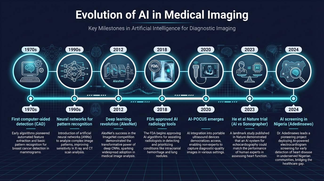 The integration of artificial intelligence into medical imaging represents one of the most transformative shifts in diagnostic medicine since the invention of ultrasound itself. To understand where we are heading with AI-enhanced POCUS, we need to understand where we've been.
**Teaching Point:** The journey from pattern recognition to clinical-grade AI has been decades in the making. The first computer-aided detection systems appeared in the 1970s, primarily for mammography screening. Neural networks showed promise in the 1990s but were limited by computational power. The deep learning revolution, sparked by AlexNet in 2012, changed everything — suddenly, machines could learn to identify patterns in medical images with accuracy approaching or exceeding human experts.
For emergency medicine, the critical inflection point came around 2020, when AI systems began to be applied specifically to point-of-care ultrasound. Unlike radiology AI, which processes stored images, POCUS-AI must work in real-time, at the bedside, often in chaotic resuscitation environments. This presents unique engineering challenges but also unique opportunities.
**Nuance:** The FDA has now approved over 800 AI/ML-enabled medical devices, with radiology comprising the largest category. However, point-of-care ultrasound AI remains in its early phases of regulatory approval. Most current AI-POCUS tools function as clinical decision support rather than autonomous diagnostic systems — an important distinction for clinical practice (Osterwalder et al., *Medicina*, 2023; PMID: 38138282).
**Audience Poll:** What is your current experience with AI in medical imaging?
- A) Never used any AI diagnostic tool
- B) Have seen AI radiology reports
- C) Have used AI-enabled ultrasound
- D) Regularly integrate AI into imaging workflow
---
## Section 2: AI in POCUS — From Basic Recognition to Advanced Interpretation
**Duration:** 12 min | **Content Tier: MUST ACT**
The integration of artificial intelligence into medical imaging represents one of the most transformative shifts in diagnostic medicine since the invention of ultrasound itself. To understand where we are heading with AI-enhanced POCUS, we need to understand where we've been.
**Teaching Point:** The journey from pattern recognition to clinical-grade AI has been decades in the making. The first computer-aided detection systems appeared in the 1970s, primarily for mammography screening. Neural networks showed promise in the 1990s but were limited by computational power. The deep learning revolution, sparked by AlexNet in 2012, changed everything — suddenly, machines could learn to identify patterns in medical images with accuracy approaching or exceeding human experts.
For emergency medicine, the critical inflection point came around 2020, when AI systems began to be applied specifically to point-of-care ultrasound. Unlike radiology AI, which processes stored images, POCUS-AI must work in real-time, at the bedside, often in chaotic resuscitation environments. This presents unique engineering challenges but also unique opportunities.
**Nuance:** The FDA has now approved over 800 AI/ML-enabled medical devices, with radiology comprising the largest category. However, point-of-care ultrasound AI remains in its early phases of regulatory approval. Most current AI-POCUS tools function as clinical decision support rather than autonomous diagnostic systems — an important distinction for clinical practice (Osterwalder et al., *Medicina*, 2023; PMID: 38138282).
**Audience Poll:** What is your current experience with AI in medical imaging?
- A) Never used any AI diagnostic tool
- B) Have seen AI radiology reports
- C) Have used AI-enabled ultrasound
- D) Regularly integrate AI into imaging workflow
---
## Section 2: AI in POCUS — From Basic Recognition to Advanced Interpretation
**Duration:** 12 min | **Content Tier: MUST ACT**
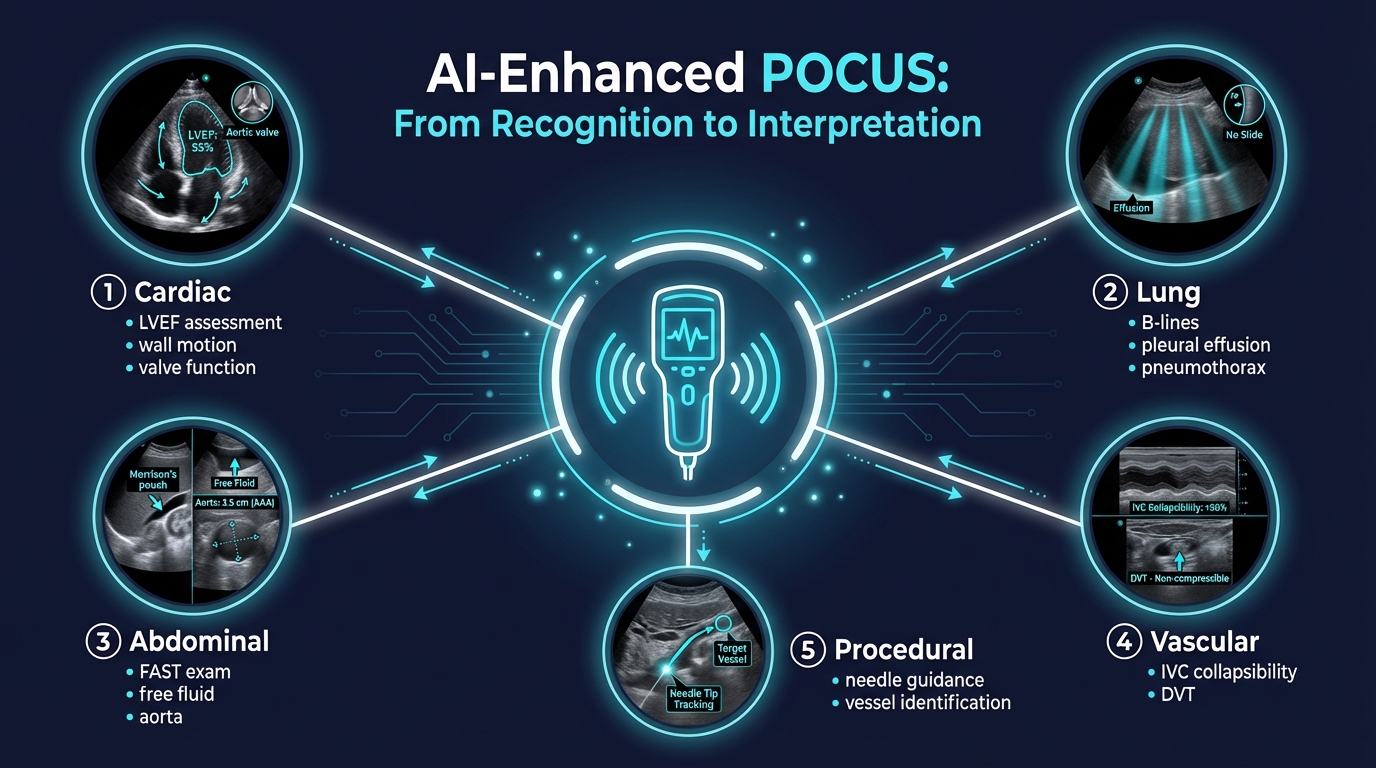 **MUST ACT:** Understand the five domains where AI is currently transforming bedside ultrasound — this will shape your practice within the next 5 years.
### Cardiac Applications
AI in cardiac POCUS has the most mature evidence base. Current systems can:
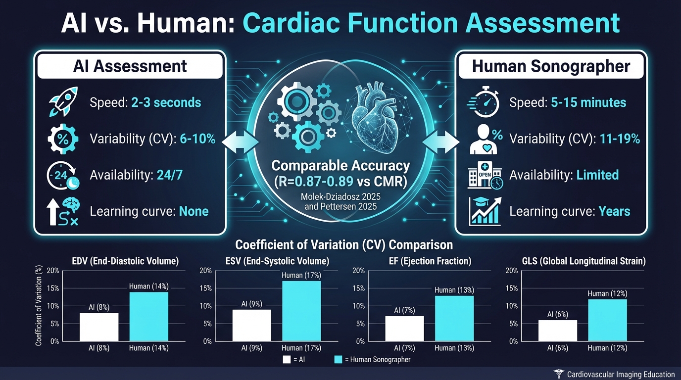
- **Automated LVEF calculation:** Deep learning models analyze 2D echocardiographic loops to calculate ejection fraction in 2-3 seconds, with accuracy comparable to expert sonographers (Mołek-Dziadosz et al., *Polish Archives of Internal Medicine*, 2025; PMID: 40888426). In their study of 118 patients, AI-based single-loop analysis achieved correlation with cardiac MRI of R=0.89 — numerically higher than expert echocardiographers (R=0.85-0.86).
- **Real-time image guidance:** Pettersen et al. demonstrated that deep learning-based guiding combined with automated measurements reduced the coefficient of variation for LV end-diastolic volume from 15% to 6% (p<0.001), and for global longitudinal strain from 11% to 7% (p=0.047) (*Open Heart*, 2025; PMID: 41360622).
- **Diastolic function assessment:** Chen et al. developed an AI system that classifies diastolic dysfunction with 90% accuracy and grades severity with 92% accuracy, processing multiple echo views simultaneously — a task that typically requires significant expertise (*JASE*, 2023; PMID: 37437669).
- **Strain imaging:** AI-derived global longitudinal strain (GLS) from ECG data correlates meaningfully with echo-derived GLS (AUROC 0.82 for detecting impaired GLS ≤12%), potentially offering a screening tool that requires no ultrasound at all (Choi et al., *Scientific Reports*, 2024; PMID: 39488646).
### Lung, Abdominal, and Vascular Applications
**Teaching Point:** Beyond cardiac assessment, AI-POCUS is expanding into:
- **Lung ultrasound:** Automated B-line counting for pulmonary edema quantification, pleural effusion detection, and pneumothorax identification
- **eFAST augmentation:** AI-enhanced free fluid detection with automated quantification
- **IVC assessment:** Real-time diameter tracking and automated collapsibility index calculation
- **Vascular access:** AI-guided needle trajectory and vessel identification
**Say Out Loud:** "The AI doesn't replace your clinical assessment — it adds a second opinion that happens to process images faster than any human can. Your job is to be the final arbiter."
**Audience Poll:** Which AI-POCUS application would be most useful in your clinical practice?
- A) Automated LVEF in undifferentiated shock
- B) AI-guided eFAST in trauma
- C) IVC collapsibility in sepsis resuscitation
- D) Lung ultrasound B-line quantification
---
## Section 3: The Evidence — AI vs. Human Performance
**Duration:** 12 min | **Content Tier: Nuance**
**MUST ACT:** Understand the five domains where AI is currently transforming bedside ultrasound — this will shape your practice within the next 5 years.
### Cardiac Applications
AI in cardiac POCUS has the most mature evidence base. Current systems can:
- **Automated LVEF calculation:** Deep learning models analyze 2D echocardiographic loops to calculate ejection fraction in 2-3 seconds, with accuracy comparable to expert sonographers (Mołek-Dziadosz et al., *Polish Archives of Internal Medicine*, 2025; PMID: 40888426). In their study of 118 patients, AI-based single-loop analysis achieved correlation with cardiac MRI of R=0.89 — numerically higher than expert echocardiographers (R=0.85-0.86).
- **Real-time image guidance:** Pettersen et al. demonstrated that deep learning-based guiding combined with automated measurements reduced the coefficient of variation for LV end-diastolic volume from 15% to 6% (p<0.001), and for global longitudinal strain from 11% to 7% (p=0.047) (*Open Heart*, 2025; PMID: 41360622).
- **Diastolic function assessment:** Chen et al. developed an AI system that classifies diastolic dysfunction with 90% accuracy and grades severity with 92% accuracy, processing multiple echo views simultaneously — a task that typically requires significant expertise (*JASE*, 2023; PMID: 37437669).
- **Strain imaging:** AI-derived global longitudinal strain (GLS) from ECG data correlates meaningfully with echo-derived GLS (AUROC 0.82 for detecting impaired GLS ≤12%), potentially offering a screening tool that requires no ultrasound at all (Choi et al., *Scientific Reports*, 2024; PMID: 39488646).
### Lung, Abdominal, and Vascular Applications
**Teaching Point:** Beyond cardiac assessment, AI-POCUS is expanding into:
- **Lung ultrasound:** Automated B-line counting for pulmonary edema quantification, pleural effusion detection, and pneumothorax identification
- **eFAST augmentation:** AI-enhanced free fluid detection with automated quantification
- **IVC assessment:** Real-time diameter tracking and automated collapsibility index calculation
- **Vascular access:** AI-guided needle trajectory and vessel identification
**Say Out Loud:** "The AI doesn't replace your clinical assessment — it adds a second opinion that happens to process images faster than any human can. Your job is to be the final arbiter."
**Audience Poll:** Which AI-POCUS application would be most useful in your clinical practice?
- A) Automated LVEF in undifferentiated shock
- B) AI-guided eFAST in trauma
- C) IVC collapsibility in sepsis resuscitation
- D) Lung ultrasound B-line quantification
---
## Section 3: The Evidence — AI vs. Human Performance
**Duration:** 12 min | **Content Tier: Nuance**
 ### The He et al. Nature Trial (2023) — The Landmark Study
**Nuance:** This is the study that changed the conversation. He et al. conducted a **blinded, randomized non-inferiority trial** — the first of its kind — comparing AI versus sonographer initial assessment of LVEF (PMID: 37020027).
**Key Details:**
- **Design:** 3,769 echocardiographic studies screened; 3,495 analyzed after excluding poor quality images
- **Primary endpoint:** Proportion of studies with >5% change between initial assessment and final cardiologist review
- **Results:** AI group had substantially fewer changes: **16.8% vs 27.2%** (difference -10.4%, 95% CI: -13.2% to -7.7%, p<0.001 for both non-inferiority AND superiority)
- **Blinding success:** Cardiologists could not distinguish AI from sonographer assessments (blinding index 0.088)
- **Time savings:** AI-guided workflow saved time for both sonographers and cardiologists
**Decision Point:** What does this mean for the ED? Consider two scenarios:
**Scenario A:** A busy overnight shift with 2 patients needing urgent cardiac assessment, one sonographer, and a 45-minute wait for formal echo. AI could provide immediate preliminary LVEF for clinical decision-making.
**Scenario B:** A community hospital without 24/7 sonographer coverage. AI-POCUS gives the emergency physician a real-time expert-level second opinion.
### The Mołek-Dziadosz Validation (2025)
The most recent validation study confirmed AI's accuracy against the gold standard — cardiac MRI. Both multiloop AI analysis (R=0.87, κ=0.68) and single-loop AI analysis (R=0.89, κ=0.75) showed strong agreement with CMR-assessed LVEF, comparable to expert echocardiographers (PMID: 40888426).
**Teaching Point:** The finding that single-loop AI analysis was numerically *better* than multiloop analysis is counterintuitive but clinically important — it suggests that AI may be better at extracting maximum information from a single high-quality view, which is exactly the clinical scenario in emergency POCUS where you often get one good window.
### The Pettersen Variability Study (2025)
**Nuance:** Perhaps even more important than absolute accuracy is *consistency*. Pettersen et al. showed that AI-assisted echocardiography reduced test-retest variability by 60% for LV volumes (CV: 6% vs 15%) (PMID: 41360622). In emergency medicine, where serial assessments guide resuscitation, this consistency could be transformative.
**Framework:**
| Metric | Standard Echo (CV%) | AI-Assisted Echo (CV%) | p-value |
|--------|-------------------|----------------------|---------|
| LV EDV | 15% | 6% | <0.001 |
| LV ESV | 19% | 10% | <0.001 |
| LV EF | 9% | 8% | 0.503 |
| GLS | 11% | 7% | 0.047 |
---
## Section 4: Automation Bias — The Critical Safety Concern
**Duration:** 10 min | **Content Tier: MUST ACT**
### The He et al. Nature Trial (2023) — The Landmark Study
**Nuance:** This is the study that changed the conversation. He et al. conducted a **blinded, randomized non-inferiority trial** — the first of its kind — comparing AI versus sonographer initial assessment of LVEF (PMID: 37020027).
**Key Details:**
- **Design:** 3,769 echocardiographic studies screened; 3,495 analyzed after excluding poor quality images
- **Primary endpoint:** Proportion of studies with >5% change between initial assessment and final cardiologist review
- **Results:** AI group had substantially fewer changes: **16.8% vs 27.2%** (difference -10.4%, 95% CI: -13.2% to -7.7%, p<0.001 for both non-inferiority AND superiority)
- **Blinding success:** Cardiologists could not distinguish AI from sonographer assessments (blinding index 0.088)
- **Time savings:** AI-guided workflow saved time for both sonographers and cardiologists
**Decision Point:** What does this mean for the ED? Consider two scenarios:
**Scenario A:** A busy overnight shift with 2 patients needing urgent cardiac assessment, one sonographer, and a 45-minute wait for formal echo. AI could provide immediate preliminary LVEF for clinical decision-making.
**Scenario B:** A community hospital without 24/7 sonographer coverage. AI-POCUS gives the emergency physician a real-time expert-level second opinion.
### The Mołek-Dziadosz Validation (2025)
The most recent validation study confirmed AI's accuracy against the gold standard — cardiac MRI. Both multiloop AI analysis (R=0.87, κ=0.68) and single-loop AI analysis (R=0.89, κ=0.75) showed strong agreement with CMR-assessed LVEF, comparable to expert echocardiographers (PMID: 40888426).
**Teaching Point:** The finding that single-loop AI analysis was numerically *better* than multiloop analysis is counterintuitive but clinically important — it suggests that AI may be better at extracting maximum information from a single high-quality view, which is exactly the clinical scenario in emergency POCUS where you often get one good window.
### The Pettersen Variability Study (2025)
**Nuance:** Perhaps even more important than absolute accuracy is *consistency*. Pettersen et al. showed that AI-assisted echocardiography reduced test-retest variability by 60% for LV volumes (CV: 6% vs 15%) (PMID: 41360622). In emergency medicine, where serial assessments guide resuscitation, this consistency could be transformative.
**Framework:**
| Metric | Standard Echo (CV%) | AI-Assisted Echo (CV%) | p-value |
|--------|-------------------|----------------------|---------|
| LV EDV | 15% | 6% | <0.001 |
| LV ESV | 19% | 10% | <0.001 |
| LV EF | 9% | 8% | 0.503 |
| GLS | 11% | 7% | 0.047 |
---
## Section 4: Automation Bias — The Critical Safety Concern
**Duration:** 10 min | **Content Tier: MUST ACT**
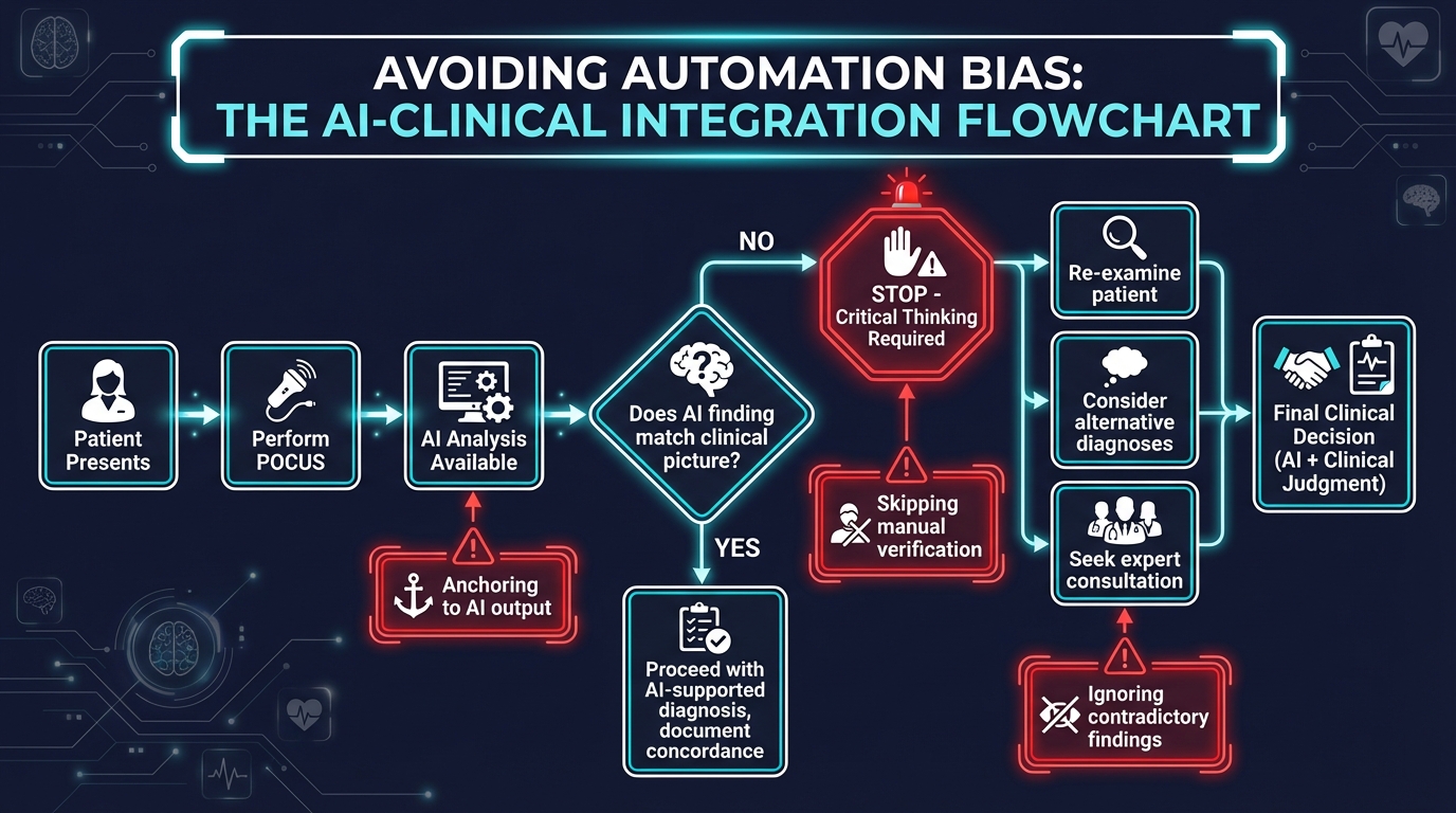 **MUST ACT:** Automation bias is the tendency to over-rely on automated systems, and it poses the single greatest safety risk in AI-assisted clinical practice. Every emergency physician who uses AI-POCUS must understand this concept.
### What Is Automation Bias?
Automation bias manifests in two dangerous patterns:
1. **Commission errors:** Acting on incorrect AI output because "the computer said so"
2. **Omission errors:** Failing to notice abnormalities because the AI didn't flag them
**Say Out Loud:** "I will always perform my own clinical assessment BEFORE looking at the AI output. The AI is my second opinion, not my first."
### The Three Safeguards
**Framework: The "AIM" Protocol for AI-Assisted POCUS**
1. **A — Assess independently first.** Form your own clinical impression from the ultrasound images before reviewing AI outputs. This prevents anchoring bias.
2. **I — Interrogate discrepancies.** When your assessment differs from AI, treat this as a critical safety signal. Neither you nor the AI is infallible — the discrepancy itself is valuable diagnostic information.
3. **M — Monitor and document.** Track concordance between your assessments and AI outputs over time. Report discrepancies. This builds institutional knowledge and improves both human and AI performance.
**Audience Poll:** A 55-year-old presents with chest pain and diaphoresis. The AI-POCUS reports "LVEF 55%, Normal." Your gestalt says something is wrong. What do you do?
- A) Trust the AI — LVEF is normal, rule out PE instead
- B) Repeat the scan yourself, looking specifically for wall motion abnormalities
- C) Order a formal echo and troponin, maintain clinical suspicion
- D) Both B and C
---
## Section 5: AI-POCUS in Global Health — The Nigerian Screening Trial
**Duration:** 10 min | **Content Tier: MUST ACT**
**MUST ACT:** Automation bias is the tendency to over-rely on automated systems, and it poses the single greatest safety risk in AI-assisted clinical practice. Every emergency physician who uses AI-POCUS must understand this concept.
### What Is Automation Bias?
Automation bias manifests in two dangerous patterns:
1. **Commission errors:** Acting on incorrect AI output because "the computer said so"
2. **Omission errors:** Failing to notice abnormalities because the AI didn't flag them
**Say Out Loud:** "I will always perform my own clinical assessment BEFORE looking at the AI output. The AI is my second opinion, not my first."
### The Three Safeguards
**Framework: The "AIM" Protocol for AI-Assisted POCUS**
1. **A — Assess independently first.** Form your own clinical impression from the ultrasound images before reviewing AI outputs. This prevents anchoring bias.
2. **I — Interrogate discrepancies.** When your assessment differs from AI, treat this as a critical safety signal. Neither you nor the AI is infallible — the discrepancy itself is valuable diagnostic information.
3. **M — Monitor and document.** Track concordance between your assessments and AI outputs over time. Report discrepancies. This builds institutional knowledge and improves both human and AI performance.
**Audience Poll:** A 55-year-old presents with chest pain and diaphoresis. The AI-POCUS reports "LVEF 55%, Normal." Your gestalt says something is wrong. What do you do?
- A) Trust the AI — LVEF is normal, rule out PE instead
- B) Repeat the scan yourself, looking specifically for wall motion abnormalities
- C) Order a formal echo and troponin, maintain clinical suspicion
- D) Both B and C
---
## Section 5: AI-POCUS in Global Health — The Nigerian Screening Trial
**Duration:** 10 min | **Content Tier: MUST ACT**
 **MUST ACT:** AI-POCUS may have its greatest impact not in well-resourced tertiary centers but in settings where expert sonographers are scarce.
### The Adedinsewo et al. Trial (Nature Medicine, 2024)
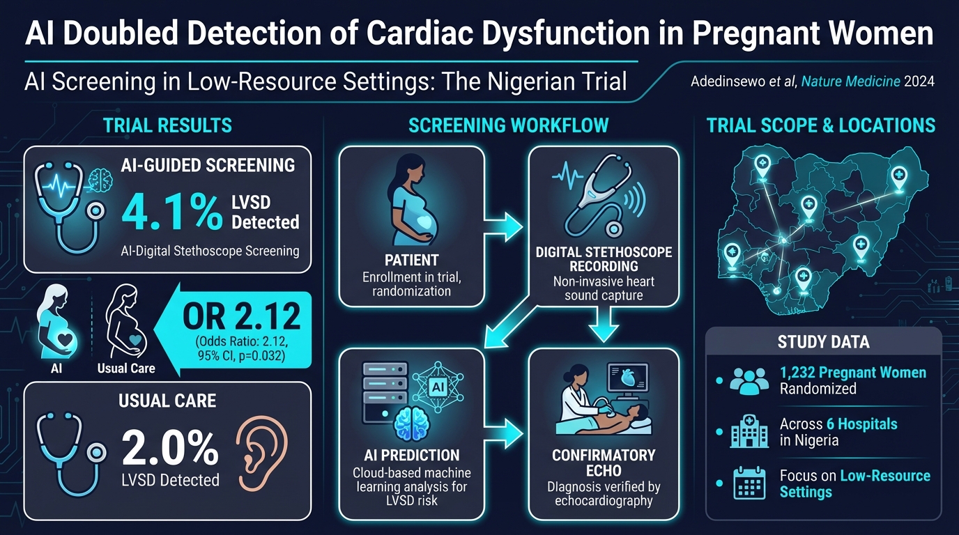
This pragmatic randomized clinical trial is the most compelling evidence for AI-POCUS's global health potential (PMID: 39223284).
**Setting:** 6 hospitals in Nigeria — a country with the highest reported incidence of peripartum cardiomyopathy worldwide.
**Design:** 1,232 pregnant and postpartum women randomized to AI-guided screening versus usual care.
**Intervention:** Digital stethoscope recordings with point-of-care AI predictions + 12-lead ECG with asynchronous AI predictions for LVSD.
**Results:**
- AI digital stethoscope screening detected LVSD in **4.1% vs 2.0%** with usual care (OR 2.12, 95% CI 1.05-4.27, p=0.032)
- AI effectively **doubled** the detection rate of left ventricular systolic dysfunction
- 12-lead AI-ECG showed a similar trend (3.4% vs 2.0%, OR 1.75, p=0.125) but did not reach significance
- No serious adverse events related to study participation
**Teaching Point:** The implications are profound. In settings where echocardiography expertise may not be available, AI can enable community health workers and non-specialist physicians to screen for life-threatening cardiac conditions using portable, affordable devices.
**Nuance:** However, generalizability remains a concern. Most AI models are trained on Western populations with different body habitus, prevalence patterns, and imaging conditions. The Nigerian trial specifically validated in its target population — this kind of population-specific validation must become standard practice.
---
## Clinical Cases
### Case 1: The Breathless Mother — Peripartum Cardiomyopathy
**MUST ACT:** AI-POCUS may have its greatest impact not in well-resourced tertiary centers but in settings where expert sonographers are scarce.
### The Adedinsewo et al. Trial (Nature Medicine, 2024)
This pragmatic randomized clinical trial is the most compelling evidence for AI-POCUS's global health potential (PMID: 39223284).
**Setting:** 6 hospitals in Nigeria — a country with the highest reported incidence of peripartum cardiomyopathy worldwide.
**Design:** 1,232 pregnant and postpartum women randomized to AI-guided screening versus usual care.
**Intervention:** Digital stethoscope recordings with point-of-care AI predictions + 12-lead ECG with asynchronous AI predictions for LVSD.
**Results:**
- AI digital stethoscope screening detected LVSD in **4.1% vs 2.0%** with usual care (OR 2.12, 95% CI 1.05-4.27, p=0.032)
- AI effectively **doubled** the detection rate of left ventricular systolic dysfunction
- 12-lead AI-ECG showed a similar trend (3.4% vs 2.0%, OR 1.75, p=0.125) but did not reach significance
- No serious adverse events related to study participation
**Teaching Point:** The implications are profound. In settings where echocardiography expertise may not be available, AI can enable community health workers and non-specialist physicians to screen for life-threatening cardiac conditions using portable, affordable devices.
**Nuance:** However, generalizability remains a concern. Most AI models are trained on Western populations with different body habitus, prevalence patterns, and imaging conditions. The Nigerian trial specifically validated in its target population — this kind of population-specific validation must become standard practice.
---
## Clinical Cases
### Case 1: The Breathless Mother — Peripartum Cardiomyopathy
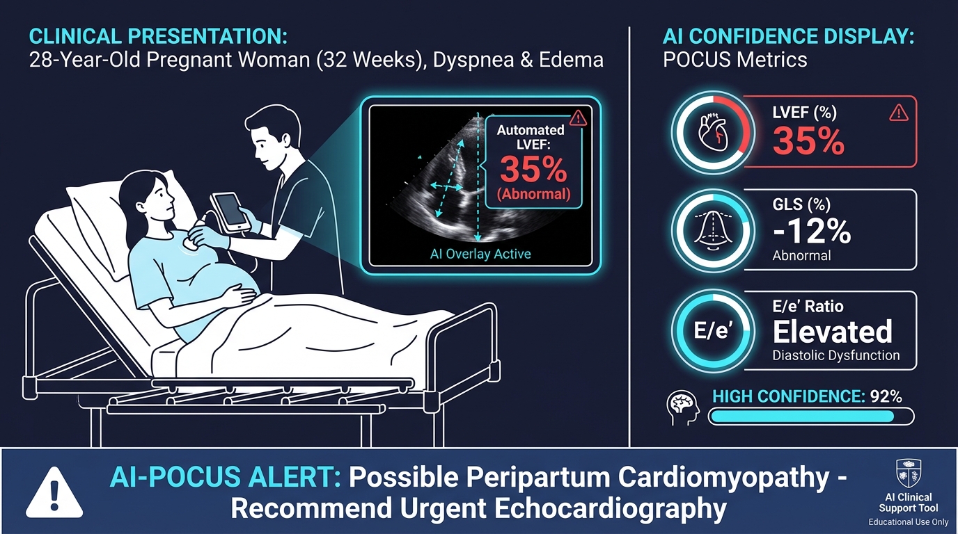 **Presentation:** A 28-year-old G2P1 woman at 32 weeks gestation presents to the ED with progressive dyspnea over 2 weeks and new bilateral lower extremity edema. She has no cardiac history. Vitals: HR 110, BP 100/65, SpO2 94% on room air, RR 24.
**Audience Poll:** What is your initial differential?
- A) Pulmonary embolism
- B) Peripartum cardiomyopathy
- C) Preeclampsia with pulmonary edema
- D) All of the above
**AI-POCUS Finding:** You perform a bedside cardiac ultrasound. The AI overlay immediately calculates LVEF at 35% (normal >55%) and flags GLS at -12% (abnormal). The AI alert reads: "Possible cardiomyopathy — recommend urgent echocardiography."
**Management Decision:** The AI finding accelerates your workup. Without AI, you might have attributed symptoms to pregnancy and deferred cardiac assessment. Instead, you activate cardiology consultation immediately, start diuresis cautiously (pregnancy-safe agents), and arrange urgent formal echo.
**Outcome:** Formal echocardiography confirms peripartum cardiomyopathy with LVEF 32%. The patient is started on hydralazine/nitrate therapy (ACE inhibitors contraindicated in pregnancy) and beta-blocker, with close maternal-fetal medicine follow-up.
**Key Teaching Point:** AI screening doubled the detection of LVSD in the Nigerian obstetric trial. This case illustrates why: pregnancy symptoms mask cardiomyopathy, and AI provides an objective trigger for further workup that a busy clinician might otherwise defer.
---
### Case 2: The Polytrauma — AI-Enhanced eFAST
**Presentation:** A 28-year-old G2P1 woman at 32 weeks gestation presents to the ED with progressive dyspnea over 2 weeks and new bilateral lower extremity edema. She has no cardiac history. Vitals: HR 110, BP 100/65, SpO2 94% on room air, RR 24.
**Audience Poll:** What is your initial differential?
- A) Pulmonary embolism
- B) Peripartum cardiomyopathy
- C) Preeclampsia with pulmonary edema
- D) All of the above
**AI-POCUS Finding:** You perform a bedside cardiac ultrasound. The AI overlay immediately calculates LVEF at 35% (normal >55%) and flags GLS at -12% (abnormal). The AI alert reads: "Possible cardiomyopathy — recommend urgent echocardiography."
**Management Decision:** The AI finding accelerates your workup. Without AI, you might have attributed symptoms to pregnancy and deferred cardiac assessment. Instead, you activate cardiology consultation immediately, start diuresis cautiously (pregnancy-safe agents), and arrange urgent formal echo.
**Outcome:** Formal echocardiography confirms peripartum cardiomyopathy with LVEF 32%. The patient is started on hydralazine/nitrate therapy (ACE inhibitors contraindicated in pregnancy) and beta-blocker, with close maternal-fetal medicine follow-up.
**Key Teaching Point:** AI screening doubled the detection of LVSD in the Nigerian obstetric trial. This case illustrates why: pregnancy symptoms mask cardiomyopathy, and AI provides an objective trigger for further workup that a busy clinician might otherwise defer.
---
### Case 2: The Polytrauma — AI-Enhanced eFAST
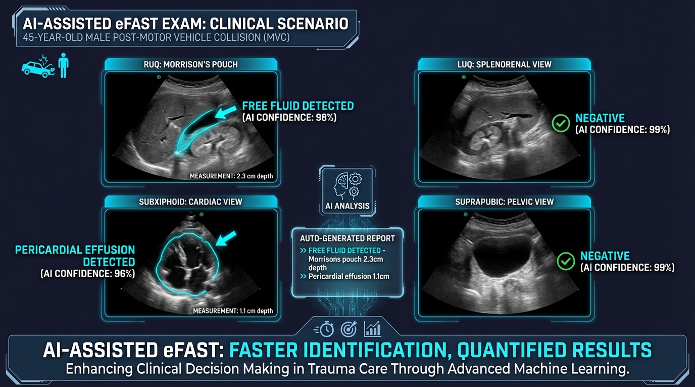 **Presentation:** A 45-year-old unrestrained driver arrives after a high-speed MVC. GCS 13 (E3V4M6), HR 125, BP 85/50, distended abdomen. The trauma team activates.
**AI-POCUS Finding:** You perform an AI-enhanced eFAST. The system highlights and quantifies free fluid in Morrison's pouch (2.3 cm depth) and detects a small pericardial effusion (1.1 cm). Automated confidence score: 97% for hemoperitoneum.
**Decision Point:** Does this patient go to the OR or CT?
- **With AI quantification:** The measured depth of 2.3 cm in Morrison's pouch combined with hemodynamic instability makes the decision straightforward — this patient needs the OR.
- **Without AI:** A novice sonographer might describe "some fluid" without quantification, potentially delaying surgical consultation.
**Outcome:** The patient goes directly to OR. Surgical exploration reveals a Grade IV splenic laceration with 1.5L hemoperitoneum. The small pericardial effusion was traumatic and self-limited.
**Key Teaching Point:** AI adds quantification to qualitative assessments. "Some free fluid" becomes "2.3 cm depth in Morrison's" — a data point that can be tracked serially and communicated precisely to surgeons.
---
### Case 3: The Septic Patient — AI-Guided Volume Assessment
**Presentation:** A 45-year-old unrestrained driver arrives after a high-speed MVC. GCS 13 (E3V4M6), HR 125, BP 85/50, distended abdomen. The trauma team activates.
**AI-POCUS Finding:** You perform an AI-enhanced eFAST. The system highlights and quantifies free fluid in Morrison's pouch (2.3 cm depth) and detects a small pericardial effusion (1.1 cm). Automated confidence score: 97% for hemoperitoneum.
**Decision Point:** Does this patient go to the OR or CT?
- **With AI quantification:** The measured depth of 2.3 cm in Morrison's pouch combined with hemodynamic instability makes the decision straightforward — this patient needs the OR.
- **Without AI:** A novice sonographer might describe "some fluid" without quantification, potentially delaying surgical consultation.
**Outcome:** The patient goes directly to OR. Surgical exploration reveals a Grade IV splenic laceration with 1.5L hemoperitoneum. The small pericardial effusion was traumatic and self-limited.
**Key Teaching Point:** AI adds quantification to qualitative assessments. "Some free fluid" becomes "2.3 cm depth in Morrison's" — a data point that can be tracked serially and communicated precisely to surgeons.
---
### Case 3: The Septic Patient — AI-Guided Volume Assessment
 **Presentation:** A 68-year-old woman with diabetes presents with fever (39.2°C), confusion, HR 118, BP 78/45. Lactate 4.8 mmol/L. You diagnose septic shock and begin resuscitation with 30 mL/kg crystalloid as per SSC guidelines.
After the initial bolus, BP improves to 88/52 but lactate remains elevated at 3.9. **Should you give more fluid?**
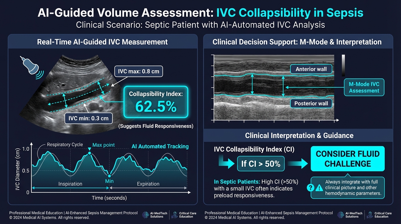
**AI-POCUS Finding:** You perform AI-guided IVC assessment. The system tracks IVC diameter in real-time over the respiratory cycle:
- IVC max: 0.8 cm
- IVC min: 0.3 cm
- Collapsibility Index: 62.5%
- AI recommendation: "High collapsibility suggests fluid responsiveness — consider additional volume challenge"
You also perform AI-assisted cardiac assessment: LVEF 55%, no RV dilation.
**Management Decision:** Based on the AI-quantified IVC collapsibility (>50%), preserved LVEF, and clinical picture, you administer another 500 mL crystalloid bolus.
**Outcome:** After the additional fluid, BP improves to 95/60, heart rate decreases to 98, and repeat lactate at 2 hours is 2.1 mmol/L. The AI's real-time IVC tracking allowed you to make a data-driven fluid decision rather than relying solely on clinical gestalt.
**Key Teaching Point:** AI excels at continuous, quantitative measurements that humans find tedious — like tracking IVC diameter variation over multiple respiratory cycles. This is where AI adds the most value: not replacing clinical judgment, but providing precise data to inform it.
---
### Case 4: The Automation Bias Trap — When AI Gets It Wrong
**Presentation:** A 68-year-old woman with diabetes presents with fever (39.2°C), confusion, HR 118, BP 78/45. Lactate 4.8 mmol/L. You diagnose septic shock and begin resuscitation with 30 mL/kg crystalloid as per SSC guidelines.
After the initial bolus, BP improves to 88/52 but lactate remains elevated at 3.9. **Should you give more fluid?**
**AI-POCUS Finding:** You perform AI-guided IVC assessment. The system tracks IVC diameter in real-time over the respiratory cycle:
- IVC max: 0.8 cm
- IVC min: 0.3 cm
- Collapsibility Index: 62.5%
- AI recommendation: "High collapsibility suggests fluid responsiveness — consider additional volume challenge"
You also perform AI-assisted cardiac assessment: LVEF 55%, no RV dilation.
**Management Decision:** Based on the AI-quantified IVC collapsibility (>50%), preserved LVEF, and clinical picture, you administer another 500 mL crystalloid bolus.
**Outcome:** After the additional fluid, BP improves to 95/60, heart rate decreases to 98, and repeat lactate at 2 hours is 2.1 mmol/L. The AI's real-time IVC tracking allowed you to make a data-driven fluid decision rather than relying solely on clinical gestalt.
**Key Teaching Point:** AI excels at continuous, quantitative measurements that humans find tedious — like tracking IVC diameter variation over multiple respiratory cycles. This is where AI adds the most value: not replacing clinical judgment, but providing precise data to inform it.
---
### Case 4: The Automation Bias Trap — When AI Gets It Wrong
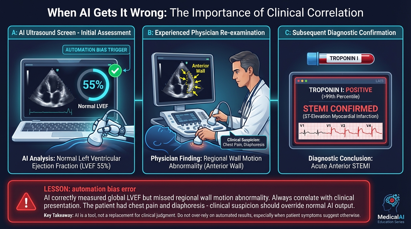 **Presentation:** A 62-year-old man with hypertension presents with substernal chest pressure for 3 hours. He appears diaphoretic and anxious. ECG shows nonspecific ST changes.
**AI-POCUS Finding:** You perform AI-assisted cardiac POCUS. The system reports: "LVEF 55% — Normal. No significant wall motion abnormality detected." The AI display shows a green checkmark.
**The Trap:** Reassured by the "normal" AI reading, a trainee considers discharging the patient with outpatient stress testing.
**What the AI Missed:** An experienced attending re-examines the same images and notices subtle anterior wall hypokinesis in the apical 4-chamber view — a regional wall motion abnormality that the AI's global LVEF algorithm averaged out. The troponin returns at 2.1 ng/mL (elevated). Serial ECG shows evolving anterior ST elevations. This is an acute STEMI.
**Outcome:** Cath lab activated, PCI to proximal LAD. The patient does well.
**Key Teaching Point:** Current AI echo models excel at global function (LVEF) but may miss regional abnormalities. Global LVEF can be preserved early in STEMI when only a single coronary territory is affected. **The AIM protocol would have prevented this error:** Assess independently first, Interrogate discrepancies (the patient's symptoms didn't match a "normal" heart), Monitor and document.
**Say Out Loud:** "A normal LVEF does NOT rule out acute coronary syndrome. Always correlate AI findings with the clinical picture. If the patient looks sick, they are sick — regardless of what the AI says."
---
### Case 5: Democratizing Diagnosis — The Rural Clinic
**Presentation:** A 62-year-old man with hypertension presents with substernal chest pressure for 3 hours. He appears diaphoretic and anxious. ECG shows nonspecific ST changes.
**AI-POCUS Finding:** You perform AI-assisted cardiac POCUS. The system reports: "LVEF 55% — Normal. No significant wall motion abnormality detected." The AI display shows a green checkmark.
**The Trap:** Reassured by the "normal" AI reading, a trainee considers discharging the patient with outpatient stress testing.
**What the AI Missed:** An experienced attending re-examines the same images and notices subtle anterior wall hypokinesis in the apical 4-chamber view — a regional wall motion abnormality that the AI's global LVEF algorithm averaged out. The troponin returns at 2.1 ng/mL (elevated). Serial ECG shows evolving anterior ST elevations. This is an acute STEMI.
**Outcome:** Cath lab activated, PCI to proximal LAD. The patient does well.
**Key Teaching Point:** Current AI echo models excel at global function (LVEF) but may miss regional abnormalities. Global LVEF can be preserved early in STEMI when only a single coronary territory is affected. **The AIM protocol would have prevented this error:** Assess independently first, Interrogate discrepancies (the patient's symptoms didn't match a "normal" heart), Monitor and document.
**Say Out Loud:** "A normal LVEF does NOT rule out acute coronary syndrome. Always correlate AI findings with the clinical picture. If the patient looks sick, they are sick — regardless of what the AI says."
---
### Case 5: Democratizing Diagnosis — The Rural Clinic
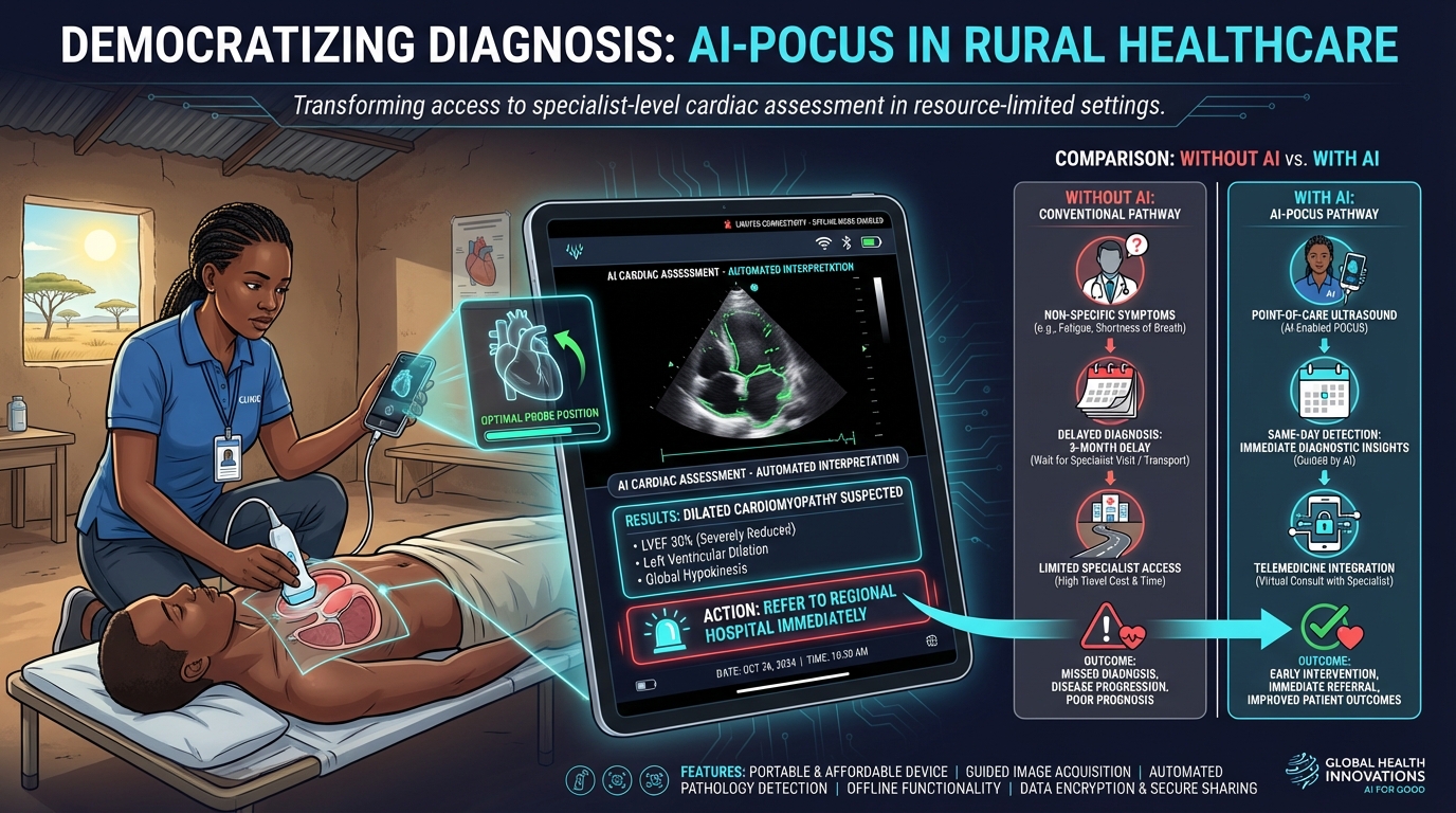 **Presentation:** A community health worker at a rural clinic in sub-Saharan Africa evaluates a 35-year-old woman who is 2 weeks postpartum. She reports progressive fatigue and dyspnea. The nearest hospital with echocardiography is 4 hours away.
**AI-POCUS Finding:** Using a handheld AI-enabled ultrasound device, the health worker obtains cardiac views following AI-guided probe positioning (green overlay shows optimal placement). The AI processes the images and reports: "LVEF 30% — Severely reduced. Dilated cardiomyopathy suspected. RECOMMEND URGENT REFERRAL."
**Without AI:** Without this technology, this patient's symptoms would likely be attributed to postpartum fatigue. She might not be referred for 2-3 months, by which time her heart failure could progress to an irreversible stage.
**With AI:** The same-day AI detection triggers immediate referral to the regional hospital, where she is diagnosed with peripartum cardiomyopathy and started on guideline-directed medical therapy.
**Outcome:** At 6-month follow-up, her LVEF has improved to 45% — a result directly attributable to early detection and treatment.
**Key Teaching Point:** This case embodies the findings of the Adedinsewo trial — AI enables non-expert operators to detect critical cardiac pathology. The technology doesn't replace cardiologists; it extends their reach to places they cannot physically be.
---
## Section 6: Skill Retention — The Balanced Sonographer
**Duration:** 8 min | **Content Tier: Teaching Point**
**Presentation:** A community health worker at a rural clinic in sub-Saharan Africa evaluates a 35-year-old woman who is 2 weeks postpartum. She reports progressive fatigue and dyspnea. The nearest hospital with echocardiography is 4 hours away.
**AI-POCUS Finding:** Using a handheld AI-enabled ultrasound device, the health worker obtains cardiac views following AI-guided probe positioning (green overlay shows optimal placement). The AI processes the images and reports: "LVEF 30% — Severely reduced. Dilated cardiomyopathy suspected. RECOMMEND URGENT REFERRAL."
**Without AI:** Without this technology, this patient's symptoms would likely be attributed to postpartum fatigue. She might not be referred for 2-3 months, by which time her heart failure could progress to an irreversible stage.
**With AI:** The same-day AI detection triggers immediate referral to the regional hospital, where she is diagnosed with peripartum cardiomyopathy and started on guideline-directed medical therapy.
**Outcome:** At 6-month follow-up, her LVEF has improved to 45% — a result directly attributable to early detection and treatment.
**Key Teaching Point:** This case embodies the findings of the Adedinsewo trial — AI enables non-expert operators to detect critical cardiac pathology. The technology doesn't replace cardiologists; it extends their reach to places they cannot physically be.
---
## Section 6: Skill Retention — The Balanced Sonographer
**Duration:** 8 min | **Content Tier: Teaching Point**
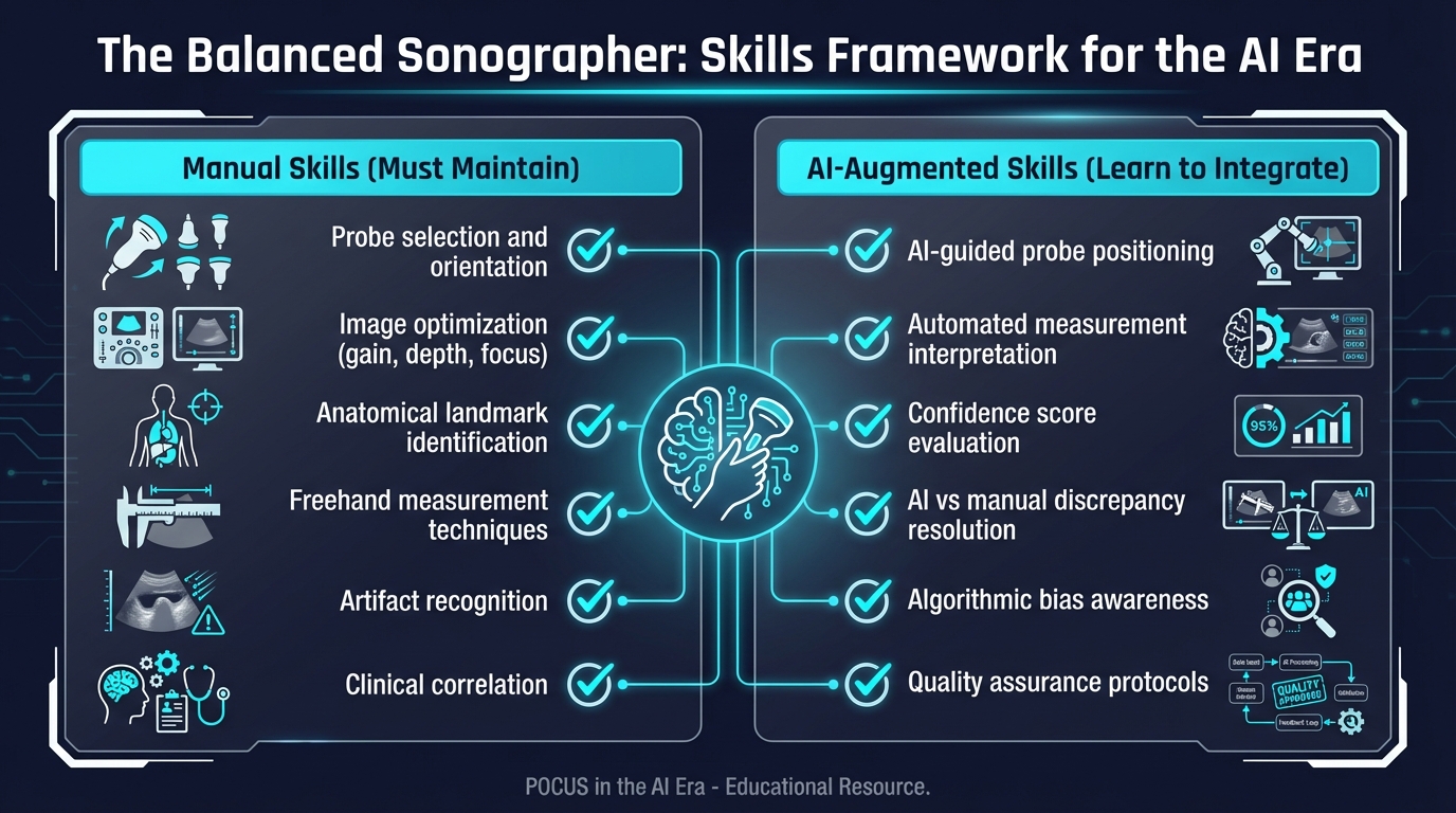 **Teaching Point:** As AI becomes standard in POCUS, there is a genuine risk that trainees will never fully develop manual ultrasound skills. This creates a dangerous dependency — what happens when the AI system goes down, loses connectivity, or encounters an edge case?
### The Skills We Must Protect
**Manual skills that must be maintained regardless of AI:**
1. Probe selection, orientation, and anatomical landmark identification
2. Image optimization (gain, depth, focus adjustment)
3. Freehand measurement technique
4. Artifact recognition (reverberation, shadowing, mirror artifacts)
5. Clinical correlation and integration with physical exam
### The New Skills We Must Develop
**AI-augmented skills to add to training:**
1. Understanding AI confidence scores and their limitations
2. Identifying when AI output conflicts with clinical assessment
3. Quality assurance — recognizing when poor image quality may compromise AI accuracy
4. Ethical considerations in AI-assisted decision making
**Audience Poll:** How should residency programs integrate AI-POCUS training?
- A) Teach manual skills first, introduce AI in senior years
- B) Teach AI-assisted and manual skills simultaneously from the start
- C) Focus primarily on AI-assisted skills since that's the future
- D) Leave AI training to post-residency CME
---
## Section 7: Algorithm Bias — The Equity Challenge
**Duration:** 8 min | **Content Tier: Nuance**
**Teaching Point:** As AI becomes standard in POCUS, there is a genuine risk that trainees will never fully develop manual ultrasound skills. This creates a dangerous dependency — what happens when the AI system goes down, loses connectivity, or encounters an edge case?
### The Skills We Must Protect
**Manual skills that must be maintained regardless of AI:**
1. Probe selection, orientation, and anatomical landmark identification
2. Image optimization (gain, depth, focus adjustment)
3. Freehand measurement technique
4. Artifact recognition (reverberation, shadowing, mirror artifacts)
5. Clinical correlation and integration with physical exam
### The New Skills We Must Develop
**AI-augmented skills to add to training:**
1. Understanding AI confidence scores and their limitations
2. Identifying when AI output conflicts with clinical assessment
3. Quality assurance — recognizing when poor image quality may compromise AI accuracy
4. Ethical considerations in AI-assisted decision making
**Audience Poll:** How should residency programs integrate AI-POCUS training?
- A) Teach manual skills first, introduce AI in senior years
- B) Teach AI-assisted and manual skills simultaneously from the start
- C) Focus primarily on AI-assisted skills since that's the future
- D) Leave AI training to post-residency CME
---
## Section 7: Algorithm Bias — The Equity Challenge
**Duration:** 8 min | **Content Tier: Nuance**
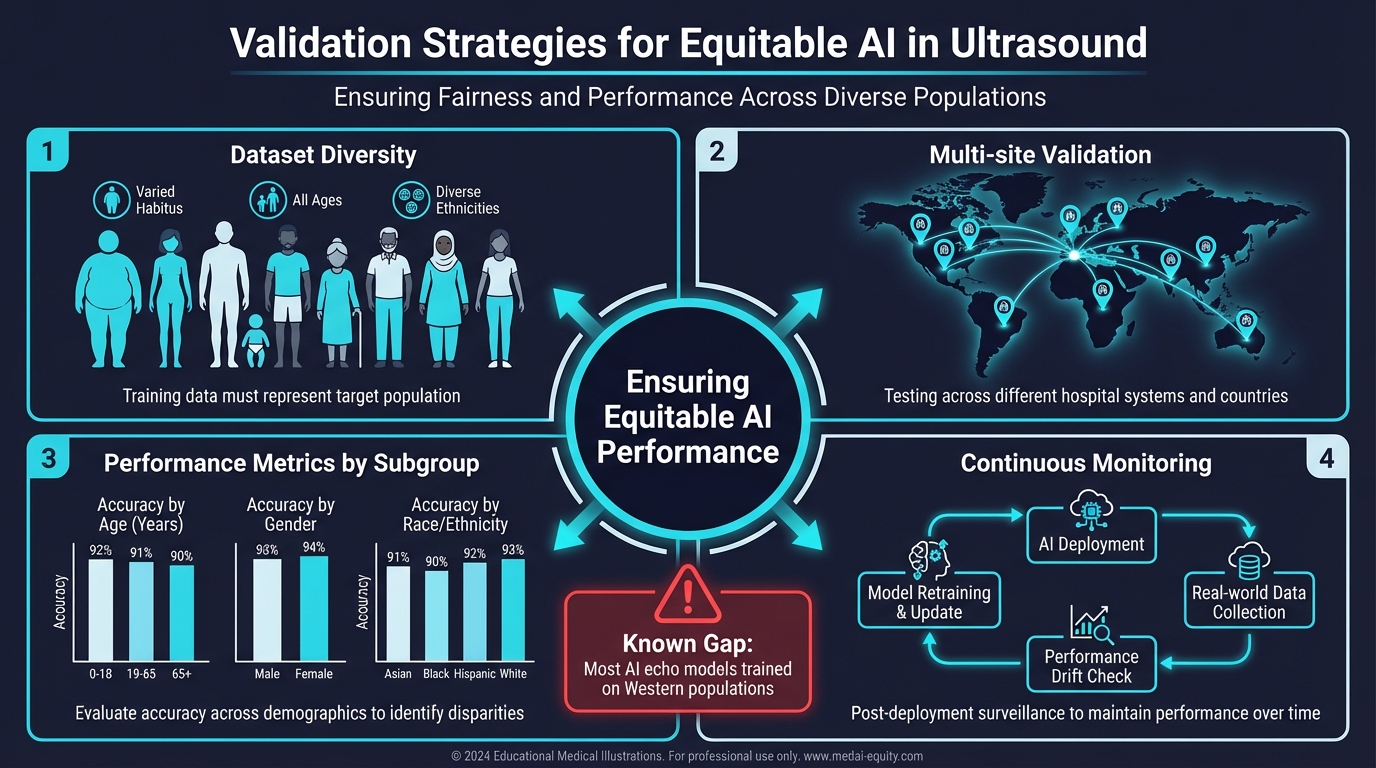 **Nuance:** AI is only as equitable as the data it was trained on. Most cardiac AI models have been developed using echocardiographic data from Western academic medical centers — predominantly white, non-obese patient populations with high-quality imaging equipment.
### Known Bias Concerns
- **Body habitus:** AI models trained on thin patients may perform poorly on obese patients where acoustic windows are limited
- **Racial/ethnic diversity:** Cardiac structure and function vary across populations. An AI model that defines "normal LVEF" based on one demographic may systematically misclassify another
- **Equipment variation:** AI trained on high-end echo machines may not generalize to portable, handheld devices used in resource-limited settings
- **Age and sex:** Diastolic function norms vary significantly with age and sex; AI must account for these physiologic differences
### The Path Forward
The Adedinsewo trial (PMID: 39223284) represents a model for responsible AI deployment — they validated specifically in the Nigerian obstetric population where the tool would be used. This kind of population-specific validation must become the standard, not the exception.
**ACC/AHA 2022 Recommendation:** The guidelines encourage using AI tools for standardized cardiac assessments (Class IIa, Level B) — but emphasize the need for validation across diverse populations before broad deployment.
---
## Section 8: Future Directions — Where Are We Heading?
**Duration:** 8 min | **Content Tier: Teaching Point**
The future of AI-POCUS extends well beyond what current systems can do:
### Near-term (2-5 years)
- **Multimodal AI:** Integration of ultrasound with ECG, vitals, and lab data for comprehensive AI-assisted clinical decision support
- **Automated protocols:** AI guiding complete exam protocols (e.g., full RUSH exam) with real-time quality feedback
- **Continuous monitoring:** AI analyzing streaming ultrasound data during resuscitation for real-time hemodynamic guidance
### Long-term (5-10 years)
- **Patient-performed ultrasound:** Wearable or self-administered devices with AI interpretation for home monitoring of heart failure patients
- **Federated learning:** AI models that learn from diverse global datasets without sharing patient data, addressing bias through inclusive training
- **Autonomous screening:** AI-driven population health screening using portable ultrasound at community health fairs, pharmacies, and primary care offices
**Audience Poll:** What future application of AI in POCUS excites you the most?
- A) Real-time hemodynamic guidance during resuscitation
- B) Home monitoring for heart failure patients
- C) Global screening with portable AI devices
- D) Multimodal integration with labs and vitals
---
## Tonight on Shift
**When you walk into the department tonight, remember these six things:**
1. **AI-POCUS is non-inferior to expert sonographers for LVEF** — the He et al. Nature trial proved this with blinding and randomization. Use it when available, but verify with your clinical assessment.
2. **Always assess independently first** — form your own impression before reviewing AI output. The AIM protocol (Assess, Interrogate, Monitor) protects you from automation bias.
3. **A normal AI LVEF does not rule out ACS** — global function can be preserved early in STEMI. Regional wall motion abnormalities are still best detected by trained human eyes.
4. **AI reduces measurement variability by 60%** — for serial assessments (IVC tracking, response to fluids), AI's consistency is its greatest strength.
5. **AI doubles cardiac detection in screening populations** — in settings without expert sonographers, AI-POCUS is a life-saving force multiplier.
6. **Maintain your manual skills** — AI systems fail, lose connectivity, and have blind spots. Your fundamental ultrasound competency is your safety net.
---
## References
1. He B, Kwan AC, Cho JH, et al. Blinded, randomized trial of sonographer versus AI cardiac function assessment. *Nature*. 2023;616:520-524. PMID: 37020027.
2. Adedinsewo DA, Morales-Lara AC, Afolabi BB, et al. Artificial intelligence guided screening for cardiomyopathies in an obstetric population: a pragmatic randomized clinical trial. *Nature Medicine*. 2024;30:3302-3310. PMID: 39223284.
3. Mołek-Dziadosz P, Woźniak A, Furman-Niedziejko A, et al. Left ventricular ejection fraction assessment: AI compared with echocardiography expert and CMR. *Polish Archives of Internal Medicine*. 2025. PMID: 40888426.
4. Pettersen H, Sabo S, Pasdeloup D, et al. Real-time deep learning-based image guiding and automated LV measurements to reduce test-retest variability. *Open Heart*. 2025;12:e003117. PMID: 41360622.
5. Chen X, Yang F, Zhang P, et al. AI-assisted LV diastolic function assessment and grading: multiview versus single view. *JASE*. 2023;36(10):1064-1078. PMID: 37437669.
6. Choi HM, Kim J, Park J, et al. AI derived ECG global longitudinal strain compared to echocardiographic measurements. *Scientific Reports*. 2024;14:26592. PMID: 39488646.
7. Yao X, Rushlow DR, Inselman JW, et al. AI-enabled electrocardiograms for identification of patients with low ejection fraction. *Nature Medicine*. 2021;27:815-819. PMID: 33958795.
8. Osterwalder J, Polyzogopoulou E, Hoffmann B. Point-of-care ultrasound — history, current and evolving clinical concepts in emergency medicine. *Medicina*. 2023;59(12):2174. PMID: 38138282.
9. Kagiyama N, Piccirilli M, Yanamala N, et al. Machine learning assessment of LV diastolic function based on ECG features. *JACC*. 2020;76(8):930-941. PMID: 32819467.
10. Kuwahara A, Iwasaki Y, Kobayashi M, et al. AI-derived LV strain in echocardiography in patients treated with chemotherapy. *Int J Cardiovasc Imaging*. 2024;40(9):1965-1974. PMID: 39042233.
**Nuance:** AI is only as equitable as the data it was trained on. Most cardiac AI models have been developed using echocardiographic data from Western academic medical centers — predominantly white, non-obese patient populations with high-quality imaging equipment.
### Known Bias Concerns
- **Body habitus:** AI models trained on thin patients may perform poorly on obese patients where acoustic windows are limited
- **Racial/ethnic diversity:** Cardiac structure and function vary across populations. An AI model that defines "normal LVEF" based on one demographic may systematically misclassify another
- **Equipment variation:** AI trained on high-end echo machines may not generalize to portable, handheld devices used in resource-limited settings
- **Age and sex:** Diastolic function norms vary significantly with age and sex; AI must account for these physiologic differences
### The Path Forward
The Adedinsewo trial (PMID: 39223284) represents a model for responsible AI deployment — they validated specifically in the Nigerian obstetric population where the tool would be used. This kind of population-specific validation must become the standard, not the exception.
**ACC/AHA 2022 Recommendation:** The guidelines encourage using AI tools for standardized cardiac assessments (Class IIa, Level B) — but emphasize the need for validation across diverse populations before broad deployment.
---
## Section 8: Future Directions — Where Are We Heading?
**Duration:** 8 min | **Content Tier: Teaching Point**
The future of AI-POCUS extends well beyond what current systems can do:
### Near-term (2-5 years)
- **Multimodal AI:** Integration of ultrasound with ECG, vitals, and lab data for comprehensive AI-assisted clinical decision support
- **Automated protocols:** AI guiding complete exam protocols (e.g., full RUSH exam) with real-time quality feedback
- **Continuous monitoring:** AI analyzing streaming ultrasound data during resuscitation for real-time hemodynamic guidance
### Long-term (5-10 years)
- **Patient-performed ultrasound:** Wearable or self-administered devices with AI interpretation for home monitoring of heart failure patients
- **Federated learning:** AI models that learn from diverse global datasets without sharing patient data, addressing bias through inclusive training
- **Autonomous screening:** AI-driven population health screening using portable ultrasound at community health fairs, pharmacies, and primary care offices
**Audience Poll:** What future application of AI in POCUS excites you the most?
- A) Real-time hemodynamic guidance during resuscitation
- B) Home monitoring for heart failure patients
- C) Global screening with portable AI devices
- D) Multimodal integration with labs and vitals
---
## Tonight on Shift
**When you walk into the department tonight, remember these six things:**
1. **AI-POCUS is non-inferior to expert sonographers for LVEF** — the He et al. Nature trial proved this with blinding and randomization. Use it when available, but verify with your clinical assessment.
2. **Always assess independently first** — form your own impression before reviewing AI output. The AIM protocol (Assess, Interrogate, Monitor) protects you from automation bias.
3. **A normal AI LVEF does not rule out ACS** — global function can be preserved early in STEMI. Regional wall motion abnormalities are still best detected by trained human eyes.
4. **AI reduces measurement variability by 60%** — for serial assessments (IVC tracking, response to fluids), AI's consistency is its greatest strength.
5. **AI doubles cardiac detection in screening populations** — in settings without expert sonographers, AI-POCUS is a life-saving force multiplier.
6. **Maintain your manual skills** — AI systems fail, lose connectivity, and have blind spots. Your fundamental ultrasound competency is your safety net.
---
## References
1. He B, Kwan AC, Cho JH, et al. Blinded, randomized trial of sonographer versus AI cardiac function assessment. *Nature*. 2023;616:520-524. PMID: 37020027.
2. Adedinsewo DA, Morales-Lara AC, Afolabi BB, et al. Artificial intelligence guided screening for cardiomyopathies in an obstetric population: a pragmatic randomized clinical trial. *Nature Medicine*. 2024;30:3302-3310. PMID: 39223284.
3. Mołek-Dziadosz P, Woźniak A, Furman-Niedziejko A, et al. Left ventricular ejection fraction assessment: AI compared with echocardiography expert and CMR. *Polish Archives of Internal Medicine*. 2025. PMID: 40888426.
4. Pettersen H, Sabo S, Pasdeloup D, et al. Real-time deep learning-based image guiding and automated LV measurements to reduce test-retest variability. *Open Heart*. 2025;12:e003117. PMID: 41360622.
5. Chen X, Yang F, Zhang P, et al. AI-assisted LV diastolic function assessment and grading: multiview versus single view. *JASE*. 2023;36(10):1064-1078. PMID: 37437669.
6. Choi HM, Kim J, Park J, et al. AI derived ECG global longitudinal strain compared to echocardiographic measurements. *Scientific Reports*. 2024;14:26592. PMID: 39488646.
7. Yao X, Rushlow DR, Inselman JW, et al. AI-enabled electrocardiograms for identification of patients with low ejection fraction. *Nature Medicine*. 2021;27:815-819. PMID: 33958795.
8. Osterwalder J, Polyzogopoulou E, Hoffmann B. Point-of-care ultrasound — history, current and evolving clinical concepts in emergency medicine. *Medicina*. 2023;59(12):2174. PMID: 38138282.
9. Kagiyama N, Piccirilli M, Yanamala N, et al. Machine learning assessment of LV diastolic function based on ECG features. *JACC*. 2020;76(8):930-941. PMID: 32819467.
10. Kuwahara A, Iwasaki Y, Kobayashi M, et al. AI-derived LV strain in echocardiography in patients treated with chemotherapy. *Int J Cardiovasc Imaging*. 2024;40(9):1965-1974. PMID: 39042233.