Advancing Anesthesia with McGRATH™ Video Laryngoscopy: Pediatric Airway Management
Pediatric Airway Management
Anesthesiology · Seminar week 16 · released July 20, 2026 · includes a discussion video
This seminar focuses on the use of McGRATH MAC Video Laryngoscopy for first-attempt intubation in infants, as detailed in the randomized controlled trial discussed by Drs.…
Learning Objectives
By the end of this seminar, participants will be able to:
- Explain the anatomical and physiological features that make pediatric tracheal intubation uniquely hazardous.
- Critically appraise the randomized evidence comparing McGRATH MAC video laryngoscopy with conventional direct laryngoscopy in infants.
- Interpret first-attempt success as a clinically meaningful but context-dependent airway outcome.
- Select and execute a pediatric intubation strategy that integrates positioning, oxygenation, pharmacology, video laryngoscopy, and rescue planning.
- Distinguish cleaning, disinfection, and sterilization requirements for reusable and single-use laryngoscope components.
- Evaluate ultraviolet-C LED disinfection through infection-prevention, engineering, and environmental-sustainability lenses.
- Anticipate how artificial intelligence, advanced imaging, simulation, and human-factors engineering may change pediatric airway management.
Introduction to Pediatric Airway Management Challenges
Duration: 10 min
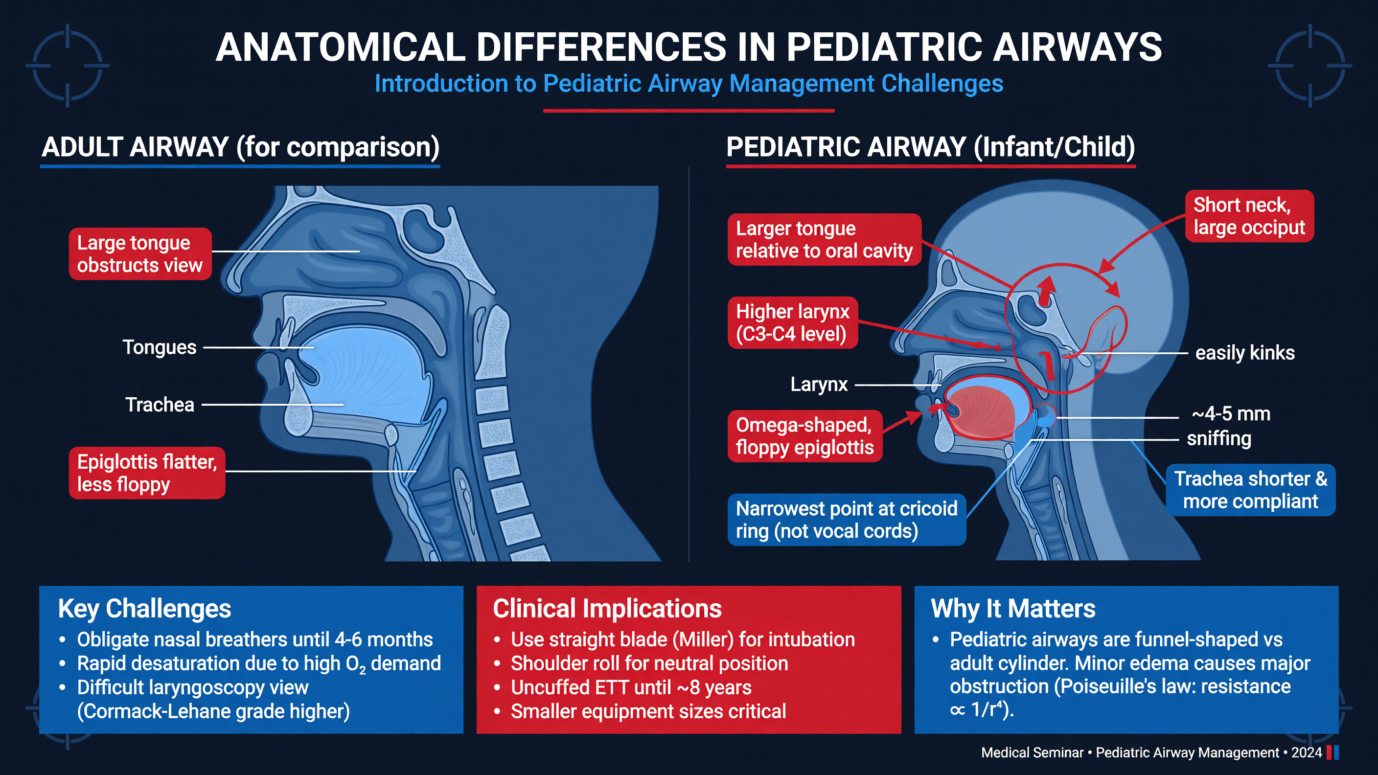
Pediatric airway management is not adult airway management performed with smaller equipment. The relevant anatomy, oxygen physiology, autonomic responses, disease patterns, and margins for error all change with age. These differences are most pronounced in neonates and infants, but they remain clinically important throughout early childhood.
Infants have a relatively large tongue, a short mandible, a large occiput, and a larynx positioned more cephalad than in adults. The epiglottis is often long, narrow, and more compliant. These features can make displacement of the tongue and exposure of the glottis difficult. The large occiput may flex the neck when the child is placed flat on an adult-sized mattress; a shoulder roll can restore a neutral or slightly extended position in an infant, whereas indiscriminate extension may worsen alignment or collapse a compliant airway. The historical description of the pediatric airway as uniformly “funnel-shaped” with the cricoid as its absolute narrowest point is an oversimplification. Contemporary imaging demonstrates an elliptical airway whose limiting dimension varies with age, plane, and functional conditions. Clinically, the subglottis remains vulnerable to edema and pressure injury.
The consequences of small changes in airway radius are substantial. Resistance to laminar flow is inversely related to the fourth power of radius. Secretions, inflammation, an oversized tube, or approximately 1 mm of circumferential edema can therefore produce a disproportionate increase in resistance. This matters in croup, bacterial tracheitis, airway burns, post-intubation edema, and congenital or acquired subglottic stenosis.
Physiology creates even greater urgency. Infants have high metabolic oxygen consumption—approximately 6–8 mL/kg/min compared with roughly 3 mL/kg/min in adults—while possessing a smaller functional residual capacity. General anesthesia, supine positioning, neuromuscular blockade, obesity, abdominal distension, and pulmonary disease further reduce oxygen reserve. Desaturation may develop within seconds after an unsuccessful attempt. Hypoxemia can provoke bradycardia through both myocardial hypoxia and strong vagal responses to laryngoscopy.
MUST ACT: In a child, oxygenation is the primary endpoint. Abandon an attempt before severe desaturation or bradycardia develops; reoxygenate, correct the mechanism of failure, and only then attempt again.
Airway assessment should identify both an anatomically difficult airway and a physiologically difficult airway. Anatomical concerns include micrognathia, limited mouth opening, macroglossia, midface hypoplasia, cervical immobility, airway masses, previous airway surgery, prior difficult intubation, or syndromes such as Pierre Robin sequence, Treacher Collins syndrome, mucopolysaccharidosis, and Down syndrome. Physiological difficulty arises from severe hypoxemia, shock, pulmonary hypertension, intracranial hypertension, metabolic acidosis, or right-ventricular failure. A technically uncomplicated laryngoscopy can still precipitate cardiovascular collapse if these conditions are ignored.
Framework: Ask five questions before induction: Can the child be oxygenated now? Will apnea be tolerated? Is laryngoscopy likely to be difficult? Can a tracheal tube be delivered through the available space? What is the rescue route if intubation and mask ventilation fail?
Preoxygenation should be adapted to the disease. A cooperative child may tolerate a tight-fitting mask with 100% oxygen. Children with shunt physiology or alveolar collapse may need positive end-expiratory pressure, noninvasive ventilation, or high-flow nasal oxygen. A two-person mask technique, oral or nasal airway, suction, and appropriate head position may be more valuable than simply increasing fresh-gas flow. Gentle pressure-limited ventilation during induction is often safer than a rigid adult-style “no ventilation” rapid-sequence technique, particularly when the anticipated safe apnea time is short.
Video laryngoscopy improves the visual pathway by placing a camera close to the blade tip and reducing the need to align oral, pharyngeal, and laryngeal axes. It does not eliminate difficult tube delivery, secretions, lens contamination, restricted mouth opening, or equipment failure. A superb screen image is not equivalent to a secured airway.
Teaching Point: Separate the task into three phases: obtaining a view, delivering the tube, and confirming ventilation. Each phase has different failure modes and may require a different corrective action.
Cuffed tracheal tubes are appropriate for most children when correctly sized and monitored. A cuffed oral tube estimate is internal diameter in millimeters = age/4 + 3.5; an uncuffed estimate is age/4 + 4. These are starting points, not guarantees. Prepare one tube 0.5 mm smaller and one 0.5 mm larger. Initial oral depth can be estimated as age/2 + 12 cm or approximately three times the tube’s internal diameter, followed by confirmation with continuous waveform capnography, bilateral ventilation assessment, and imaging when indicated. Maintain the minimum cuff pressure that produces an effective seal, generally no more than 20–25 cm H₂O.
Decision Point: Use the device that maximizes first-attempt success in the hands of the available team, while ensuring that a second technique, supraglottic airway, effective mask ventilation, and the institution’s pediatric cannot-intubate/cannot-oxygenate pathway are immediately available.
Audience Poll: Which problem most often limits your pediatric intubations: rapid desaturation, glottic visualization, tube delivery, secretions, or team coordination?
Details of the Randomized Controlled Trial on McGRATH MAC Efficacy
Duration: 10 min
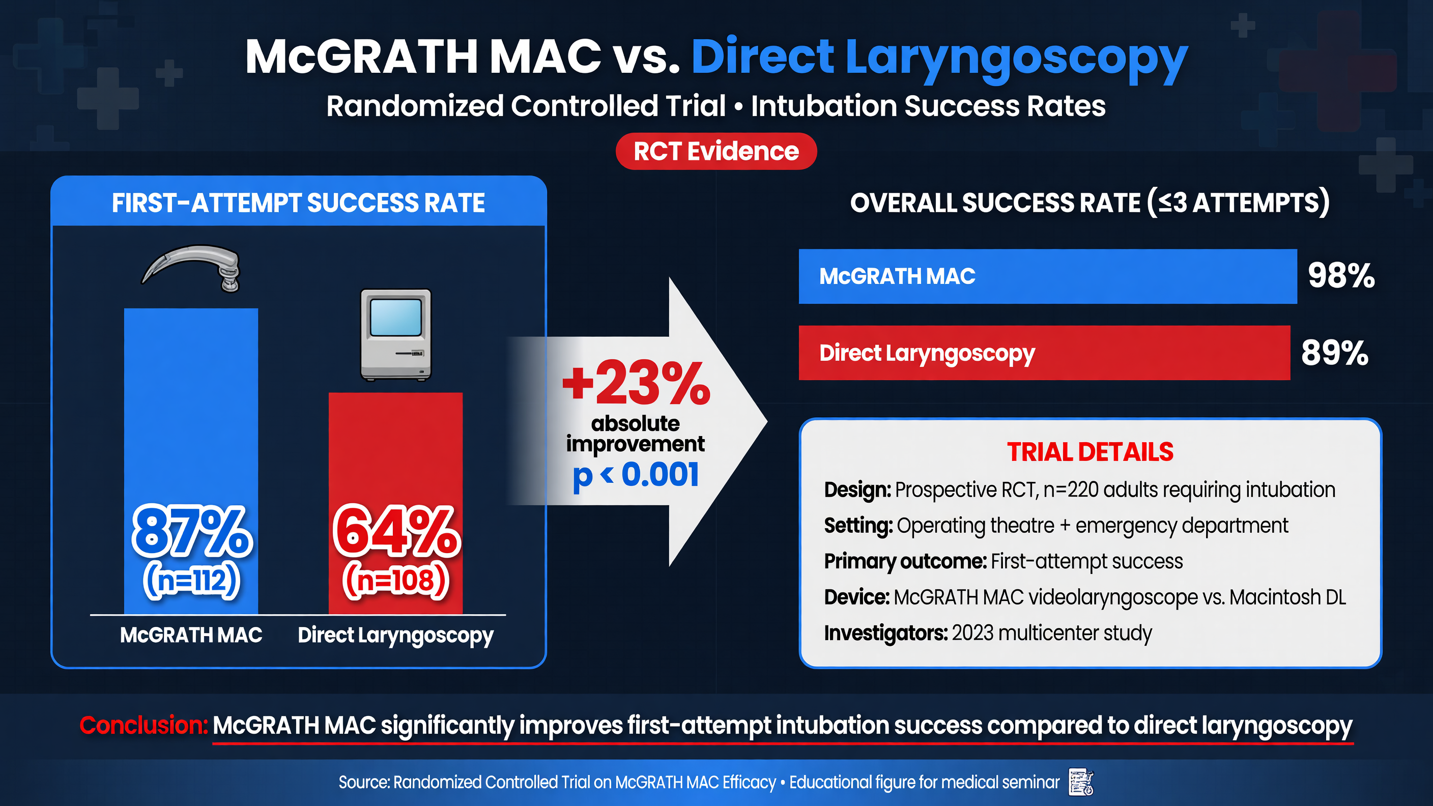
The randomized trial reported by Jagannathan and Uchinami in Anesthesia & Analgesia in 2026 compared McGRATH MAC video laryngoscopy with conventional direct laryngoscopy for tracheal intubation in infants. The key result supplied in the research brief is a first-attempt success rate of 86.9% with McGRATH MAC versus 74.6% with conventional laryngoscopy.
That 12.3-percentage-point absolute difference is clinically meaningful. Expressed another way, failure on the first attempt decreased from 25.4% to 13.1%, an approximate 48% relative reduction in first-attempt failure. The crude number needed to treat is 1/0.123, or 8.1; conventionally rounded, approximately nine infants would need initial use of McGRATH MAC rather than the comparator to produce one additional first-attempt success, assuming the trial effect is reproducible in the local population. Confidence intervals and the prespecified statistical analysis remain essential for deciding how precisely that benefit has been estimated.
MUST ACT: Do not translate a percentage difference into policy without reviewing the full trial’s population, endpoint definition, operator experience, crossover rules, confidence interval, adverse events, and funding disclosures.
The mechanism supporting the result is plausible. The McGRATH MAC places a camera near the distal blade while retaining familiar Macintosh-style geometry. The operator can insert and lift in a broadly conventional manner but view the larynx without achieving a direct line of sight. The screen also creates a shared visual field. A supervisor can see whether the blade is too deep, whether the epiglottis is folded, and whether laryngeal manipulation improves the view. This makes real-time coaching possible without relying on a trainee’s description of an unseen airway.
Shared visualization probably explains part of the educational value seen in pediatric and neonatal video-laryngoscopy research. In a randomized neonatal teaching trial, O’Shea and colleagues found that allowing the instructor to view the video image improved trainee success compared with an instructor unable to see that image (PMID: 26459647). That study was not a McGRATH efficacy trial, but it supports the broader mechanism: video laryngoscopy converts an otherwise private procedural view into a team-visible task.
Nuance: A better laryngeal view does not guarantee easy tube delivery. The camera may produce an excellent image while the tube impacts the anterior tracheal wall, arytenoid cartilage, or right laryngeal structure. Blade depth, lifting vector, stylet curvature, and the point at which the tube is introduced remain critical.
A Macintosh-style video blade should generally be advanced under direct vision until the tongue and epiglottic region are safely approached; the operator then transitions attention to the screen. Watching only the monitor from the moment the blade enters the mouth risks lip, palate, tonsillar, or pharyngeal injury outside the camera’s field. Once the vallecula is engaged, a controlled lift—not levering on the teeth or gums—should expose the glottis. An excessive “close-up” view can hinder tube delivery. Withdrawing the blade slightly often creates more working space and improves the approach angle.
Trial interpretation requires attention to co-interventions. Neuromuscular blockade, preoxygenation, use of a stylet, blade size, operator grade, external laryngeal manipulation, suction readiness, and an elective versus emergency setting can materially affect first-attempt success. Results from fasting, fully monitored infants in an operating room may not transfer unchanged to a child with bronchiolitis, shock, copious secretions, or active resuscitation in an emergency department.
Framework: Appraise the trial using PICO plus technique: the infants enrolled, the exact McGRATH configuration, the conventional comparator, the definition and adjudication of an attempt, and the standardized intubation protocol.
A first-attempt endpoint should ideally require tracheal placement confirmed by sustained waveform capnography. Investigators should define whether an attempt begins when the blade crosses the lips, whether temporary blade withdrawal counts as a new attempt, and how device crossover is handled. Secondary outcomes should include time to ventilation, oxygen saturation nadir, bradycardia, esophageal placement, airway trauma, rescue interventions, and severe tracheal-intubation-associated events. A slower successful attempt accompanied by profound hypoxemia is not necessarily superior to a rapid, safely aborted attempt.
The findings should therefore support—not replace—clinical judgment. They strengthen the case for age-appropriate video laryngoscopy as an initial device, especially where operators use it regularly and supervisors can exploit the shared screen. They do not establish that every blade geometry, every video platform, or every operator will produce the same benefit.
Decision Point: If McGRATH MAC is selected as the initial device, match the blade to the child and anticipated technique, shape the stylet before induction, position the monitor for both operator and supervisor, and state the bailout trigger aloud.
Audience Poll: Would a 12.3-percentage-point improvement change your unit’s default first-line device, or would training, device availability, and local performance data outweigh the trial result?
Interpreting First-Attempt Success Rates in Infants
Duration: 10 min
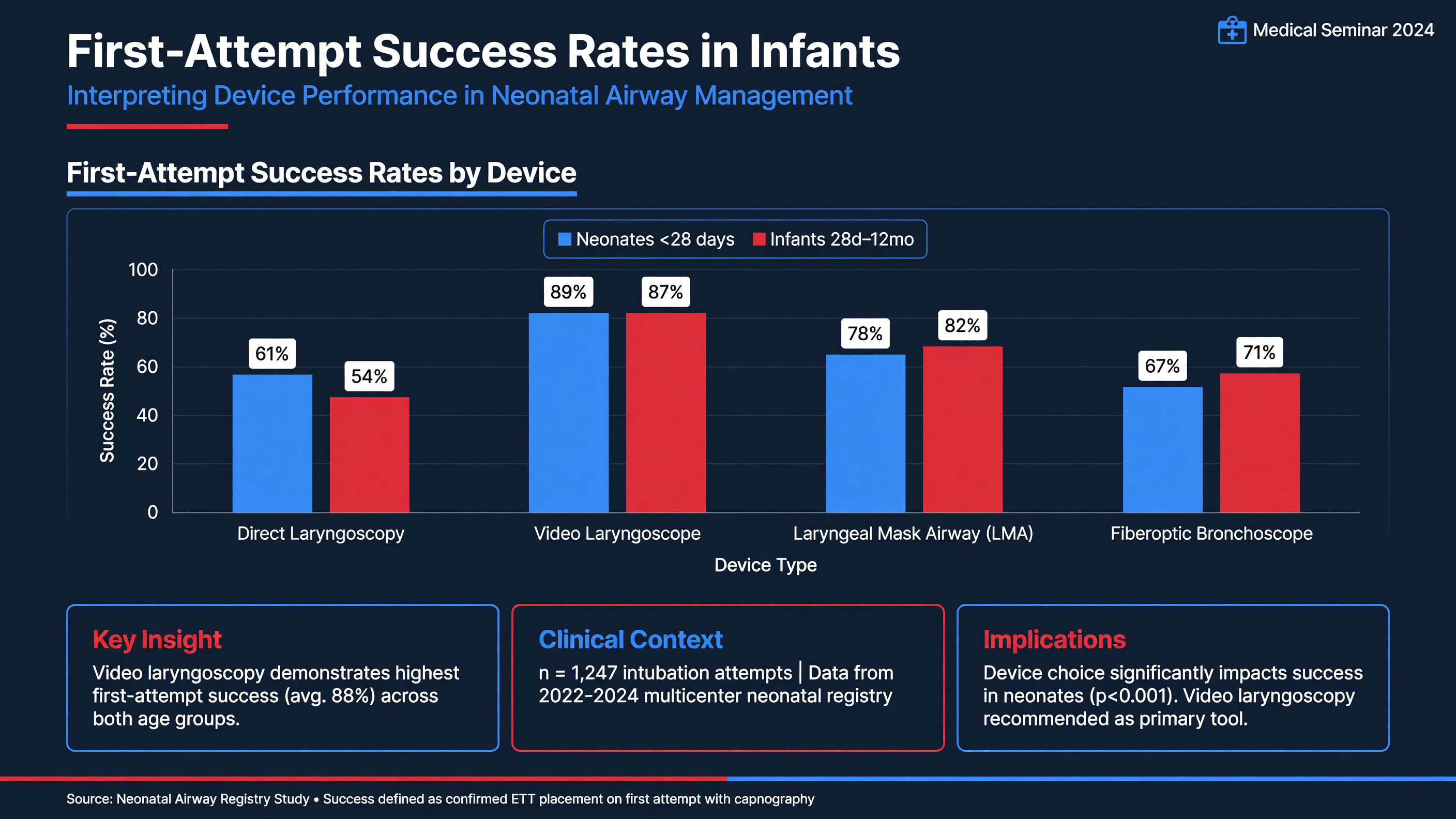
First-attempt success is more than a procedural score. In an infant, each additional attempt adds apnea time, airway manipulation, edema, bleeding, gastric insufflation, aspiration risk, and the possibility that an initially manageable airway becomes progressively more difficult. Pediatric registry evidence demonstrates that repeated attempts are associated with more severe complications. The Pediatric Difficult Intubation Registry showed a strong relationship between multiple attempts and major adverse outcomes in children with difficult tracheal intubation (PMID: 26705976).
The first challenge is definitional. “First attempt,” “first pass,” and “successful intubation” are not always interchangeable. One study may count an attempt when the blade enters the mouth; another may count only passage of a tube toward the glottis. Repositioning the blade without removing it may constitute one prolonged attempt in one dataset and several maneuvers in another. Success may be determined by direct visualization, colorimetric carbon dioxide, auscultation, or continuous waveform capnography. Comparisons are only credible when these definitions are aligned.
Teaching Point: The clinically relevant outcome is not merely tube placement on the first pass. It is first-attempt placement achieved within a safe physiological window, without severe hypoxemia, bradycardia, trauma, or unrecognized esophageal intubation.
Baseline risk also matters. Raising success from 95% to 98% produces a small absolute benefit, even if statistically significant. Raising success from 60% to 80% prevents one failure for every five patients treated. The Jagannathan and Uchinami rates—86.9% versus 74.6%—suggest a large absolute opportunity for improvement because the comparator group’s first-attempt failure rate was substantial. Local results may differ if the conventional-laryngoscopy group has exceptional expertise or if clinicians rarely use the video device.
Operator experience is a major effect modifier. A trainee may benefit greatly from a shared display and real-time coaching. An expert who performs direct laryngoscopy frequently may show a smaller difference, although expertise does not eliminate the infant’s physiological vulnerability. Conversely, introducing a video device without deliberate training can temporarily worsen performance. Familiarity with direct laryngoscopy does not automatically transfer to screen-guided depth perception or video-guided tube delivery.
Nuance: Device comparisons can be confounded by blade geometry. A standard Macintosh-shaped video blade, a hyperangulated blade, and a straight blade are different techniques, even when all are described as video laryngoscopes.
A standard-geometry blade allows both direct and indirect viewing and may facilitate a rapid transition for clinicians trained with conventional laryngoscopy. A hyperangulated blade can expose a very anterior glottis without axis alignment but usually requires a matching stylet and a deliberate withdrawal-and-rotate sequence to advance the tube. A straight blade may be advantageous when a long, floppy epiglottis must be lifted directly. Device choice should be based on the anticipated anatomical problem rather than the generic label “video.”
When the glottis is visible but the tube will not advance, repeating the same motion is rarely useful. Optimize blade depth, reduce the magnified close-up view, apply external laryngeal manipulation, rotate the tube, alter the stylet curvature, or use a bougie only if its size and design are appropriate for the child. Withdraw a rigid stylet carefully once the tube tip has entered the glottis. Forceful advancement risks arytenoid, vocal-cord, cricoid, or tracheal injury.
Framework: Classify failure immediately: oxygenation failure, visualization failure, tube-delivery failure, equipment failure, or physiological collapse. Each category demands a different response.
Time-to-intubation must be interpreted alongside first-attempt success. A device that increases success but substantially prolongs apnea could be harmful in a critically hypoxemic infant. Report induction-to-ventilation time, blade-in-mouth time, saturation nadir, area under a clinically relevant saturation threshold, heart-rate change, and need for rescue ventilation. These measures expose clinically important differences that a binary endpoint can conceal.
Institutions should monitor their own performance. Useful quality metrics include first-attempt success by location and operator level, frequency of video-laryngoscope use, hypoxemia and bradycardia rates, number of attempts, rescue-device use, and whether the planned device was immediately available. Review should be nonpunitive and video may be used for education only under approved privacy, consent, and data-governance policies.
Decision Point: After one failed attempt, ask whether the next attempt includes a meaningful change—more experienced operator, different blade, improved position, better suction, altered stylet, or a different route. If not, repeating the attempt is difficult to justify.
MUST ACT: Set an attempt limit before induction. Escalate early, maintain oxygenation with mask ventilation or a supraglottic airway, and activate the pediatric difficult-airway pathway before fixation on tracheal intubation causes preventable harm.
Audience Poll: Which metric would most change your practice: first-attempt success, time to ventilation, oxygen saturation nadir, severe adverse events, or successful rescue after a failed attempt?
Sustainable Disinfection with Ultraviolet-C LED Methods
Duration: 10 min
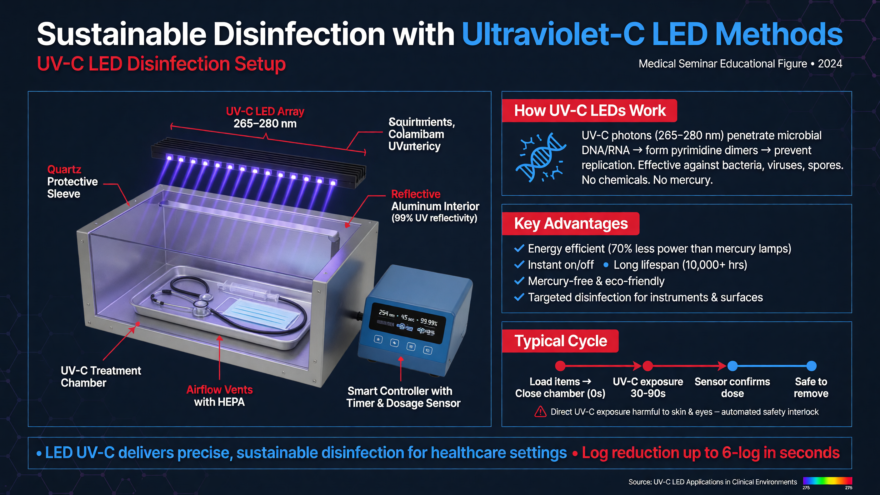
Ultraviolet-C disinfection is attractive because it can inactivate microorganisms without liquid chemical disinfectants, extensive water use, or high-temperature processing. UV-C generally spans wavelengths from 200 to 280 nm; germicidal LED systems commonly operate near 265–280 nm. Photons are absorbed by microbial nucleic acids, producing photochemical lesions that interfere with replication and transcription. Adequate exposure can reduce viable bacteria, viruses, and fungi.
The proposed British Journal of Anaesthesia UV-C LED approach referenced in the research brief should be understood as a promising engineering strategy, not an automatic substitute for validated reprocessing. Laryngoscope blades contact mucous membranes and are classified as semicritical devices under the Spaulding framework. Reusable blades therefore require at least high-level disinfection between patients, unless the manufacturer or institutional policy requires sterilization. Whether a UV-C system meets that standard depends on demonstrated microbial reduction, geometry, dose, device compatibility, quality assurance, and regulatory authorization.
MUST ACT: Remove blood, mucus, and proteinaceous material before UV-C exposure. UV light is not cleaning, and organic soil can shield microorganisms even when the surface appears adequately illuminated.
Delivered dose is the product of irradiance and exposure time, usually expressed in mJ/cm². The dose displayed by a machine may not equal the dose reaching the least-exposed surface. Laryngoscope blades contain curves, joints, channels, recesses, camera windows, and attachment interfaces that create shadows. Distance and angle alter irradiance, while fingerprints, dried secretions, scratches, and surface reflectivity can reduce penetration. A cabinet validated with a flat test coupon cannot automatically be assumed effective for an assembled laryngoscope.
Framework: A safe UV-C process requires four verified stages: physical cleaning, complete exposure of all relevant surfaces, delivery of a validated minimum dose, and documented release of the device back into clean storage.
Engineering controls should include a closed chamber, door interlocks, cycle monitoring, fault alarms, and prevention of direct exposure to staff. UV-C can injure eyes and skin. LEDs at conventional germicidal wavelengths are less likely than shorter-wavelength sources to generate ozone, but occupational and environmental safety must still be evaluated for the particular system. Irradiance sensors or dosimeters should be placed at worst-case locations during validation, not only at the chamber center.
Microbial testing should challenge the process with organisms appropriate to high-level disinfection claims and with clinically relevant soil. Testing only easily inactivated vegetative bacteria on pristine surfaces provides insufficient assurance. Repeated-cycle studies should also examine lens clarity, adhesive integrity, plastics, elastomers, labels, seals, and electronic components. UV exposure may cause yellowing, embrittlement, or loss of optical performance over time.
Nuance: UV-C can be sustainable only if it replaces a more resource-intensive validated step or measurably reduces chemicals, water, energy, packaging, or device turnover. Adding UV-C after unchanged conventional reprocessing may add cost and energy without reducing the dominant environmental burden.
A credible sustainability assessment should consider the entire life cycle: manufacture of the UV-C cabinet, electricity, cleaning materials, transport, consumables, device damage, staff time, and end-of-life disposal. It should compare reusable and single-use blades using local infection-control requirements and actual use volumes. Reusable equipment is not inherently greener if reprocessing is energy intensive or causes frequent premature replacement; disposable equipment is not inherently safer if storage, packaging, and waste streams are poorly controlled.
For McGRATH systems, the exact pathway depends on the component. A blade labeled single-use must not be reprocessed or reused. The display and handle should be cleaned and disinfected only with agents and methods permitted by the manufacturer’s instructions for use. Immersion, high heat, abrasive cleaning, or unapproved UV exposure can damage seals, optics, electronics, or surface coatings. A validated UV-C protocol for one model cannot be extrapolated to another merely because both are called video laryngoscopes.
Decision Point: Before implementation, infection prevention, sterile processing, biomedical engineering, occupational health, sustainability leadership, and frontline anesthesia staff should jointly determine whether UV-C is a validated primary process, a supplemental terminal step, or an inappropriate use for that component.
Operational validation should define loading position, whether the blade is disassembled, permissible materials, cycle duration, minimum dose, cleaning method, drying requirements, acceptance criteria, and response to an interrupted cycle. Every processed device needs a clear transition from contaminated to clean workflow; a successfully disinfected blade placed on a contaminated counter has immediately defeated the process.
Teaching Point: Sustainable disinfection is not achieved by choosing the newest technology. It is achieved by reliably preventing infection with the lowest total material, chemical, water, energy, and waste burden.
Audience Poll: In your institution, who has final authority to approve a new UV-C reprocessing pathway: anesthesia, infection prevention, sterile processing, biomedical engineering, or a multidisciplinary committee?
Discussion on Pediatric Laryngoscope Maintenance
Duration: 10 min
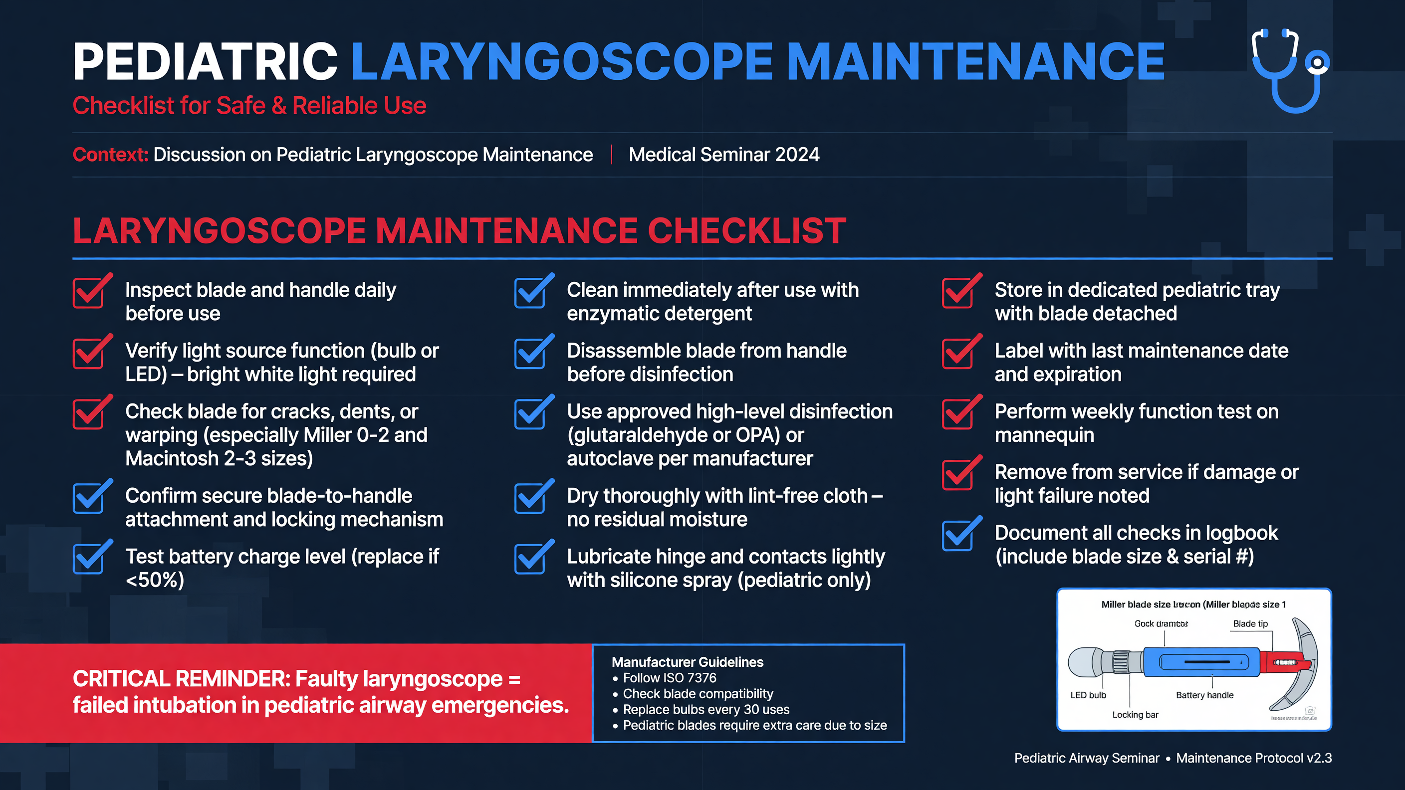
Pediatric laryngoscope maintenance is a patient-safety system, not a housekeeping task. A camera that intermittently fails, a depleted battery, an incompatible blade, or an inadequately disinfected handle may remain unnoticed until a rapidly desaturating child is anesthetized. Maintenance must therefore encompass pre-use readiness, post-use decontamination, scheduled technical inspection, inventory control, and clear accountability.
Before every planned intubation, power on the device and inspect image brightness, focus, color, field of view, and screen stability. Lock the selected blade into place and verify that the image does not flicker when the blade or handle is gently moved. Check the remaining battery indication according to the manufacturer’s system. Ensure that an immediately usable backup device is present; knowing that another laryngoscope exists elsewhere in the department is not an adequate rescue plan.
Confirm that the chosen blade fits the patient and the intended technique. Pediatric carts should stock age- and weight-appropriate options rather than a single generic “small” blade. Inspect packaging and expiration dates for single-use blades. Prepare the primary cuffed tube plus tubes 0.5 mm smaller and larger, a suitable stylet, functioning suction, mask, oral and nasal airways, supraglottic airway, capnography, and the next planned intubation device.
MUST ACT: Complete the equipment check before administering sedative or neuromuscular-blocking medication. If the primary device fails after induction, switch immediately to the verified backup rather than troubleshooting electronics during apnea.
Point-of-use cleaning should begin before secretions dry. Contaminated components must be transported in a closed, labeled container through a workflow that physically separates dirty and clean equipment. Staff should follow the manufacturer’s instructions and institutional infection-prevention policy for detergents, contact times, wipes, rinsing, drying, immersion, and high-level disinfection. “Wiped until it looks clean” is neither a reproducible process nor evidence of disinfection.
Video-laryngoscope handles and displays deserve particular attention. Gloves contaminated during blade removal can transfer material to the handle, screen, cart, keyboard, drug drawer, or clean replacement blade. The handle may also be directly contaminated by saliva or blood during use. Clean from less contaminated to more contaminated areas while preventing fluid entry into ports, battery compartments, hinges, or nonimmersible seams.
Framework: Maintain a one-way chain: bedside use, safe blade removal, contained transport, cleaning, validated disinfection, drying, inspection, protected storage, and documented readiness.
After reprocessing, inspect for cracks, surface crazing, loose joints, corrosion, residue, damaged seals, clouded optics, scratches, and degradation of printed size markings. A blurred or discolored image should not be accepted simply because the device still powers on. Quarantine damaged equipment, label it clearly, and route it to biomedical engineering. Do not return a questionable device to the airway cart “for later checking.”
Storage conditions matter. Clean equipment should be protected from dust, splash, handling, and contact with contaminated devices. Blades should be organized so staff can identify size and type quickly without opening multiple packages. Battery management should include scheduled checks, rotation by expiration date, and compliance with the manufacturer’s long-term storage instructions. Emergency carts require documented inspection after use and at a locally determined interval even when sealed.
Nuance: The apparent convenience of single-use blades shifts maintenance rather than eliminating it. The reusable display still requires cleaning, while procurement, packaging integrity, expiration control, supply shortages, and waste disposal become critical dependencies.
Maintenance records can reveal latent system problems. Track device identifier, date of service, cleaning or disinfection cycle where applicable, battery replacement, repair, failed pre-use checks, and image-quality complaints. Recurrent failures involving the same component should trigger engineering review rather than repeated informal workarounds. If UV-C is used, its cycle record and dosimetry requirements belong in the same quality system.
A standardized cart layout reduces cognitive load. The McGRATH unit, blades, tubes, stylets, suction, capnography adapters, and rescue supraglottic airways should occupy consistent locations. In mixed adult-pediatric areas, clearly separate pediatric blade and tube sizes. Label components that are not interchangeable between device generations or manufacturers.
Decision Point: If a device passes a power check but has an intermittent image, scratched lens, uncertain reprocessing history, or unverified blade compatibility, remove it from service. “Probably usable” is an unacceptable category for emergency airway equipment.
Competency should include maintenance as well as insertion technique. Simulation drills can deliberately introduce a dead battery, fogged lens, absent pediatric blade, or contaminated backup device. The team should demonstrate that it can identify the problem, preserve oxygenation, transition to another device, and report the equipment defect.
Teaching Point: Reliability is multiplicative. Excellent technique cannot compensate for a missing blade, failed screen, blocked suction catheter, or unavailable capnography.
Audience Poll: When was the last time your pediatric airway simulation included equipment failure or contamination rather than anatomical difficulty alone?
Future Directions and Innovations in Pediatric Airway Management
Duration: 10 min
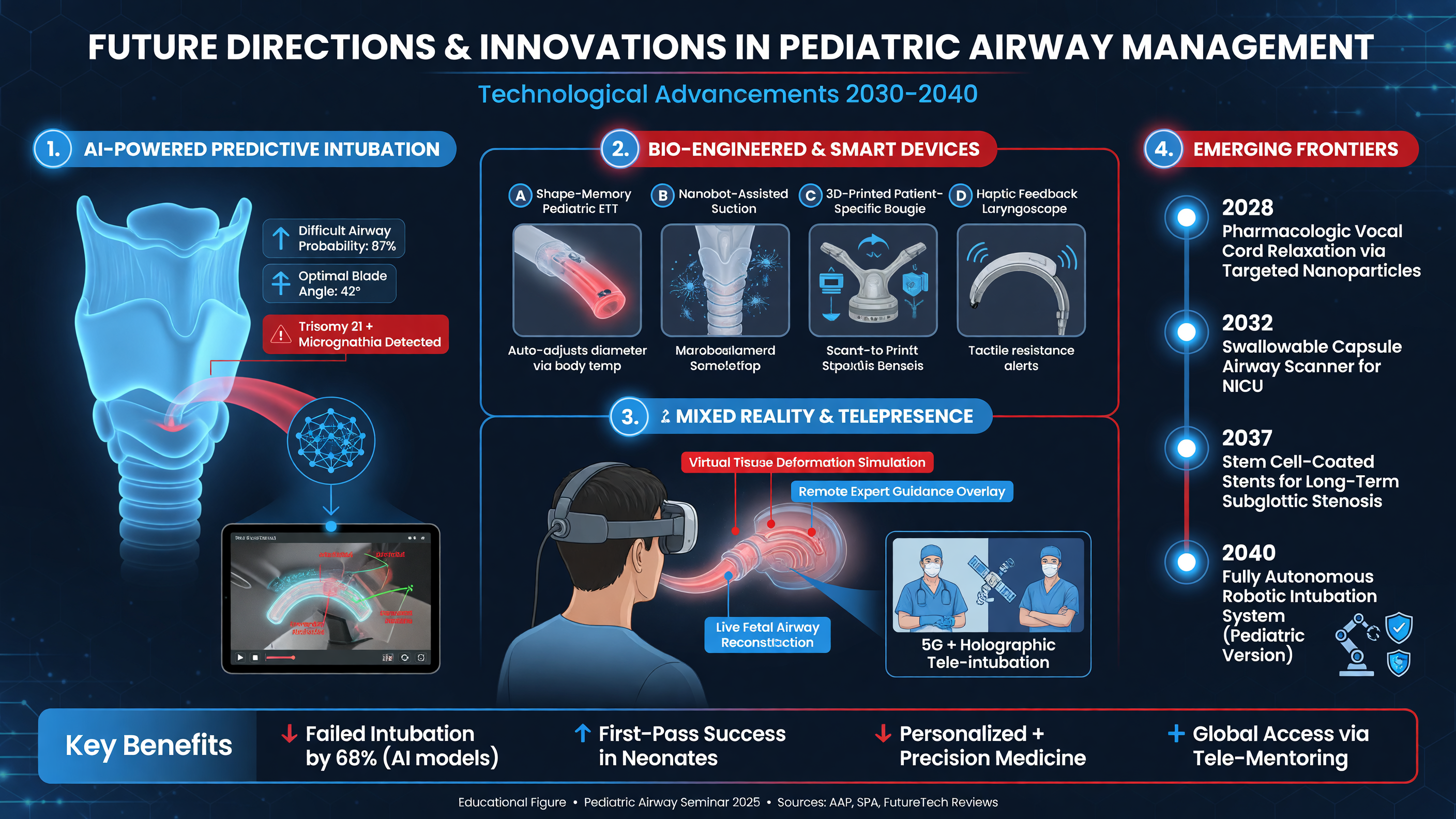
The next generation of pediatric airway technology will likely combine improved optics, physiological monitoring, decision support, and team communication. The most useful innovations will not merely produce sharper images; they will shorten the interval from recognition of difficulty to an effective corrective action while preserving oxygenation.
Artificial intelligence may identify the epiglottis, arytenoids, vocal cords, tracheal opening, and tube tip in real time. A system could quantify the percentage of glottic opening, detect when the blade is too deep, highlight secretion-obscured anatomy, or warn when the tube is approaching the esophagus. Automated timestamps could record attempt duration and relate it to oxygen saturation and heart-rate changes. Such tools may be particularly helpful during training, when the clinician has not yet developed stable visual pattern recognition.
Nuance: AI guidance is probabilistic. Blood, secretions, congenital anatomy, airway masses, low light, motion artifact, and rare syndromes may differ from the system’s training data. A confident overlay can still be wrong.
Clinical validation must therefore test more than image-label accuracy. Studies should evaluate first-attempt success, time to ventilation, hypoxemia, bradycardia, rescue interventions, cognitive load, and inappropriate reliance on erroneous prompts. Performance should be reported across age, size, skin pigmentation where external imaging is involved, craniofacial abnormalities, clinical location, and operator experience. Algorithms require monitoring after deployment because software updates, camera changes, and evolving practice can alter performance.
Future devices may incorporate antifog technology, improved depth of field, smaller camera modules, more durable displays, force sensing, and ergonomics designed for neonatal hands and mouths rather than miniaturized adult instruments. Force sensors could warn against excessive pressure on the tongue, teeth, or soft tissue. However, alarms must be carefully designed; excessive alerts during a hypoxemic emergency can increase rather than decrease cognitive load.
Framework: Judge an innovation across five domains: clinical efficacy, human factors, infection prevention, environmental impact, and resilience during power, network, or supply failure.
Integration with oxygenation is another priority. High-flow nasal oxygen, noninvasive ventilation, optimized mask interfaces, and closed-loop oxygen delivery may extend the safe window for laryngoscopy. These systems should not be marketed as creating unlimited apnea time. Airway obstruction, severe shunt, low cardiac output, and complete loss of ventilation can sharply limit their effect. Gentle ventilation during induction and timely reoxygenation will remain central skills.
Point-of-care ultrasound may contribute to tube-depth assessment, bilateral lung ventilation, detection of pneumothorax, and evaluation of gastric insufflation. It can support difficult-airway planning by identifying anterior neck anatomy, but it should not replace continuous waveform capnography as the primary confirmation of tracheal placement in a perfusing patient. Flexible endoscopy, optical stylets, and video-enabled supraglottic devices will increasingly be used as complementary components rather than competing technologies.
Training will shift from procedure counts toward demonstrated competency. Video recordings can enable frame-by-frame review of blade insertion, laryngeal exposure, tube trajectory, and decision timing. Simulation can recreate rapid desaturation, bradycardia, secretions, equipment failure, and team fixation. Data governance is essential: recordings may contain identifiers, staff performance information, or sensitive anatomy and must not be stored or shared casually.
Decision Point: Adopt video recording for education only after defining consent or institutional authority, access control, retention, de-identification, medicolegal handling, and whether recordings enter the health record.
Research should move beyond device-versus-device superiority trials in low-risk elective cases. Pragmatic studies should include neonates, infants with anticipated difficult airways, emergency intubations, children with critical illness, and resource-limited environments. Core outcomes should include first-attempt success, severe desaturation, bradycardia, airway trauma, time to effective ventilation, rescue success, and neurodevelopmentally relevant outcomes when feasible. Environmental life-cycle analysis and cost-effectiveness should be prespecified rather than added as promotional afterthoughts.
The future is also organizational. Shared airway registries can identify where attempts fail, which rescue plans work, and whether disparities exist across locations or patient groups. Just-in-time cognitive aids can display tube sizes, drug doses, equipment locations, and escalation pathways based on entered weight. These aids need independent double-checking, particularly when weight is estimated or entered in pounds rather than kilograms.
MUST ACT: Preserve fundamental airway competence. No AI overlay, robotic aid, or advanced camera substitutes for effective mask ventilation, suction, positioning, pharmacological preparation, supraglottic rescue, and timely escalation.
Teaching Point: The ideal pediatric airway platform is not the device with the most features. It is the system that helps a trained team secure ventilation on the first attempt, recognizes failure early, and remains reliable under real clinical conditions.
Audience Poll: Which innovation would most improve pediatric airway safety in your setting: AI guidance, better oxygenation technology, force sensing, universal video review, smarter dosing aids, or more realistic team simulation?
Case Study: Urgent Intubation of a 3-Year-Old
A 3-year-old child weighing 14 kg presents with severe pneumonia and progressive respiratory failure. Despite high-flow nasal oxygen, the child has an oxygen saturation of 88–91%, respiratory rate of 58 breaths/min, heart rate of 168 beats/min, blood pressure of 76/42 mm Hg, and declining mental status. There are copious secretions and bilateral crackles. The child has no known craniofacial syndrome or previous difficult airway.
This is a physiologically difficult airway even if the anatomy proves straightforward. The immediate differential includes alveolar shunt from pneumonia, mucus plugging, evolving acute respiratory distress syndrome, fatigue, sepsis-associated myocardial dysfunction, pneumothorax, and aspiration. Bedside assessment should determine whether suction, recruitment, bronchodilation, pleural decompression, or hemodynamic resuscitation can improve stability before induction. Preparation must occur simultaneously because delayed intubation may lead to arrest.
Framework: Separate the plan into physiology, anatomy, equipment, pharmacology, personnel, and rescue. Name one team member for each critical function: primary operator, airway assistant, medication administration, physiological monitoring, and overall leadership.
The child is positioned with the external auditory canal approximately aligned with the sternal notch while avoiding excessive neck extension. Preoxygenation continues using a tight-fitting mask or noninvasive support with positive end-expiratory pressure. A two-person mask seal, suction, oral airway, and capnography are prepared. Nasal oxygen may remain in place during laryngoscopy if it does not obstruct the procedure.
McGRATH MAC with an age-appropriate blade—often size 2 for a child of this age, subject to patient anatomy and manufacturer guidance—is selected as Plan A. The device is powered on and tested before medications are given. A cuffed 4.0-mm tube is prepared, with 3.5- and 4.5-mm tubes immediately available. The starting estimate from age/4 + 3.5 is 4.25 mm, demonstrating why adjacent sizes are necessary. A stylet is shaped to support the intended blade path without allowing its tip to protrude beyond the tube.
Two suction systems should be available because secretions can rapidly obscure both the camera and the airway. Plan B is reoxygenation followed by a changed laryngoscopy strategy with the most experienced operator. Plan C is a size-appropriate supraglottic airway, potentially serving as a conduit for flexible bronchoscopic intubation. The institutional pediatric cannot-intubate/cannot-oxygenate pathway is reviewed and specialist help summoned early.
Decision Point: Because this child is hypotensive and hypoxemic, a prolonged no-ventilation rapid-sequence induction is hazardous. Use gentle, pressure-limited ventilation during drug onset unless a specific contraindication outweighs the risk of desaturation.
For a hemodynamically stable child, ketamine 1–2 mg/kg IV is a common induction range. In catecholamine-depleted shock, even ketamine can reveal myocardial depression; a reduced, titrated dose such as 0.5–1 mg/kg may be appropriate while resuscitation continues. Rocuronium 1.0–1.2 mg/kg IV provides rapid neuromuscular blockade; at 1.2 mg/kg, this child would receive approximately 17 mg. Propofol may cause profound vasodilation and hypotension in this context. Etomidate 0.2–0.3 mg/kg is another hemodynamically considered option, with drug selection determined by local practice and the child’s physiology.
Atropine is not required routinely for every pediatric intubation. A dose of 0.02 mg/kg IV may be considered when clinically important bradycardia is present or anticipated, including repeated vagal stimulation or selected use with succinylcholine. In this 14-kg child, 0.02 mg/kg equals 0.28 mg. Medication doses, concentration, route, and syringe labeling should be independently verified.
MUST ACT: Do not let drug preparation delay correction of hypotension. Establish vascular access, have appropriately diluted vasoactive medication ready, and anticipate that positive-pressure ventilation will reduce venous return.
During laryngoscopy, the operator inserts the blade while looking into the mouth, then transitions to the display as the epiglottis approaches. Secretions are suctioned before they cover the lens. If the view is too close, the blade is withdrawn slightly. External laryngeal manipulation is directed by the operator watching the screen. The tube is advanced under continuous visualization without force.
If the first attempt fails, the blade is removed and the child is reoxygenated. The team identifies whether failure resulted from poor view, secretions, tube trajectory, equipment, or physiology. A second attempt occurs only after a meaningful change. Persistent ineffective ventilation prompts supraglottic rescue and activation of the emergency pathway rather than repeated laryngoscopy.
After placement, sustained waveform capnography confirms tracheal ventilation. Initial depth is approximately 13.5 cm at the lips using age/2 + 12, but auscultation, chest movement, capnographic waveform, ventilator pressures, and imaging determine final position. Cuff pressure is maintained at the minimum effective seal, generally no more than 20–25 cm H₂O. Sedation, analgesia, hemodynamic support, lung-protective ventilation, and secure tube fixation are addressed immediately.
Teaching Point: A successful intubation is a complete transition from spontaneous breathing to stable mechanical ventilation—not merely passage of a tube through the vocal cords.
Following the procedure, the single-use blade is discarded according to policy. The reusable display and handle are cleaned and disinfected using the approved manufacturer-compatible process. Any image dropout, lens contamination, battery concern, or physical damage encountered during the case is documented before the device is returned to service.
Audience Poll: In this case, which intervention is most likely to prevent harm: choosing McGRATH first, applying positive pressure during preoxygenation, reducing the induction dose, preparing two suction systems, or limiting repeated attempts?
Tonight on Shift
- [ ] Build the plan before induction: identify the primary operator, shared-display supervisor, attempt limit, backup device, supraglottic rescue, and pediatric emergency-airway pathway.
- [ ] Optimize physiology: preoxygenate with an effective seal and appropriate PEEP, treat hypotension, keep suction ready, and use gentle ventilation during induction when apnea is poorly tolerated.
- [ ] Verify every component: power on McGRATH, attach the correct blade, inspect the image and battery, prepare three adjacent tube sizes, shape the stylet, and confirm capnography availability.
- [ ] Calculate and cross-check medications: use weight-based induction and neuromuscular-blocking doses, adjust for shock, label syringes, and avoid routine atropine without a defined indication.
- [ ] Make each attempt different and safer: stop before severe hypoxemia or bradycardia, reoxygenate, identify the failure mechanism, and escalate operator or technique rather than repeating the same maneuver.
- [ ] Close the safety loop: confirm sustained waveform capnography and cuff pressure, document airway difficulty, discard single-use blades, and return reusable equipment only after validated cleaning, disinfection, inspection, and storage.
Read this seminar as Markdown · All seminars · Lecture library · Question bank